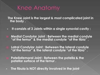
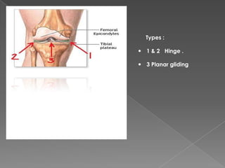
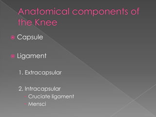
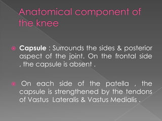
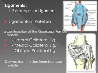
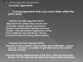
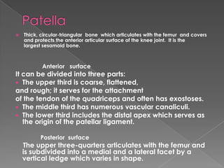
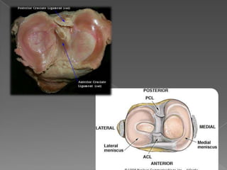
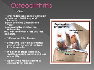
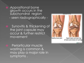
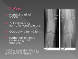
The Knee Joint is the largest & most complicated joint in
the body .
- It consists of 3 Joints within a single synovial cavity :
 Medial Condylar Joint : Between the medial condyle
“of the femur” & the medial condyle “of the tibia” .
 Latral Condylar Joint : Between the lateral condyle
“of the femur” & the lateral condyle “of the tibia” .
 Patellofemoral Joint : Between the patella & the
patellar surface of the femur .
- The fibula is NOT directly involved in the joint
Types :
 1 & 2 Hinge .
 3 Planar gliding
 Capsule
 Ligament
1. Extracapsular
2. Intracapsular
 Cruciate ligament
 Mensci
 Capsule : Surrounds the sides & posterior
aspect of the joint. On the frontal side
, the capsule is absent .
 On each side of the patella , the
capsule is strengthened by the tendons
of Vastus Lateralis & Vastus Medialis .
Ligaments :
1 .Extracapsular Ligaments :
 Ligamentum Patellea
a continuation of the Quariceps Femoris
muscle
 - Lateral Collateral Lig.
 - Medial Collateral Lig.
 - Oblique Popliteal Lig
Derived from the Semimembranosus
muscle
2. Intracapsular Ligaments :
Cruciate Ligaments :
2 strong ligaments that cross each other within the
joint cavity .
 Anterior Cruciate Ligament (ACL)
Attached to the anterior intercondylar area
of the tibia , passes upward , backward &
laterally to get attached to the lateral femoral
condyle . Prevents posterior displacement of the
femur (( With the knee joint flexed , the ACL
prevents the tibia from being pulled anteriorly
 Posterior Cruciate Ligament (PCL)
Attached to the posterior intercondylar area of the tibia , passes
upward , forward , & medially to get attached to the medial
femoral condyle .
Prevents anterior displacement of the femur With the knee joint
flexed , the PCL prevents the tibia from being pulled posteriorly .
 Thick, circular-triangular bone which articulates with the femur and covers
and protects the anterior articular surface of the knee joint. It is the
largest sesamoid bone.
Anterior surface
It can be divided into three parts:
 The upper third is coarse, flattened,
and rough; it serves for the attachment
of the tendon of the quadriceps and often has exostoses.
 The middle third has numerous vascular canaliculi.
 The lower third includes the distal apex which serves as
the origin of the patellar ligament.
Posterior surface
The upper three-quarters articulates with the femur and
is subdivided into a medial and a lateral facet by a
vertical ledge which varies in shape.
 It is attached to the tendon of the quadriceps femoris
muscle, which contracts to extend/straighten the knee.
The vastus intermedialis muscle is attached to the base of
patella. Thevastus latus lateralis and vastus medialis are
attached to lateral and medial borders of patella respectively.
 The knee is normally in slight valgus so there is a natural
tendency for the patella to pulled to the lateral side when the
quadriceps muscle is contracted
 The patella is stabilized by the insertion of vastus medialis and
the prominence of the anterior femoral condyles, which
prevent lateral dislocation during flexion.
 When injuries occur, all structures are simultaneously
affected.
These ligaments hold the patella in place
during static and dynamic phases.
 Femoral nerve :
› Fibular nerve
› Tibial nerve
Flexion : these muscles produce flexion :
Biceps femoris, Semitendinosus, Semimembranosus,
Gracilis, Sartorius, Popliteus .
Flexion is limited by the contact of the back of the leg with
the thigh .
- Extension by the Quadriceps femoris .
 Extension is limited by the tension of all the ligaments of
the joint .
- Medial Rotation : by the Sartorius , Gracilis , Semtendinosus.
 Lateral Rotation : by the Biceps femoris .
Clinic
 c/o: middle age patient complain
of pain starts insidiously and
increase
slowly over time ( months and
years)
aggravated by exertion and
relieved by
rest, with time relief is less and less
complete.
 Stiffness :mainly after rest
 Symptoms follow an intermittent
course with periods of remission
lasts for months
 In advance stage : deformity
,swelling, muscle wasting and loss
of mobility .
 No systemic manifestations in
contrast to inf. diseases.
 Osteoarthritis (OA) : a chronic inflammatory
joint disorder in which theres progressive
softening & destruction of the articular
cartilage, accompanied by new growth of
cartilage and bone at the joint margins
(osteophytes) and capsular fibrosis...
leading to bone exposure & severe pain .
 OA is the most common joint dis.
 The knee is the most common
 It can be primary or secondary :
Usually it’s Primary ( Idiopathic ) &
affecting both knee joints (Bilateral)
 Secondary causes might be :
Trauma , localized or metabolic
diseases , mechanical factors,
Bone Dysplasia , etc
 Trauma
 Congenital or developmental
 Metabolic
 Endocrine
 Calcium deposition diseases
 Other bone and joint diseases
 Neuropathic (Charcot joints)
 Endemic
 Miscellaneous
1. OA results from a
disparity between the stress
applied to the articular
cartilage & the ability of the
cartilage to withstand that
stress , due to :
› Weakening of the articular cartilage ( genetic defect in collagen type ll
or inflammatory disorder “RA” ) .
› Increased mechanical stress in some parts of the articular surface.
2. The abraded bone under a cartilage ulcer may take on the
appearance of ivory (eburnation = the bony sclerosis which
occurs at the areas of cartilage loss.). Growth of cartilage and
bone at the joint margins leads to osteophytes (spurs), which alter
the contour of the joint and may restrict movement
 Appositional bone
growth occurs in the
subchondral region
- seen radiographically -
 Synovitis & thickening of
the joint capsule may
occur & further restrict
movement
 Periarticular muscle
wasting is common &
may play a major role in
symptoms .
 Narrowing of joint
space.
 Subarticular cyst
formation and sclerosis.
 Osteophyte formation.
 Evidences of 2ndry
causes e.g. old
fracture.
The first two are restricted initially to the
major load-bearing part of the joint but
later the entire joint is affected.
Thanks for Attention

Knee muscles & movement

  • 2.
    The Knee Jointis the largest & most complicated joint in the body . - It consists of 3 Joints within a single synovial cavity :  Medial Condylar Joint : Between the medial condyle “of the femur” & the medial condyle “of the tibia” .  Latral Condylar Joint : Between the lateral condyle “of the femur” & the lateral condyle “of the tibia” .  Patellofemoral Joint : Between the patella & the patellar surface of the femur . - The fibula is NOT directly involved in the joint
  • 3.
    Types :  1& 2 Hinge .  3 Planar gliding
  • 4.
     Capsule  Ligament 1.Extracapsular 2. Intracapsular  Cruciate ligament  Mensci
  • 5.
     Capsule :Surrounds the sides & posterior aspect of the joint. On the frontal side , the capsule is absent .  On each side of the patella , the capsule is strengthened by the tendons of Vastus Lateralis & Vastus Medialis .
  • 6.
    Ligaments : 1 .ExtracapsularLigaments :  Ligamentum Patellea a continuation of the Quariceps Femoris muscle  - Lateral Collateral Lig.  - Medial Collateral Lig.  - Oblique Popliteal Lig Derived from the Semimembranosus muscle
  • 7.
    2. Intracapsular Ligaments: Cruciate Ligaments : 2 strong ligaments that cross each other within the joint cavity .  Anterior Cruciate Ligament (ACL) Attached to the anterior intercondylar area of the tibia , passes upward , backward & laterally to get attached to the lateral femoral condyle . Prevents posterior displacement of the femur (( With the knee joint flexed , the ACL prevents the tibia from being pulled anteriorly  Posterior Cruciate Ligament (PCL) Attached to the posterior intercondylar area of the tibia , passes upward , forward , & medially to get attached to the medial femoral condyle . Prevents anterior displacement of the femur With the knee joint flexed , the PCL prevents the tibia from being pulled posteriorly .
  • 8.
     Thick, circular-triangularbone which articulates with the femur and covers and protects the anterior articular surface of the knee joint. It is the largest sesamoid bone. Anterior surface It can be divided into three parts:  The upper third is coarse, flattened, and rough; it serves for the attachment of the tendon of the quadriceps and often has exostoses.  The middle third has numerous vascular canaliculi.  The lower third includes the distal apex which serves as the origin of the patellar ligament. Posterior surface The upper three-quarters articulates with the femur and is subdivided into a medial and a lateral facet by a vertical ledge which varies in shape.
  • 9.
     It isattached to the tendon of the quadriceps femoris muscle, which contracts to extend/straighten the knee. The vastus intermedialis muscle is attached to the base of patella. Thevastus latus lateralis and vastus medialis are attached to lateral and medial borders of patella respectively.  The knee is normally in slight valgus so there is a natural tendency for the patella to pulled to the lateral side when the quadriceps muscle is contracted  The patella is stabilized by the insertion of vastus medialis and the prominence of the anterior femoral condyles, which prevent lateral dislocation during flexion.  When injuries occur, all structures are simultaneously affected. These ligaments hold the patella in place during static and dynamic phases.
  • 11.
     Femoral nerve: › Fibular nerve › Tibial nerve
  • 12.
    Flexion : thesemuscles produce flexion : Biceps femoris, Semitendinosus, Semimembranosus, Gracilis, Sartorius, Popliteus . Flexion is limited by the contact of the back of the leg with the thigh . - Extension by the Quadriceps femoris .  Extension is limited by the tension of all the ligaments of the joint . - Medial Rotation : by the Sartorius , Gracilis , Semtendinosus.  Lateral Rotation : by the Biceps femoris .
  • 14.
    Clinic  c/o: middleage patient complain of pain starts insidiously and increase slowly over time ( months and years) aggravated by exertion and relieved by rest, with time relief is less and less complete.  Stiffness :mainly after rest  Symptoms follow an intermittent course with periods of remission lasts for months  In advance stage : deformity ,swelling, muscle wasting and loss of mobility .  No systemic manifestations in contrast to inf. diseases.
  • 15.
     Osteoarthritis (OA): a chronic inflammatory joint disorder in which theres progressive softening & destruction of the articular cartilage, accompanied by new growth of cartilage and bone at the joint margins (osteophytes) and capsular fibrosis... leading to bone exposure & severe pain .  OA is the most common joint dis.  The knee is the most common
  • 16.
     It canbe primary or secondary : Usually it’s Primary ( Idiopathic ) & affecting both knee joints (Bilateral)  Secondary causes might be : Trauma , localized or metabolic diseases , mechanical factors, Bone Dysplasia , etc
  • 17.
     Trauma  Congenitalor developmental  Metabolic  Endocrine  Calcium deposition diseases  Other bone and joint diseases  Neuropathic (Charcot joints)  Endemic  Miscellaneous
  • 18.
    1. OA resultsfrom a disparity between the stress applied to the articular cartilage & the ability of the cartilage to withstand that stress , due to : › Weakening of the articular cartilage ( genetic defect in collagen type ll or inflammatory disorder “RA” ) . › Increased mechanical stress in some parts of the articular surface. 2. The abraded bone under a cartilage ulcer may take on the appearance of ivory (eburnation = the bony sclerosis which occurs at the areas of cartilage loss.). Growth of cartilage and bone at the joint margins leads to osteophytes (spurs), which alter the contour of the joint and may restrict movement
  • 19.
     Appositional bone growthoccurs in the subchondral region - seen radiographically -  Synovitis & thickening of the joint capsule may occur & further restrict movement  Periarticular muscle wasting is common & may play a major role in symptoms .
  • 20.
     Narrowing ofjoint space.  Subarticular cyst formation and sclerosis.  Osteophyte formation.  Evidences of 2ndry causes e.g. old fracture. The first two are restricted initially to the major load-bearing part of the joint but later the entire joint is affected.
  • 21.