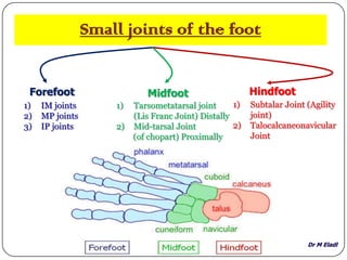
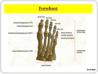
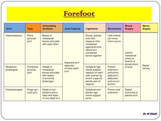
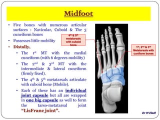
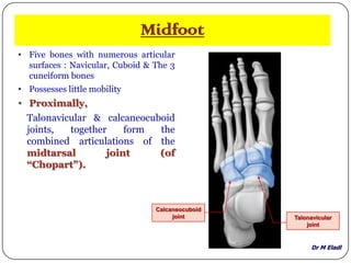
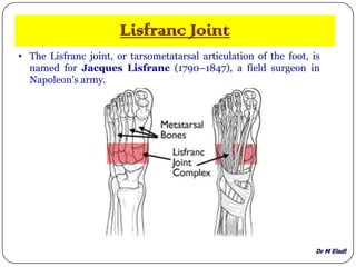
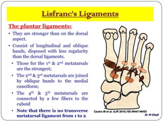
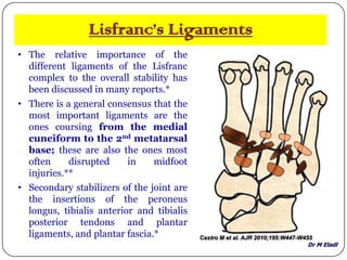
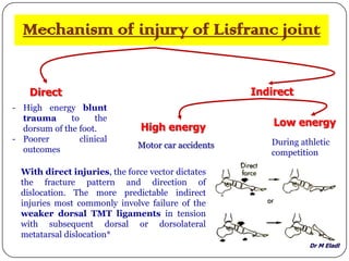
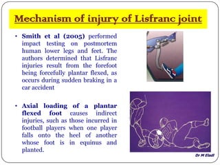
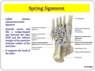
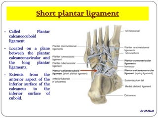
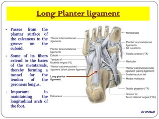
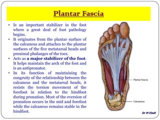
SlideShare a Scribd company logo
MOHAMED ELADL
01/02/2013
Dr M Eladl
Small joints of the foot
Forefoot Midfoot Hindfoot
1) IM joints
2) MP joints
3) IP joints
1) Tarsometatarsal joint
(Lis Franc Joint) Distally
2) Mid-tarsal Joint
(of chopart) Proximally
1) Subtalar Joint (Agility
joint)
2) Talocalcaneonavicular
Joint
Dr M Eladl
Forefoot
Dr M Eladl
Forefoot
Dr M Eladl
Midfoot
• Five bones with numerous articular
surfaces : Navicular, Cuboid & The 3
cuneiform bones
• Possesses little mobility
• Distally,
• The 1st MT with the medial
cuneiform (with 6 degrees mobility)
• The 2nd & 3rd MT with the
intermediate & lateral cuneiform
(firmly fixed).
• The 4th & 5th metatarsals articulate
with cuboid bone (Mobile).
• Each of these has an individual
joint capsule but all are wrapped
in one big capsule as well to form
the tarso-metatarsal joint
“LisFranc joint”.
1st, 2nd & 3rd
Metatarsals with
cuniform bones
4th & 5th
metatarsals
with cuboid
bone
Dr M Eladl
Midfoot
• Five bones with numerous articular
surfaces : Navicular, Cuboid & The 3
cuneiform bones
• Possesses little mobility
• Proximally,
Talonavicular & calcaneocuboid
joints, together form the
combined articulations of the
midtarsal joint (of
“Chopart”).
Talonavicular
joint
Calcaneocuboid
joint
Dr M Eladl
Lisfranc Joint
• The Lisfranc joint, or tarsometatarsal articulation of the foot, is
named for Jacques Lisfranc (1790–1847), a field surgeon in
Napoleon's army.
Dr M Eladl
Lisfranc Joint
• The Lisfranc joint, or tarsometatarsal articulation of the foot, is
named for Jacques Lisfranc (1790–1847), a field surgeon in
Napoleon's army.
• Lisfranc described an
amputation performed
through this joint because
of gangrene that developed
after an injury incurred
when a soldier fell off a
horse with his foot caught in
the stirrup.
• (their foot stuck in the
stirrup, they dislocated
portions of the mid-foot).
Dr M Eladl
Lisfranc Joint
• Some investigators have suggested
that the term “Lisfranc joint
complex” should be used to refer
to tarsometatarsal articulations
and that the term “Lisfranc
joint” should be applied to medial
articulation involving medial
(first) and middle (second)
cuneiforms with the first and
second metatarsals .*
• The Lisfranc joint complex has
three separate articular
capsules, creating three distinct
articular compartments: medial,
central, and lateral lacking
communication with one another
(**).
Dr M Eladl
Lisfranc’s Ligaments
• The joint capsule and TMT ligaments
provide limited soft-tissue support to the
Lisfranc joint.
• Ligaments are grouped according to
anatomic location (dorsal, plantar, and
interosseous)
The dorsal ligaments:
Are strong, flat bands.
• The medial cuneiform is joined to the 1st
metatarsal the by a broad, thin band;
• The 2nd metatarsal has 3 ligaments; one
from each cuneiform
• The 3rd metatarsal has one ligament from
the lateral cuneiform;
• The 4th has one from the cuboid; and
• The 5th has one from the cuboid.
Castro M et al. AJR 2010;195:W447-W455
Dr M Eladl
Lisfranc’s Ligaments
Castro M et al. AJR 2010;195:W447-W455
Dorsal ligaments of Lisfranc joint complex. Sagittal proton density MR image after
intraarticular injection of gadolinium solution (B) and photograph of
corresponding gross anatomic section (C) in cadaveric specimen show short and
flat dorsal tarsometatarsal ligament between medial cuniform and 1st metatarsal
(arrows). Note homogeneous low signal intensity at MR image.
Dr M Eladl
Lisfranc’s Ligaments
The plantar ligaments:
• They are stronger than on the dorsal
aspect.
• Consist of longitudinal and oblique
bands, disposed with less regularity
than the dorsal ligaments.
• Those for the 1st & 2nd metatarsals
are the strongest;
• The 2nd & 3rd metatarsals are joined
by oblique bands to the medial
cuneiform;
• The 4th & 5th metatarsals are
connected by a few fibers to the
cuboid
• Note that there is no transverse
metatarsal ligament from 1 to 2
Castro M et al. AJR 2010;195:W447-W455
Dr M Eladl
Lisfranc’s Ligaments
• The relative importance of the
different ligaments of the Lisfranc
complex to the overall stability has
been discussed in many reports.*
• There is a general consensus that the
most important ligaments are the
ones coursing from the medial
cuneiform to the 2nd metatarsal
base; these are also the ones most
often disrupted in midfoot
injuries.**
• Secondary stabilizers of the joint are
the insertions of the peroneus
longus, tibialis anterior and tibialis
posterior tendons and plantar
ligaments, and plantar fascia.* Castro M et al. AJR 2010;195:W447-W455
Dr M Eladl
Mechanism of injury of Lisfranc joint
Direct Indirect
- High energy blunt
trauma to the
dorsum of the foot.
- Poorer clinical
outcomes Motor car accidents
High energy Low energy
During athletic
competition
With direct injuries, the force vector dictates
the fracture pattern and direction of
dislocation. The more predictable indirect
injuries most commonly involve failure of the
weaker dorsal TMT ligaments in tension
with subsequent dorsal or dorsolateral
metatarsal dislocation*
Dr M Eladl
Mechanism of injury of Lisfranc joint
• Smith et al (2005) performed
impact testing on postmortem
human lower legs and feet. The
authors determined that Lisfranc
injuries result from the forefoot
being forcefully plantar flexed, as
occurs during sudden braking in a
car accident
• Axial loading of a plantar
flexed foot causes indirect
injuries, such as those incurred in
football players when one player
falls onto the heel of another
whose foot is in equinus and
planted.
Dr M Eladl
Midtarsal joint (of “Chopart”).
Is a compound joint formed by 2
separate joints aligned
transversely:
1) Talonavicular part of the
talo-calcaneo-navicular joint
(Ball & Socket) and
2) Calcaneo-cuboid joint
(Saddle).
Dr M Eladl
Midtarsal joint (of “Chopart”).
- At this joint, the midfoot and
forefoot rotate as a unit on the
hindfoot around a longitudinal
(AP) axis, augmenting the
inversion and eversion
movements occurring at the
clinical subtalar joint.
- Transection across the transverse
tarsal joint is a standard method
for surgical amputation of the
foot.
- The complex anatomy of the
Chopart joint, optimally adapting
to uneven surfaces upon first heel
contact & forcefully pushing the
foot off the ground at the end of
the walk cycle.
Dr M Eladl
Subtalar Joint
• known as the “Agility joint”.
• Is between: the large posterior
calcaneal facet on the inferior
surface of the talus & the
corresponding posterior talar
facet on superior surface of
calcaneus
• The articular cavity is enclosed by
synovial membrane, which is
covered by a fibrous capsule.
• Allows gliding and rotation, which
are involved in inversion and
eversion of the foot.
Dr M Eladl
Subtalar Joint ligaments
Lateral, medial, posterior, and interosseous talocalcaneal ligaments
stabilize the joint.
Daftary A et al. Radiographics 2005;25:1215-1226
Drawing of the lateral surface of the calcaneus shows the peroneal tubercle (P), as well as the
lateral talocalcaneal (LTL), interosseous (IOL), and bifurcate (B) ligaments.
Dr M Eladl
TaloCalcaneoNavicular Joint
• A complex joint in which the head of
talus articulates with calcaneus &
plantar calcaneonavicular ligament
(spring ligament) below & the
navicular in front
Dr M Eladl
TaloCalcaneoNavicular Joint
• A complex joint in which the head of
talus articulates with calcaneus &
plantar calcaneonavicular ligament
(spring ligament) below & the
navicular in front
• Allows gliding & rotation movements,
which together with subtalar joint are
involved with inversion & eversion
of the foot.
Dr M Eladl
TALOCALCANEONAVICULAR JOINT
LIGAMENTS
• Talonavicular ligament: superiorly, which passes between the
neck of the talus and adjacent regions of the navicular;
• Interosseous talocalcaneal ligament: posteriorly;
Dr M Eladl
TALOCALCANEONAVICULAR JOINT
LIGAMENTS
• Talonavicular ligament: superiorly by the, which passes between
the neck of the talus and adjacent regions of the navicular;
• Interosseous talocalcaneal ligament: posteriorly;
• Plantar calcaneonavicular ligament (Spring): inferiorly
Dr M Eladl
Spring ligament
- Called plantar
calcaneonavicular
ligament
- Extends across and
fills a wedge-shaped
gap between the talar
shelf and the inferior
margin of the posterior
articular surface of the
navicular
- It supports the head of
the talus
Dr M Eladl
Short plantar ligament
- Called Plantar
calcaneocuboid
ligament
- Located on a plane
between the plantar
calcaneonavicular and
the long plantar
ligaments.
- Extends from the
anterior aspect of the
inferior surface of the
calcaneus to the
inferior surface of
cuboid.
Dr M Eladl
Long Planter ligament
- Passes from the
plantar surface of
the calcaneus to the
groove on the
cuboid.
- Some of its fibers
extend to the bases
of the metatarsals,
thereby forming a
tunnel for the
tendon of the
peroneus longus.
- Important in
maintaining the
longitudinal arch of
the foot.
Dr M Eladl
Plantar Fascia
• Is an important stabilizer in the foot
where a great deal of foot pathology
begins.
• It originates from the plantar surface of
the calcaneus and attaches to the plantar
surfaces of the five metatarsal heads and
proximal phalanges of the toes.
• Acts as a major stabilizer of the foot.
It helps maintain the arch of the foot and
is an antipronator.
• In its function of maintaining the
congruity of the relationship between the
calcaneus and the metatarsal heads, it
resists the torsion movement of the
forefoot in relation to the hindfoot
during pronation. Most of the eversion of
pronation occurs in the mid and forefoot
while the calcaneus remains stable in the
hindfoot.
Dr M Eladl
References
1) Moore KL et al. (2010): Clinically Orientated Anatomy. 6th Ed. Lippincott, Williams & Wilkins. Philadelphia.
2) Castro, M., L. Melao, et al.(2010) "Lisfranc joint ligamentous complex: MRI with anatomic correlation in
cadavers." AJR Am J Roentgenol 195(6): W447-55.
3) Vuori JP, Aro HT. Lisfranc joint injuries: trauma mechanisms and associated injuries. J
Trauma. 1993;35:40–5.
4) Englanoff G, Anglin D, Hutson HR. Lisfranc fracture-dislocation: a frequently missed diagnosis in the
emergency department. Ann Emerg Med. 1995;26:229–33.
5) Myerson M. The diagnosis and treatment of injuries to the Lisfranc joint complex. Orthop Clin North
Am. 1989;20:655–64.
6) Burroughs, K. E., C. D. Reimer, et al. (1998). "Lisfranc injury of the foot: a commonly missed diagnosis." Am
Fam Physician 58(1): 118-24.
7) Schulze, W; Richter, J; Russe, O; Ingelfinger, P; Muhr, G: Surgical treatment of talus fractures: a retrospective
study of 80 cases followed for 1–15 years. Acta Orthop. Scand. 73:344 –351, 2002.
8) Suckel, A., O. Muller, et al. (2007). "Changes in Chopart joint load following tibiotalar arthrodesis: in vitro
analysis of 8 cadaver specimens in a dynamic model." BMC Musculoskelet Disord 8: 80.
9) http://www.sportsinjuryclinic.net/anatomy/ankle-anatomy
10) http://www.footeducation.com/ligaments-of-foot-and-ankle-overview
11) Preidler KW, Wang YC, Brossmann J, Trudell D, Daenen B, Resnick D. Tarsometatarsal joint: anatomic details
on MR images. Radiology 1996; 99:733–736
12) Delfaut EM, Rosenberg ZS, Demondion X. Malalignment at Lisfranc joint: MR features in asymptomatic
patients and cadaveric specimens. Skeletal Radiol 2002; 31:499–504
13) Chiodo CP, Myerson M. Developments and advances in the diagnosis and treatment of injuries to the
tarsometatarsal joint. Orthop Clin North Am 2001; 32:11–20
14) Kura H, Luo Z, Kitaoka HB, Smutz WP, An K. Mechanical behavior of Lisfranc and dorsal cuneometatarsal
ligaments: in vitro biomechanical study. J Orthop Trauma 2001; 15:107–110

More Related Content

What's hot

Ankle joint Anatomy
Ankle joint AnatomyAnkle joint Anatomy
Ankle joint Anatomy
dr.supriti verma bhatnagar
 
Slideshow: Rotator Cuff
Slideshow: Rotator CuffSlideshow: Rotator Cuff
Slideshow: Rotator Cuff
The Funky Professor
 
The ankle joint
The  ankle jointThe  ankle joint
The ankle joint
Idris Siddiqui
 
Tarsal, Metatarsal and Phalanges of the Foot
Tarsal, Metatarsal and Phalanges of the FootTarsal, Metatarsal and Phalanges of the Foot
Tarsal, Metatarsal and Phalanges of the Foot
Sado Anatomist
 
Posterior compartment of leg
Posterior compartment of legPosterior compartment of leg
Posterior compartment of leg
Aunum Iqbal
 
5.posterior compartment of the thigh
5.posterior compartment of the thigh5.posterior compartment of the thigh
5.posterior compartment of the thigh
Dr. Mohammad Mahmoud
 
Slideshow: The Foot
Slideshow: The FootSlideshow: The Foot
Slideshow: The Foot
The Funky Professor
 
9. anatomy of the foot
9. anatomy of the foot9. anatomy of the foot
9. anatomy of the foot
Dr. Mohammad Mahmoud
 
4. wrist and hand joints
4. wrist and hand joints4. wrist and hand joints
4. wrist and hand joints
Dr. Mohammad Mahmoud
 
Knee Joint Anatomy
Knee Joint AnatomyKnee Joint Anatomy
Knee Joint Anatomy
Kommireddy Kumar
 
Arches of foot
Arches  of  footArches  of  foot
Arches of foot
harsha2063
 
The wrist joint
The wrist jointThe wrist joint
The wrist joint
Idris Siddiqui
 
Anatomy of knee joint
Anatomy of knee jointAnatomy of knee joint
Anatomy of knee joint
BipulBorthakur
 
Muscles of thigh
Muscles of thighMuscles of thigh
Muscles of thigh
Idris Siddiqui
 
Knee joint
Knee jointKnee joint
Knee joint
Idris Siddiqui
 
Hip joint
Hip jointHip joint
Hip joint
Idris Siddiqui
 
Sciatic nerve
Sciatic nerveSciatic nerve
Sciatic nerve
Idris Siddiqui
 
Hip joint anatomy
Hip joint anatomyHip joint anatomy
Hip joint anatomy
Ruhul Amin
 
Ankle joint
Ankle jointAnkle joint
Ankle joint
Rahul Jha
 

What's hot (20)

Ankle joint Anatomy
Ankle joint AnatomyAnkle joint Anatomy
Ankle joint Anatomy
 
Slideshow: Rotator Cuff
Slideshow: Rotator CuffSlideshow: Rotator Cuff
Slideshow: Rotator Cuff
 
The ankle joint
The  ankle jointThe  ankle joint
The ankle joint
 
Tarsal, Metatarsal and Phalanges of the Foot
Tarsal, Metatarsal and Phalanges of the FootTarsal, Metatarsal and Phalanges of the Foot
Tarsal, Metatarsal and Phalanges of the Foot
 
Posterior compartment of leg
Posterior compartment of legPosterior compartment of leg
Posterior compartment of leg
 
5.posterior compartment of the thigh
5.posterior compartment of the thigh5.posterior compartment of the thigh
5.posterior compartment of the thigh
 
Thigh
ThighThigh
Thigh
 
Slideshow: The Foot
Slideshow: The FootSlideshow: The Foot
Slideshow: The Foot
 
9. anatomy of the foot
9. anatomy of the foot9. anatomy of the foot
9. anatomy of the foot
 
4. wrist and hand joints
4. wrist and hand joints4. wrist and hand joints
4. wrist and hand joints
 
Knee Joint Anatomy
Knee Joint AnatomyKnee Joint Anatomy
Knee Joint Anatomy
 
Arches of foot
Arches  of  footArches  of  foot
Arches of foot
 
The wrist joint
The wrist jointThe wrist joint
The wrist joint
 
Anatomy of knee joint
Anatomy of knee jointAnatomy of knee joint
Anatomy of knee joint
 
Muscles of thigh
Muscles of thighMuscles of thigh
Muscles of thigh
 
Knee joint
Knee jointKnee joint
Knee joint
 
Hip joint
Hip jointHip joint
Hip joint
 
Sciatic nerve
Sciatic nerveSciatic nerve
Sciatic nerve
 
Hip joint anatomy
Hip joint anatomyHip joint anatomy
Hip joint anatomy
 
Ankle joint
Ankle jointAnkle joint
Ankle joint
 

Viewers also liked

Anatomy of the ankle and joints of foot
Anatomy of the ankle and joints of footAnatomy of the ankle and joints of foot
Anatomy of the ankle and joints of foot
Akram Jaffar
 
1. Biomechanics of ankle joint subtalar joint and foot
1. Biomechanics of ankle joint subtalar joint and foot1. Biomechanics of ankle joint subtalar joint and foot
1. Biomechanics of ankle joint subtalar joint and foot
Saurab Sharma
 
Anatomy of Feet
Anatomy of Feet Anatomy of Feet
Anatomy of Feet
Ammedicine Medicine
 
Lis Franc Injury
Lis Franc InjuryLis Franc Injury
Lis Franc Injuryjfreshour
 
Lisfranc injury
Lisfranc injuryLisfranc injury
Lisfranc injury
Subodh Pathak
 
Anatomy of foot and ankle
Anatomy of foot and ankleAnatomy of foot and ankle
Anatomy of foot and ankle
Junaid Ahmad
 
Lisfranc
LisfrancLisfranc
Lisfranc
vickydeepu05
 
Foot and Ankle Anatomy
Foot and Ankle AnatomyFoot and Ankle Anatomy
Foot and Ankle Anatomy
northcoastfootcare
 
Lisfranc fractures
Lisfranc fracturesLisfranc fractures
Lisfranc fracturesorthoprince
 
Kienbock’s disease
Kienbock’s diseaseKienbock’s disease
Kienbock’s diseaseorthoprince
 
Mid foot lisfranc fracture
Mid foot lisfranc fractureMid foot lisfranc fracture
Mid foot lisfranc fractureAbhishek Sachdev
 
Pudendal Neuralgia and Lower Extremity (Feet) Biomechanics - ICS 2011, Glasg...
Pudendal Neuralgia and Lower Extremity (Feet) Biomechanics - ICS  2011, Glasg...Pudendal Neuralgia and Lower Extremity (Feet) Biomechanics - ICS  2011, Glasg...
Pudendal Neuralgia and Lower Extremity (Feet) Biomechanics - ICS 2011, Glasg...
Cristina Shupe
 
L16 lisfranc & midfoot inj
L16 lisfranc & midfoot injL16 lisfranc & midfoot inj
L16 lisfranc & midfoot inj
Claudiu Cucu
 
Dance
DanceDance
Saphenous Vein Harvesting
Saphenous Vein HarvestingSaphenous Vein Harvesting
Saphenous Vein Harvesting
Mohamad Fadzly Mohamad Yusof
 

Viewers also liked (20)

Anatomy of the ankle and joints of foot
Anatomy of the ankle and joints of footAnatomy of the ankle and joints of foot
Anatomy of the ankle and joints of foot
 
Anatomy of the foot
Anatomy of the footAnatomy of the foot
Anatomy of the foot
 
Foot anatomy ppt
Foot anatomy pptFoot anatomy ppt
Foot anatomy ppt
 
1. Biomechanics of ankle joint subtalar joint and foot
1. Biomechanics of ankle joint subtalar joint and foot1. Biomechanics of ankle joint subtalar joint and foot
1. Biomechanics of ankle joint subtalar joint and foot
 
Anatomy of Feet
Anatomy of Feet Anatomy of Feet
Anatomy of Feet
 
Lis Franc Injury
Lis Franc InjuryLis Franc Injury
Lis Franc Injury
 
Lisfranc injury
Lisfranc injuryLisfranc injury
Lisfranc injury
 
Anatomy of foot and ankle
Anatomy of foot and ankleAnatomy of foot and ankle
Anatomy of foot and ankle
 
Lisfranc
LisfrancLisfranc
Lisfranc
 
Lecture 15 parekh lisfranc
Lecture 15 parekh lisfrancLecture 15 parekh lisfranc
Lecture 15 parekh lisfranc
 
Foot and Ankle Anatomy
Foot and Ankle AnatomyFoot and Ankle Anatomy
Foot and Ankle Anatomy
 
Lisfranc fractures
Lisfranc fracturesLisfranc fractures
Lisfranc fractures
 
Kienbock’s disease
Kienbock’s diseaseKienbock’s disease
Kienbock’s disease
 
Mid foot lisfranc fracture
Mid foot lisfranc fractureMid foot lisfranc fracture
Mid foot lisfranc fracture
 
Pudendal Neuralgia and Lower Extremity (Feet) Biomechanics - ICS 2011, Glasg...
Pudendal Neuralgia and Lower Extremity (Feet) Biomechanics - ICS  2011, Glasg...Pudendal Neuralgia and Lower Extremity (Feet) Biomechanics - ICS  2011, Glasg...
Pudendal Neuralgia and Lower Extremity (Feet) Biomechanics - ICS 2011, Glasg...
 
L16 lisfranc & midfoot inj
L16 lisfranc & midfoot injL16 lisfranc & midfoot inj
L16 lisfranc & midfoot inj
 
Dance
DanceDance
Dance
 
Saphenous Vein Harvesting
Saphenous Vein HarvestingSaphenous Vein Harvesting
Saphenous Vein Harvesting
 
Talar Fracture
Talar FractureTalar Fracture
Talar Fracture
 
Esophagitis
EsophagitisEsophagitis
Esophagitis
 

Similar to Anatomy of small joints of the foot

Anatomy of the anterior & lateral compartments of the leg
Anatomy of the anterior & lateral compartments of the legAnatomy of the anterior & lateral compartments of the leg
Anatomy of the anterior & lateral compartments of the legMohamed Ahmed Eladl
 
The foot
The footThe foot
The foot
Buyungo Steven
 
805_Front-of-Forearm.pptx
805_Front-of-Forearm.pptx805_Front-of-Forearm.pptx
805_Front-of-Forearm.pptx
GUNASEKARANM20
 
ARTICULAR DISC OF TEMPOROMANDIBULAR JOINT
ARTICULAR DISC OF TEMPOROMANDIBULAR JOINTARTICULAR DISC OF TEMPOROMANDIBULAR JOINT
ARTICULAR DISC OF TEMPOROMANDIBULAR JOINT
DR. SNEHA NAIR
 
Bones of hand and anatomical spaceas
Bones of hand and anatomical spaceasBones of hand and anatomical spaceas
Bones of hand and anatomical spaceas
Eimaan Ktk
 
Ankel joint
Ankel jointAnkel joint
04 ankle fractures ppt
04 ankle fractures ppt04 ankle fractures ppt
04 ankle fractures ppt
yashavardhan yashu
 
Arches of the foot and plantar fascitiis
Arches of the foot and plantar fascitiisArches of the foot and plantar fascitiis
Arches of the foot and plantar fascitiis
Dr Nirav Mungalpara
 
Biomechanics of ankle and foot
Biomechanics of ankle and footBiomechanics of ankle and foot
Biomechanics of ankle and foot
AragyaKhadka
 
Lisfranc injuries
Lisfranc injuriesLisfranc injuries
Lisfranc injuries
Praveen Kumar Reddy Gorantla
 
Ankle joint
Ankle  jointAnkle  joint
Hand anatomy 2
Hand anatomy 2 Hand anatomy 2
Hand anatomy 2
Mahmoud Gouda
 
Foot biomechanics
Foot biomechanicsFoot biomechanics
Foot biomechanics
deepakanap
 
Dr Anuj Dr Tarun LISFRANC INJURY.pdf
Dr Anuj Dr Tarun LISFRANC INJURY.pdfDr Anuj Dr Tarun LISFRANC INJURY.pdf
Dr Anuj Dr Tarun LISFRANC INJURY.pdf
anuj700257
 
Foot and ankle anatomy & biomechanics.pptx
Foot and ankle anatomy & biomechanics.pptxFoot and ankle anatomy & biomechanics.pptx
Foot and ankle anatomy & biomechanics.pptx
NtambaraNelson
 
Distal femur fracture
Distal femur fractureDistal femur fracture
Distal femur fracture
Dr Sharanprasad Hongal
 
distalfemur-170720141254.pdf
distalfemur-170720141254.pdfdistalfemur-170720141254.pdf
distalfemur-170720141254.pdf
DrShubhamNagdev
 
Anatomy and imaging of knee joint
Anatomy and imaging of knee jointAnatomy and imaging of knee joint
Anatomy and imaging of knee joint
Kajal Jha
 
Foot Anatomy.ppt
Foot Anatomy.pptFoot Anatomy.ppt
Foot Anatomy.ppt
JosephBirch5
 

Similar to Anatomy of small joints of the foot (20)

Anatomy of the anterior & lateral compartments of the leg
Anatomy of the anterior & lateral compartments of the legAnatomy of the anterior & lateral compartments of the leg
Anatomy of the anterior & lateral compartments of the leg
 
The foot
The footThe foot
The foot
 
805_Front-of-Forearm.pptx
805_Front-of-Forearm.pptx805_Front-of-Forearm.pptx
805_Front-of-Forearm.pptx
 
ARTICULAR DISC OF TEMPOROMANDIBULAR JOINT
ARTICULAR DISC OF TEMPOROMANDIBULAR JOINTARTICULAR DISC OF TEMPOROMANDIBULAR JOINT
ARTICULAR DISC OF TEMPOROMANDIBULAR JOINT
 
Bones of hand and anatomical spaceas
Bones of hand and anatomical spaceasBones of hand and anatomical spaceas
Bones of hand and anatomical spaceas
 
Ankel joint
Ankel jointAnkel joint
Ankel joint
 
04 ankle fractures ppt
04 ankle fractures ppt04 ankle fractures ppt
04 ankle fractures ppt
 
Arches of the foot and plantar fascitiis
Arches of the foot and plantar fascitiisArches of the foot and plantar fascitiis
Arches of the foot and plantar fascitiis
 
Biomechanics of ankle and foot
Biomechanics of ankle and footBiomechanics of ankle and foot
Biomechanics of ankle and foot
 
Lisfranc injuries
Lisfranc injuriesLisfranc injuries
Lisfranc injuries
 
Ankle joint
Ankle  jointAnkle  joint
Ankle joint
 
Hand anatomy 2
Hand anatomy 2 Hand anatomy 2
Hand anatomy 2
 
Foot biomechanics
Foot biomechanicsFoot biomechanics
Foot biomechanics
 
Anatomy of the Knee Joint
Anatomy of the Knee JointAnatomy of the Knee Joint
Anatomy of the Knee Joint
 
Dr Anuj Dr Tarun LISFRANC INJURY.pdf
Dr Anuj Dr Tarun LISFRANC INJURY.pdfDr Anuj Dr Tarun LISFRANC INJURY.pdf
Dr Anuj Dr Tarun LISFRANC INJURY.pdf
 
Foot and ankle anatomy & biomechanics.pptx
Foot and ankle anatomy & biomechanics.pptxFoot and ankle anatomy & biomechanics.pptx
Foot and ankle anatomy & biomechanics.pptx
 
Distal femur fracture
Distal femur fractureDistal femur fracture
Distal femur fracture
 
distalfemur-170720141254.pdf
distalfemur-170720141254.pdfdistalfemur-170720141254.pdf
distalfemur-170720141254.pdf
 
Anatomy and imaging of knee joint
Anatomy and imaging of knee jointAnatomy and imaging of knee joint
Anatomy and imaging of knee joint
 
Foot Anatomy.ppt
Foot Anatomy.pptFoot Anatomy.ppt
Foot Anatomy.ppt
 

Recently uploaded

Cambridge International AS A Level Biology Coursebook - EBook (MaryFosbery J...
Cambridge International AS  A Level Biology Coursebook - EBook (MaryFosbery J...Cambridge International AS  A Level Biology Coursebook - EBook (MaryFosbery J...
Cambridge International AS A Level Biology Coursebook - EBook (MaryFosbery J...
AzmatAli747758
 
The approach at University of Liverpool.pptx
The approach at University of Liverpool.pptxThe approach at University of Liverpool.pptx
The approach at University of Liverpool.pptx
Jisc
 
Home assignment II on Spectroscopy 2024 Answers.pdf
Home assignment II on Spectroscopy 2024 Answers.pdfHome assignment II on Spectroscopy 2024 Answers.pdf
Home assignment II on Spectroscopy 2024 Answers.pdf
Tamralipta Mahavidyalaya
 
MARUTI SUZUKI- A Successful Joint Venture in India.pptx
MARUTI SUZUKI- A Successful Joint Venture in India.pptxMARUTI SUZUKI- A Successful Joint Venture in India.pptx
MARUTI SUZUKI- A Successful Joint Venture in India.pptx
bennyroshan06
 
Unit 8 - Information and Communication Technology (Paper I).pdf
Unit 8 - Information and Communication Technology (Paper I).pdfUnit 8 - Information and Communication Technology (Paper I).pdf
Unit 8 - Information and Communication Technology (Paper I).pdf
Thiyagu K
 
ESC Beyond Borders _From EU to You_ InfoPack general.pdf
ESC Beyond Borders _From EU to You_ InfoPack general.pdfESC Beyond Borders _From EU to You_ InfoPack general.pdf
ESC Beyond Borders _From EU to You_ InfoPack general.pdf
Fundacja Rozwoju Społeczeństwa Przedsiębiorczego
 
Ethnobotany and Ethnopharmacology ......
Ethnobotany and Ethnopharmacology ......Ethnobotany and Ethnopharmacology ......
Ethnobotany and Ethnopharmacology ......
Ashokrao Mane college of Pharmacy Peth-Vadgaon
 
2024.06.01 Introducing a competency framework for languag learning materials ...
2024.06.01 Introducing a competency framework for languag learning materials ...2024.06.01 Introducing a competency framework for languag learning materials ...
2024.06.01 Introducing a competency framework for languag learning materials ...
Sandy Millin
 
Overview on Edible Vaccine: Pros & Cons with Mechanism
Overview on Edible Vaccine: Pros & Cons with MechanismOverview on Edible Vaccine: Pros & Cons with Mechanism
Overview on Edible Vaccine: Pros & Cons with Mechanism
DeeptiGupta154
 
The Challenger.pdf DNHS Official Publication
The Challenger.pdf DNHS Official PublicationThe Challenger.pdf DNHS Official Publication
The Challenger.pdf DNHS Official Publication
Delapenabediema
 
Students, digital devices and success - Andreas Schleicher - 27 May 2024..pptx
Students, digital devices and success - Andreas Schleicher - 27 May 2024..pptxStudents, digital devices and success - Andreas Schleicher - 27 May 2024..pptx
Students, digital devices and success - Andreas Schleicher - 27 May 2024..pptx
EduSkills OECD
 
Chapter 3 - Islamic Banking Products and Services.pptx
Chapter 3 - Islamic Banking Products and Services.pptxChapter 3 - Islamic Banking Products and Services.pptx
Chapter 3 - Islamic Banking Products and Services.pptx
Mohd Adib Abd Muin, Senior Lecturer at Universiti Utara Malaysia
 
Phrasal Verbs.XXXXXXXXXXXXXXXXXXXXXXXXXX
Phrasal Verbs.XXXXXXXXXXXXXXXXXXXXXXXXXXPhrasal Verbs.XXXXXXXXXXXXXXXXXXXXXXXXXX
Phrasal Verbs.XXXXXXXXXXXXXXXXXXXXXXXXXX
MIRIAMSALINAS13
 
Synthetic Fiber Construction in lab .pptx
Synthetic Fiber Construction in lab .pptxSynthetic Fiber Construction in lab .pptx
Synthetic Fiber Construction in lab .pptx
Pavel ( NSTU)
 
Supporting (UKRI) OA monographs at Salford.pptx
Supporting (UKRI) OA monographs at Salford.pptxSupporting (UKRI) OA monographs at Salford.pptx
Supporting (UKRI) OA monographs at Salford.pptx
Jisc
 
The French Revolution Class 9 Study Material pdf free download
The French Revolution Class 9 Study Material pdf free downloadThe French Revolution Class 9 Study Material pdf free download
The French Revolution Class 9 Study Material pdf free download
Vivekanand Anglo Vedic Academy
 
Instructions for Submissions thorugh G- Classroom.pptx
Instructions for Submissions thorugh G- Classroom.pptxInstructions for Submissions thorugh G- Classroom.pptx
Instructions for Submissions thorugh G- Classroom.pptx
Jheel Barad
 
How libraries can support authors with open access requirements for UKRI fund...
How libraries can support authors with open access requirements for UKRI fund...How libraries can support authors with open access requirements for UKRI fund...
How libraries can support authors with open access requirements for UKRI fund...
Jisc
 
How to Break the cycle of negative Thoughts
How to Break the cycle of negative ThoughtsHow to Break the cycle of negative Thoughts
How to Break the cycle of negative Thoughts
Col Mukteshwar Prasad
 
Basic phrases for greeting and assisting costumers
Basic phrases for greeting and assisting costumersBasic phrases for greeting and assisting costumers
Basic phrases for greeting and assisting costumers
PedroFerreira53928
 

Recently uploaded (20)

Cambridge International AS A Level Biology Coursebook - EBook (MaryFosbery J...
Cambridge International AS  A Level Biology Coursebook - EBook (MaryFosbery J...Cambridge International AS  A Level Biology Coursebook - EBook (MaryFosbery J...
Cambridge International AS A Level Biology Coursebook - EBook (MaryFosbery J...
 
The approach at University of Liverpool.pptx
The approach at University of Liverpool.pptxThe approach at University of Liverpool.pptx
The approach at University of Liverpool.pptx
 
Home assignment II on Spectroscopy 2024 Answers.pdf
Home assignment II on Spectroscopy 2024 Answers.pdfHome assignment II on Spectroscopy 2024 Answers.pdf
Home assignment II on Spectroscopy 2024 Answers.pdf
 
MARUTI SUZUKI- A Successful Joint Venture in India.pptx
MARUTI SUZUKI- A Successful Joint Venture in India.pptxMARUTI SUZUKI- A Successful Joint Venture in India.pptx
MARUTI SUZUKI- A Successful Joint Venture in India.pptx
 
Unit 8 - Information and Communication Technology (Paper I).pdf
Unit 8 - Information and Communication Technology (Paper I).pdfUnit 8 - Information and Communication Technology (Paper I).pdf
Unit 8 - Information and Communication Technology (Paper I).pdf
 
ESC Beyond Borders _From EU to You_ InfoPack general.pdf
ESC Beyond Borders _From EU to You_ InfoPack general.pdfESC Beyond Borders _From EU to You_ InfoPack general.pdf
ESC Beyond Borders _From EU to You_ InfoPack general.pdf
 
Ethnobotany and Ethnopharmacology ......
Ethnobotany and Ethnopharmacology ......Ethnobotany and Ethnopharmacology ......
Ethnobotany and Ethnopharmacology ......
 
2024.06.01 Introducing a competency framework for languag learning materials ...
2024.06.01 Introducing a competency framework for languag learning materials ...2024.06.01 Introducing a competency framework for languag learning materials ...
2024.06.01 Introducing a competency framework for languag learning materials ...
 
Overview on Edible Vaccine: Pros & Cons with Mechanism
Overview on Edible Vaccine: Pros & Cons with MechanismOverview on Edible Vaccine: Pros & Cons with Mechanism
Overview on Edible Vaccine: Pros & Cons with Mechanism
 
The Challenger.pdf DNHS Official Publication
The Challenger.pdf DNHS Official PublicationThe Challenger.pdf DNHS Official Publication
The Challenger.pdf DNHS Official Publication
 
Students, digital devices and success - Andreas Schleicher - 27 May 2024..pptx
Students, digital devices and success - Andreas Schleicher - 27 May 2024..pptxStudents, digital devices and success - Andreas Schleicher - 27 May 2024..pptx
Students, digital devices and success - Andreas Schleicher - 27 May 2024..pptx
 
Chapter 3 - Islamic Banking Products and Services.pptx
Chapter 3 - Islamic Banking Products and Services.pptxChapter 3 - Islamic Banking Products and Services.pptx
Chapter 3 - Islamic Banking Products and Services.pptx
 
Phrasal Verbs.XXXXXXXXXXXXXXXXXXXXXXXXXX
Phrasal Verbs.XXXXXXXXXXXXXXXXXXXXXXXXXXPhrasal Verbs.XXXXXXXXXXXXXXXXXXXXXXXXXX
Phrasal Verbs.XXXXXXXXXXXXXXXXXXXXXXXXXX
 
Synthetic Fiber Construction in lab .pptx
Synthetic Fiber Construction in lab .pptxSynthetic Fiber Construction in lab .pptx
Synthetic Fiber Construction in lab .pptx
 
Supporting (UKRI) OA monographs at Salford.pptx
Supporting (UKRI) OA monographs at Salford.pptxSupporting (UKRI) OA monographs at Salford.pptx
Supporting (UKRI) OA monographs at Salford.pptx
 
The French Revolution Class 9 Study Material pdf free download
The French Revolution Class 9 Study Material pdf free downloadThe French Revolution Class 9 Study Material pdf free download
The French Revolution Class 9 Study Material pdf free download
 
Instructions for Submissions thorugh G- Classroom.pptx
Instructions for Submissions thorugh G- Classroom.pptxInstructions for Submissions thorugh G- Classroom.pptx
Instructions for Submissions thorugh G- Classroom.pptx
 
How libraries can support authors with open access requirements for UKRI fund...
How libraries can support authors with open access requirements for UKRI fund...How libraries can support authors with open access requirements for UKRI fund...
How libraries can support authors with open access requirements for UKRI fund...
 
How to Break the cycle of negative Thoughts
How to Break the cycle of negative ThoughtsHow to Break the cycle of negative Thoughts
How to Break the cycle of negative Thoughts
 
Basic phrases for greeting and assisting costumers
Basic phrases for greeting and assisting costumersBasic phrases for greeting and assisting costumers
Basic phrases for greeting and assisting costumers
 

Anatomy of small joints of the foot

  • 2. Dr M Eladl Small joints of the foot Forefoot Midfoot Hindfoot 1) IM joints 2) MP joints 3) IP joints 1) Tarsometatarsal joint (Lis Franc Joint) Distally 2) Mid-tarsal Joint (of chopart) Proximally 1) Subtalar Joint (Agility joint) 2) Talocalcaneonavicular Joint
  • 5. Dr M Eladl Midfoot • Five bones with numerous articular surfaces : Navicular, Cuboid & The 3 cuneiform bones • Possesses little mobility • Distally, • The 1st MT with the medial cuneiform (with 6 degrees mobility) • The 2nd & 3rd MT with the intermediate & lateral cuneiform (firmly fixed). • The 4th & 5th metatarsals articulate with cuboid bone (Mobile). • Each of these has an individual joint capsule but all are wrapped in one big capsule as well to form the tarso-metatarsal joint “LisFranc joint”. 1st, 2nd & 3rd Metatarsals with cuniform bones 4th & 5th metatarsals with cuboid bone
  • 6. Dr M Eladl Midfoot • Five bones with numerous articular surfaces : Navicular, Cuboid & The 3 cuneiform bones • Possesses little mobility • Proximally, Talonavicular & calcaneocuboid joints, together form the combined articulations of the midtarsal joint (of “Chopart”). Talonavicular joint Calcaneocuboid joint
  • 7. Dr M Eladl Lisfranc Joint • The Lisfranc joint, or tarsometatarsal articulation of the foot, is named for Jacques Lisfranc (1790–1847), a field surgeon in Napoleon's army.
  • 8. Dr M Eladl Lisfranc Joint • The Lisfranc joint, or tarsometatarsal articulation of the foot, is named for Jacques Lisfranc (1790–1847), a field surgeon in Napoleon's army. • Lisfranc described an amputation performed through this joint because of gangrene that developed after an injury incurred when a soldier fell off a horse with his foot caught in the stirrup. • (their foot stuck in the stirrup, they dislocated portions of the mid-foot).
  • 9. Dr M Eladl Lisfranc Joint • Some investigators have suggested that the term “Lisfranc joint complex” should be used to refer to tarsometatarsal articulations and that the term “Lisfranc joint” should be applied to medial articulation involving medial (first) and middle (second) cuneiforms with the first and second metatarsals .* • The Lisfranc joint complex has three separate articular capsules, creating three distinct articular compartments: medial, central, and lateral lacking communication with one another (**).
  • 10. Dr M Eladl Lisfranc’s Ligaments • The joint capsule and TMT ligaments provide limited soft-tissue support to the Lisfranc joint. • Ligaments are grouped according to anatomic location (dorsal, plantar, and interosseous) The dorsal ligaments: Are strong, flat bands. • The medial cuneiform is joined to the 1st metatarsal the by a broad, thin band; • The 2nd metatarsal has 3 ligaments; one from each cuneiform • The 3rd metatarsal has one ligament from the lateral cuneiform; • The 4th has one from the cuboid; and • The 5th has one from the cuboid. Castro M et al. AJR 2010;195:W447-W455
  • 11. Dr M Eladl Lisfranc’s Ligaments Castro M et al. AJR 2010;195:W447-W455 Dorsal ligaments of Lisfranc joint complex. Sagittal proton density MR image after intraarticular injection of gadolinium solution (B) and photograph of corresponding gross anatomic section (C) in cadaveric specimen show short and flat dorsal tarsometatarsal ligament between medial cuniform and 1st metatarsal (arrows). Note homogeneous low signal intensity at MR image.
  • 12. Dr M Eladl Lisfranc’s Ligaments The plantar ligaments: • They are stronger than on the dorsal aspect. • Consist of longitudinal and oblique bands, disposed with less regularity than the dorsal ligaments. • Those for the 1st & 2nd metatarsals are the strongest; • The 2nd & 3rd metatarsals are joined by oblique bands to the medial cuneiform; • The 4th & 5th metatarsals are connected by a few fibers to the cuboid • Note that there is no transverse metatarsal ligament from 1 to 2 Castro M et al. AJR 2010;195:W447-W455
  • 13. Dr M Eladl Lisfranc’s Ligaments • The relative importance of the different ligaments of the Lisfranc complex to the overall stability has been discussed in many reports.* • There is a general consensus that the most important ligaments are the ones coursing from the medial cuneiform to the 2nd metatarsal base; these are also the ones most often disrupted in midfoot injuries.** • Secondary stabilizers of the joint are the insertions of the peroneus longus, tibialis anterior and tibialis posterior tendons and plantar ligaments, and plantar fascia.* Castro M et al. AJR 2010;195:W447-W455
  • 14. Dr M Eladl Mechanism of injury of Lisfranc joint Direct Indirect - High energy blunt trauma to the dorsum of the foot. - Poorer clinical outcomes Motor car accidents High energy Low energy During athletic competition With direct injuries, the force vector dictates the fracture pattern and direction of dislocation. The more predictable indirect injuries most commonly involve failure of the weaker dorsal TMT ligaments in tension with subsequent dorsal or dorsolateral metatarsal dislocation*
  • 15. Dr M Eladl Mechanism of injury of Lisfranc joint • Smith et al (2005) performed impact testing on postmortem human lower legs and feet. The authors determined that Lisfranc injuries result from the forefoot being forcefully plantar flexed, as occurs during sudden braking in a car accident • Axial loading of a plantar flexed foot causes indirect injuries, such as those incurred in football players when one player falls onto the heel of another whose foot is in equinus and planted.
  • 16. Dr M Eladl Midtarsal joint (of “Chopart”). Is a compound joint formed by 2 separate joints aligned transversely: 1) Talonavicular part of the talo-calcaneo-navicular joint (Ball & Socket) and 2) Calcaneo-cuboid joint (Saddle).
  • 17. Dr M Eladl Midtarsal joint (of “Chopart”). - At this joint, the midfoot and forefoot rotate as a unit on the hindfoot around a longitudinal (AP) axis, augmenting the inversion and eversion movements occurring at the clinical subtalar joint. - Transection across the transverse tarsal joint is a standard method for surgical amputation of the foot. - The complex anatomy of the Chopart joint, optimally adapting to uneven surfaces upon first heel contact & forcefully pushing the foot off the ground at the end of the walk cycle.
  • 18. Dr M Eladl Subtalar Joint • known as the “Agility joint”. • Is between: the large posterior calcaneal facet on the inferior surface of the talus & the corresponding posterior talar facet on superior surface of calcaneus • The articular cavity is enclosed by synovial membrane, which is covered by a fibrous capsule. • Allows gliding and rotation, which are involved in inversion and eversion of the foot.
  • 19. Dr M Eladl Subtalar Joint ligaments Lateral, medial, posterior, and interosseous talocalcaneal ligaments stabilize the joint. Daftary A et al. Radiographics 2005;25:1215-1226 Drawing of the lateral surface of the calcaneus shows the peroneal tubercle (P), as well as the lateral talocalcaneal (LTL), interosseous (IOL), and bifurcate (B) ligaments.
  • 20. Dr M Eladl TaloCalcaneoNavicular Joint • A complex joint in which the head of talus articulates with calcaneus & plantar calcaneonavicular ligament (spring ligament) below & the navicular in front
  • 21. Dr M Eladl TaloCalcaneoNavicular Joint • A complex joint in which the head of talus articulates with calcaneus & plantar calcaneonavicular ligament (spring ligament) below & the navicular in front • Allows gliding & rotation movements, which together with subtalar joint are involved with inversion & eversion of the foot.
  • 22. Dr M Eladl TALOCALCANEONAVICULAR JOINT LIGAMENTS • Talonavicular ligament: superiorly, which passes between the neck of the talus and adjacent regions of the navicular; • Interosseous talocalcaneal ligament: posteriorly;
  • 23. Dr M Eladl TALOCALCANEONAVICULAR JOINT LIGAMENTS • Talonavicular ligament: superiorly by the, which passes between the neck of the talus and adjacent regions of the navicular; • Interosseous talocalcaneal ligament: posteriorly; • Plantar calcaneonavicular ligament (Spring): inferiorly
  • 24. Dr M Eladl Spring ligament - Called plantar calcaneonavicular ligament - Extends across and fills a wedge-shaped gap between the talar shelf and the inferior margin of the posterior articular surface of the navicular - It supports the head of the talus
  • 25. Dr M Eladl Short plantar ligament - Called Plantar calcaneocuboid ligament - Located on a plane between the plantar calcaneonavicular and the long plantar ligaments. - Extends from the anterior aspect of the inferior surface of the calcaneus to the inferior surface of cuboid.
  • 26. Dr M Eladl Long Planter ligament - Passes from the plantar surface of the calcaneus to the groove on the cuboid. - Some of its fibers extend to the bases of the metatarsals, thereby forming a tunnel for the tendon of the peroneus longus. - Important in maintaining the longitudinal arch of the foot.
  • 27. Dr M Eladl Plantar Fascia • Is an important stabilizer in the foot where a great deal of foot pathology begins. • It originates from the plantar surface of the calcaneus and attaches to the plantar surfaces of the five metatarsal heads and proximal phalanges of the toes. • Acts as a major stabilizer of the foot. It helps maintain the arch of the foot and is an antipronator. • In its function of maintaining the congruity of the relationship between the calcaneus and the metatarsal heads, it resists the torsion movement of the forefoot in relation to the hindfoot during pronation. Most of the eversion of pronation occurs in the mid and forefoot while the calcaneus remains stable in the hindfoot.
  • 28. Dr M Eladl References 1) Moore KL et al. (2010): Clinically Orientated Anatomy. 6th Ed. Lippincott, Williams & Wilkins. Philadelphia. 2) Castro, M., L. Melao, et al.(2010) "Lisfranc joint ligamentous complex: MRI with anatomic correlation in cadavers." AJR Am J Roentgenol 195(6): W447-55. 3) Vuori JP, Aro HT. Lisfranc joint injuries: trauma mechanisms and associated injuries. J Trauma. 1993;35:40–5. 4) Englanoff G, Anglin D, Hutson HR. Lisfranc fracture-dislocation: a frequently missed diagnosis in the emergency department. Ann Emerg Med. 1995;26:229–33. 5) Myerson M. The diagnosis and treatment of injuries to the Lisfranc joint complex. Orthop Clin North Am. 1989;20:655–64. 6) Burroughs, K. E., C. D. Reimer, et al. (1998). "Lisfranc injury of the foot: a commonly missed diagnosis." Am Fam Physician 58(1): 118-24. 7) Schulze, W; Richter, J; Russe, O; Ingelfinger, P; Muhr, G: Surgical treatment of talus fractures: a retrospective study of 80 cases followed for 1–15 years. Acta Orthop. Scand. 73:344 –351, 2002. 8) Suckel, A., O. Muller, et al. (2007). "Changes in Chopart joint load following tibiotalar arthrodesis: in vitro analysis of 8 cadaver specimens in a dynamic model." BMC Musculoskelet Disord 8: 80. 9) http://www.sportsinjuryclinic.net/anatomy/ankle-anatomy 10) http://www.footeducation.com/ligaments-of-foot-and-ankle-overview 11) Preidler KW, Wang YC, Brossmann J, Trudell D, Daenen B, Resnick D. Tarsometatarsal joint: anatomic details on MR images. Radiology 1996; 99:733–736 12) Delfaut EM, Rosenberg ZS, Demondion X. Malalignment at Lisfranc joint: MR features in asymptomatic patients and cadaveric specimens. Skeletal Radiol 2002; 31:499–504 13) Chiodo CP, Myerson M. Developments and advances in the diagnosis and treatment of injuries to the tarsometatarsal joint. Orthop Clin North Am 2001; 32:11–20 14) Kura H, Luo Z, Kitaoka HB, Smutz WP, An K. Mechanical behavior of Lisfranc and dorsal cuneometatarsal ligaments: in vitro biomechanical study. J Orthop Trauma 2001; 15:107–110

Editor's Notes

  1. http://www.footandanklesurgery.info/images/backgr1.jpg
  2. +Tibialtalar joint (Ankle Joint)
  3. Moore KL et al. (2010): Clinically Orientated Anatomy. 6th Ed. Lippincott, Williams & Wilkins. Philadelphia.
  4. To lessen ambiguity, some investigators (3) have suggested that the term “Lisfranc joint complex” should be used to refer to tarsometatarsal articulations and that the term “Lisfranc joint” should be applied to medial articulation involving the first and second metatarsals with the medial (first) and middle (second) cuneiforms
  5. Myerson  M.  The diagnosis and treatment of injuries to the Lisfranc joint complex.  OrthopClin North Am.  1989;20:655–64.
  6. Dorsal ligaments of Lisfranc joint complex. Schematic representation of dorsal ligaments (brown)of Lisfranc joint complex. Dorsal intertarsal(orange) and intermetatarsal(peach) ligaments are also represented
  7. Castro, M., L. Melao, et al. 2010 "Lisfranc joint ligamentous complex: MRI with anatomic correlation in cadavers." AJR Am J Roentgenol195(6): W447-55.OBJECTIVE: The aim of our study was to clarify the ligamentous anatomy of the Lisfranc joint complex and show the diagnostic capability of MRI in the assessment of the Lisfranc joint complex with detailed anatomic correlation in cadavers. MATERIALS AND METHODS: Ten fresh cadaveric feet were studied with high-spatial-resolution MRI before and after the intraarticular injection of a gadopentetatedimeglumine solution. MR images were evaluated by two readers in consensus, with emphasis on the visibility of the ligamentous structures and their appearance. Readers also measured the dimensions (length, width, and thickness) of the Lisfranc ligament and of the first plantar tarsometatarsal ligament, or plantar Lisfranc ligament. For anatomic analysis, nine cadaveric specimens were sectioned in 3-mm-thick slices in the same planes used during MRI. One additional foot specimen was used for dissection. RESULTS: In all 10 cadaveric specimens we were able to identify and characterize with MRI the different ligamentous elements that contribute to the overall stability of the Lisfranc joint complex. CONCLUSION: By clearly defining the normal ligaments that contribute to the stability of the Lisfranc joint, MRI allows a more precise and correct diagnosis of the origin of the Lisfranc joint instability, perhaps permitting a more specific surgical management. MRI also allows a better understanding of the normal imaging anatomy of the different ligamentous components of the Lisfranc joint, mainly of the Lisfranc and plantar Lisfranc ligaments.
  8. The Lisfranc ligament is a large band of plantar collagenous tissue that spans the articulation of the medial cuneiform and the second metatarsal base.4,6,7 While transverse ligaments connect the bases of the lateral four metatarsals, no transverse ligament exists between the first and second metatarsal bases. The joint capsule and dorsal ligaments form the only minimal support on the dorsal surface of the Lisfranc joint.3,6,7 The bony architecture of this joint, specifically the “keystone” wedging of the second metatarsal into the cuneiform, forms the focal point that supports the entire tarsometatarsal articulation.2 This anatomy establishes a “weak link” that, with stress, is prone to injury.
  9. * Preidler KW, Wang YC, Brossmann J, Trudell D, Daenen B, Resnick D. Tarsometatarsal joint: anatomic details on MR images. Radiology 1996; 99:733–736* Delfaut EM, Rosenberg ZS, Demondion X. Malalignment at Lisfranc joint: MR features in asymptomatic patients and cadaveric specimens. Skeletal Radiol2002; 31:499–504** Chiodo CP, Myerson M. Developments and advances in the diagnosis and treatment of injuries to the tarsometatarsal joint. OrthopClin North Am 2001; 32:11–20** Kura H, Luo Z, Kitaoka HB, Smutz WP, An K. Mechanical behavior of Lisfranc and dorsal cuneometatarsal ligaments: in vitro biomechanical study. J Orthop Trauma 2001; 15:107–110
  10.  Smith BR, Begeman PC, Leland R, et al. A mechanism of injury to the forefoot in car crashes. Traffic InjPrev 2005;6(2):156-169.
  11. * Thompson MC, Mormino MA. Injury to the tarsometatarsal joint complex. J Am AcadOrthopSurg 2003;11(4):260-267.
  12. Elftman H: The transverse tarsal joint and its control. ClinOrthop1960, 16:41-46.