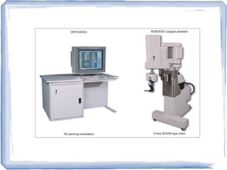
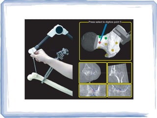
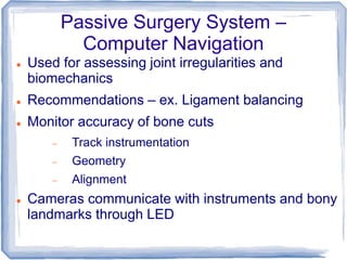
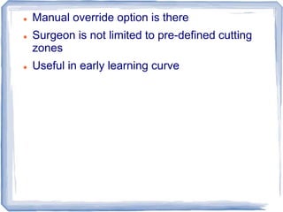
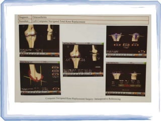
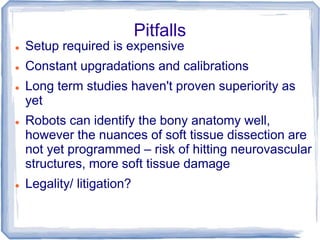
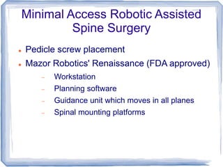
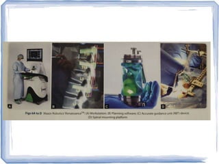
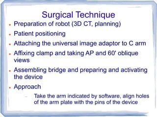
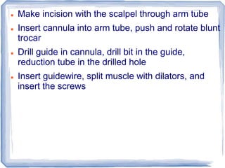
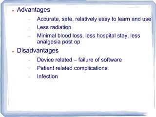
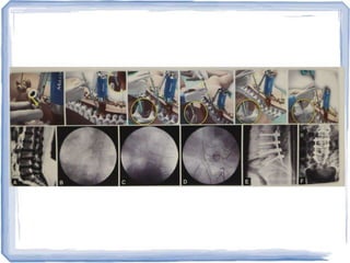
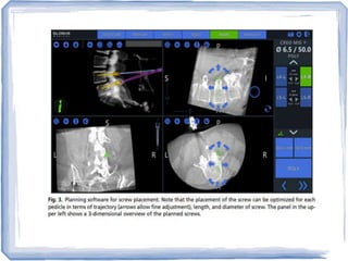
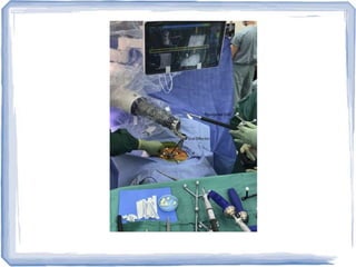
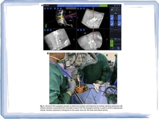
Robots in Orthopaedics
Introduction
 Was introduced to achieve higher speed and
accuracy in surgery
 Examples of their use - UKR, THR, Spine
pedicle screw placement
 Reduction of error
 Improved surgical technique
 Minimally invasive surgery may be done
 However, due to high on-table setup time for
the machines, time taken may be the same, if
not more
 Updates to software and equipment
 Cost
 Learning curve
 Ultimate results in robots v.s. Manual....is there a
significant difference?
Principle of Use
 Pre op templating with software, creation of
patient-specific model an surgical plan
 Orienting the robot to the patient's anatomy
through uploading of CT scans
 Imageless systems – do the same intra-op
 Intraoperative registration of the model and
plan
 Employment of the robot for bone-cuts and
carrying out the pre-operative plan
Robotic Systems
 Autonomous
 Surgeon performs approach, then sets up the
machine and engages the robot
 Robot works by itself
 Haptic (tactile)
 Surgeon drives the robot's arms
 Constant input from surgeon
 Image vs. Imageless
 Closed vs. Open platforms – company specific
 Soft Tissue – DaVinci Robot (used since 2000)
Haptic Robotic System
 Unicondylar Knee Replacement – Robotic arm
interactive orthopedic system – RIO made by
Mako Surgical Corp.
 Pre op CT used for 3D model of knee
 Surgeon marks the bony surfaces of femur
and tibia intra op, allowing the pre op model to
be merged into the active anatomy
 Knee ROM is seen, flex/ex gaps assessed,
component sizing and placement finalized,
and cutting zone is created
 3D model can be viewed which cutting with the
burr
 Resection of bone is confined to the pre-defined
space on the force controlled tip of the burr
 If the surgeon exceeds the cutting zone, the burr
automatically stops
 Roche et al. (2010) measured 344 radiological
parameters in 43 cases...only 3 were off
 Cobb et al. Used the Acrobot robot
 Robots gave better American Knee Society
scores, increased operative time
 Better alignment and positioning
 Accuracy within 2 degrees of pre op plan....only
40% of manual cases had within 2 degree
accuracy
Autonomous Robotic Systems
 Surgeon is in control of an emergency shut-off
switch
 ROBODOC – introduced in 1992 (Curexo Tech,
California)
 MBARS (Mini bone attached robotic system)
Hybrid system – Carnegie Mellon University,
Pittsburg, PA
 ROBODOC – showed promise in THR, but had
safety concerns
 Praxiteles (Praxim, France) also developed
Passive Surgery System –
Computer Navigation
 Used for assessing joint irregularities and
biomechanics
 Recommendations – ex. Ligament balancing
 Monitor accuracy of bone cuts
 Track instrumentation
 Geometry
 Alignment
 Cameras communicate with instruments and bony
landmarks through LED
 Manual override option is there
 Surgeon is not limited to pre-defined cutting
zones
 Useful in early learning curve
Pitfalls
 Setup required is expensive
 Constant upgradations and calibrations
 Long term studies haven't proven superiority as
yet
 Robots can identify the bony anatomy well,
however the nuances of soft tissue dissection are
not yet programmed – risk of hitting neurovascular
structures, more soft tissue damage
 Legality/ litigation?
Minimal Access Robotic Assisted
Spine Surgery
 Pedicle screw placement
 Mazor Robotics' Renaissance (FDA approved)
 Workstation
 Planning software
 Guidance unit which moves in all planes
 Spinal mounting platforms
Surgical Technique
 Preparation of robot (3D CT, planning)
 Patient positioning
 Attaching the universal image adaptor to C arm
 Affixing clamp and taking AP and 60' oblique
views
 Assembling bridge and preparing and activating
the device
 Approach
 Take the arm indicated by software, align holes
of the arm plate with the pins of the device
 Make incision with the scalpel through arm tube
 Insert cannula into arm tube, push and rotate blunt
trocar
 Drill guide in cannula, drill bit in the guide,
reduction tube in the drilled hole
 Insert guidewire, split muscle with dilators, and
insert the screws
 Advantages
 Accurate, safe, relatively easy to learn and use
 Less radiation
 Minimal blood loss, less hospital stay, less
analgesia post op
 Disadvantages
 Device related – failure of software
 Patient related complications
 Infection
 I'll be back
Thank You

Robotics in orthopedics

  • 1.
  • 2.
    Introduction  Was introducedto achieve higher speed and accuracy in surgery  Examples of their use - UKR, THR, Spine pedicle screw placement  Reduction of error  Improved surgical technique  Minimally invasive surgery may be done  However, due to high on-table setup time for the machines, time taken may be the same, if not more
  • 3.
     Updates tosoftware and equipment  Cost  Learning curve  Ultimate results in robots v.s. Manual....is there a significant difference?
  • 4.
    Principle of Use Pre op templating with software, creation of patient-specific model an surgical plan  Orienting the robot to the patient's anatomy through uploading of CT scans  Imageless systems – do the same intra-op  Intraoperative registration of the model and plan  Employment of the robot for bone-cuts and carrying out the pre-operative plan
  • 5.
    Robotic Systems  Autonomous Surgeon performs approach, then sets up the machine and engages the robot  Robot works by itself  Haptic (tactile)  Surgeon drives the robot's arms  Constant input from surgeon  Image vs. Imageless  Closed vs. Open platforms – company specific  Soft Tissue – DaVinci Robot (used since 2000)
  • 6.
    Haptic Robotic System Unicondylar Knee Replacement – Robotic arm interactive orthopedic system – RIO made by Mako Surgical Corp.  Pre op CT used for 3D model of knee  Surgeon marks the bony surfaces of femur and tibia intra op, allowing the pre op model to be merged into the active anatomy  Knee ROM is seen, flex/ex gaps assessed, component sizing and placement finalized, and cutting zone is created  3D model can be viewed which cutting with the burr
  • 7.
     Resection ofbone is confined to the pre-defined space on the force controlled tip of the burr  If the surgeon exceeds the cutting zone, the burr automatically stops  Roche et al. (2010) measured 344 radiological parameters in 43 cases...only 3 were off  Cobb et al. Used the Acrobot robot  Robots gave better American Knee Society scores, increased operative time  Better alignment and positioning  Accuracy within 2 degrees of pre op plan....only 40% of manual cases had within 2 degree accuracy
  • 8.
    Autonomous Robotic Systems Surgeon is in control of an emergency shut-off switch  ROBODOC – introduced in 1992 (Curexo Tech, California)  MBARS (Mini bone attached robotic system) Hybrid system – Carnegie Mellon University, Pittsburg, PA  ROBODOC – showed promise in THR, but had safety concerns  Praxiteles (Praxim, France) also developed
  • 12.
    Passive Surgery System– Computer Navigation  Used for assessing joint irregularities and biomechanics  Recommendations – ex. Ligament balancing  Monitor accuracy of bone cuts  Track instrumentation  Geometry  Alignment  Cameras communicate with instruments and bony landmarks through LED
  • 13.
     Manual overrideoption is there  Surgeon is not limited to pre-defined cutting zones  Useful in early learning curve
  • 15.
    Pitfalls  Setup requiredis expensive  Constant upgradations and calibrations  Long term studies haven't proven superiority as yet  Robots can identify the bony anatomy well, however the nuances of soft tissue dissection are not yet programmed – risk of hitting neurovascular structures, more soft tissue damage  Legality/ litigation?
  • 16.
    Minimal Access RoboticAssisted Spine Surgery  Pedicle screw placement  Mazor Robotics' Renaissance (FDA approved)  Workstation  Planning software  Guidance unit which moves in all planes  Spinal mounting platforms
  • 18.
    Surgical Technique  Preparationof robot (3D CT, planning)  Patient positioning  Attaching the universal image adaptor to C arm  Affixing clamp and taking AP and 60' oblique views  Assembling bridge and preparing and activating the device  Approach  Take the arm indicated by software, align holes of the arm plate with the pins of the device
  • 19.
     Make incisionwith the scalpel through arm tube  Insert cannula into arm tube, push and rotate blunt trocar  Drill guide in cannula, drill bit in the guide, reduction tube in the drilled hole  Insert guidewire, split muscle with dilators, and insert the screws
  • 20.
     Advantages  Accurate,safe, relatively easy to learn and use  Less radiation  Minimal blood loss, less hospital stay, less analgesia post op  Disadvantages  Device related – failure of software  Patient related complications  Infection
  • 26.
     I'll beback Thank You