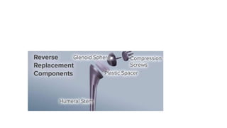
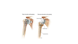
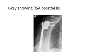
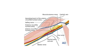
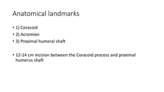
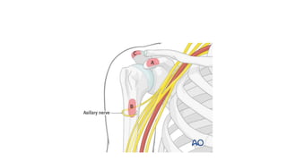
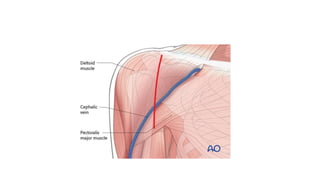
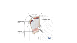
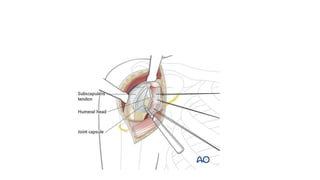
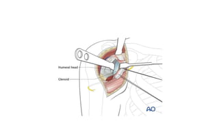
Reverse shoulder
arthroplasty
Biomechanics
• Reverses the orientation of the shoulder joint by replacing the glenoid
fossa with the glenoid base plate and glenosphere and the humeral
head With a shaft and Concave cup.This prosthesis alters the center
of rotation of shoulder by moving it medially and inferiorly.This
subsequently increases deltoid tension which enhances torque and
line of pull of the deltoid.This results in an improved shoulder
elevation.
X-ray showing RSA prosthesis
Indications
Most important is non functional rotator cuff
1)Cuff tear arthropathy
2)3 and 4 part proximal humerus fractures in elderly
3)Non union proximal humerus
4)Severe inflammatory arthritis with cuff tear
5)Failed shoulder hemiarthroplasty with anterosuperior instability.
6) Reconstruction after tumor removal.
Pre requisites
• 1)Intact deltoid
• 2) Adequate bone stock to support the glenoid.
• 3)No neurological deficiency-Parkinsons, Syringomyelia.
• 4)No excessive demands
Surgical procedure
The preferred approach is the deltopectoral approach.
The course of the following neurovascular structures should be kept in mind
1)Cephalic vein
2) Anterior circumflex humeral artery
3) Ascending arcuate branch of Anterior circumflex humeral artery.
4) Posterior circumflex humeral artery
5) Musculocutaneous nerve
6) Axillary nerve
7) Suprascapular nerve and artery
Anatomical landmarks
• 1) Coracoid
• 2) Acromion
• 3) Proximal humeral shaft
• 12-14 cm incision between the Coracoid process and proximal
humerus shaft
• Deltopectoral interval-Between deltoid and pectoralis major is
developed.
Dissection down to the deltopectoral groove
• Blunt dissection between deltoid and pectoralis major is done to
expose clavipectoralfascia.
Exposure
• Identify the Coracoid process and conjoined tendon
• Incise the clavipectoralfascia lateral to conjoint tendon and I ferior to
coracoacromial ligament.
• Retract the deltoid muscle laterally
• Take care to avoid injury to Musculocutaneous nerve
• Subscapularis tendon is identified and divided vertically lateral to
musculotendinous junction.
• Reflect the subscapularis and enter the joint theough a vertical
capsulotomy medial to lateral stump of subscapularis.
• Humeral head is retracted in order to visualise the glenoid.
• Guidepin placed in 10-15 degree of inferior tilt to prevent scapular
notching.
• Team glenoid till smiley face appearance seen IE Hard sclerotic bone
superiorly and bleeding Cancellous bone inferiorly.This confirms
inferior tilt of base plate.
• Impact the base plate and secure it with the screws.Peripheral screws
are ideally placed in the pillars of densest cortical bone.
• Impact the glenosphere I to position
• Place the humeral stem using trial components to test for motion and
stability And trial reduction is done
• To dislocate glenhumeral joint place instrument between bearing
surface and glenosphere.
• Once proper bearing position is chosen dry the Morse taper and
impact it and reduce joint for final time.
• Thorough wash
• Closure with subscapularis repair.
Outcomes
• Post traumatic conditions and revision procedures have worse
outcomes.
• Post operative protocol
• Immobilizer for 6 weeks
• Active range of motion At 6 weeks
• Deltoid strengthening at 12 weeks
Complications
• 1)Infection
• 2) Neurovascular injury
• 3)Loosening or breakage of Implant
• 4)Acromial stress fracture
• 5)Notching of inferior scapula
How to prevent
• Increase size of glenosphere
• 10-15 degree inferior tilt
• Inferior base plate placement
• Thank you

Reverse shoulder arthroplasty

  • 1.
  • 3.
    Biomechanics • Reverses theorientation of the shoulder joint by replacing the glenoid fossa with the glenoid base plate and glenosphere and the humeral head With a shaft and Concave cup.This prosthesis alters the center of rotation of shoulder by moving it medially and inferiorly.This subsequently increases deltoid tension which enhances torque and line of pull of the deltoid.This results in an improved shoulder elevation.
  • 5.
  • 6.
    Indications Most important isnon functional rotator cuff 1)Cuff tear arthropathy 2)3 and 4 part proximal humerus fractures in elderly 3)Non union proximal humerus 4)Severe inflammatory arthritis with cuff tear 5)Failed shoulder hemiarthroplasty with anterosuperior instability. 6) Reconstruction after tumor removal.
  • 7.
    Pre requisites • 1)Intactdeltoid • 2) Adequate bone stock to support the glenoid. • 3)No neurological deficiency-Parkinsons, Syringomyelia. • 4)No excessive demands
  • 8.
    Surgical procedure The preferredapproach is the deltopectoral approach. The course of the following neurovascular structures should be kept in mind 1)Cephalic vein 2) Anterior circumflex humeral artery 3) Ascending arcuate branch of Anterior circumflex humeral artery. 4) Posterior circumflex humeral artery 5) Musculocutaneous nerve 6) Axillary nerve 7) Suprascapular nerve and artery
  • 10.
    Anatomical landmarks • 1)Coracoid • 2) Acromion • 3) Proximal humeral shaft • 12-14 cm incision between the Coracoid process and proximal humerus shaft
  • 13.
    • Deltopectoral interval-Betweendeltoid and pectoralis major is developed.
  • 14.
    Dissection down tothe deltopectoral groove • Blunt dissection between deltoid and pectoralis major is done to expose clavipectoralfascia.
  • 16.
    Exposure • Identify theCoracoid process and conjoined tendon • Incise the clavipectoralfascia lateral to conjoint tendon and I ferior to coracoacromial ligament. • Retract the deltoid muscle laterally • Take care to avoid injury to Musculocutaneous nerve
  • 17.
    • Subscapularis tendonis identified and divided vertically lateral to musculotendinous junction.
  • 19.
    • Reflect thesubscapularis and enter the joint theough a vertical capsulotomy medial to lateral stump of subscapularis.
  • 21.
    • Humeral headis retracted in order to visualise the glenoid.
  • 23.
    • Guidepin placedin 10-15 degree of inferior tilt to prevent scapular notching. • Team glenoid till smiley face appearance seen IE Hard sclerotic bone superiorly and bleeding Cancellous bone inferiorly.This confirms inferior tilt of base plate. • Impact the base plate and secure it with the screws.Peripheral screws are ideally placed in the pillars of densest cortical bone. • Impact the glenosphere I to position
  • 24.
    • Place thehumeral stem using trial components to test for motion and stability And trial reduction is done • To dislocate glenhumeral joint place instrument between bearing surface and glenosphere. • Once proper bearing position is chosen dry the Morse taper and impact it and reduce joint for final time. • Thorough wash • Closure with subscapularis repair.
  • 25.
    Outcomes • Post traumaticconditions and revision procedures have worse outcomes. • Post operative protocol • Immobilizer for 6 weeks • Active range of motion At 6 weeks • Deltoid strengthening at 12 weeks
  • 26.
    Complications • 1)Infection • 2)Neurovascular injury • 3)Loosening or breakage of Implant • 4)Acromial stress fracture • 5)Notching of inferior scapula
  • 29.
    How to prevent •Increase size of glenosphere • 10-15 degree inferior tilt • Inferior base plate placement
  • 30.