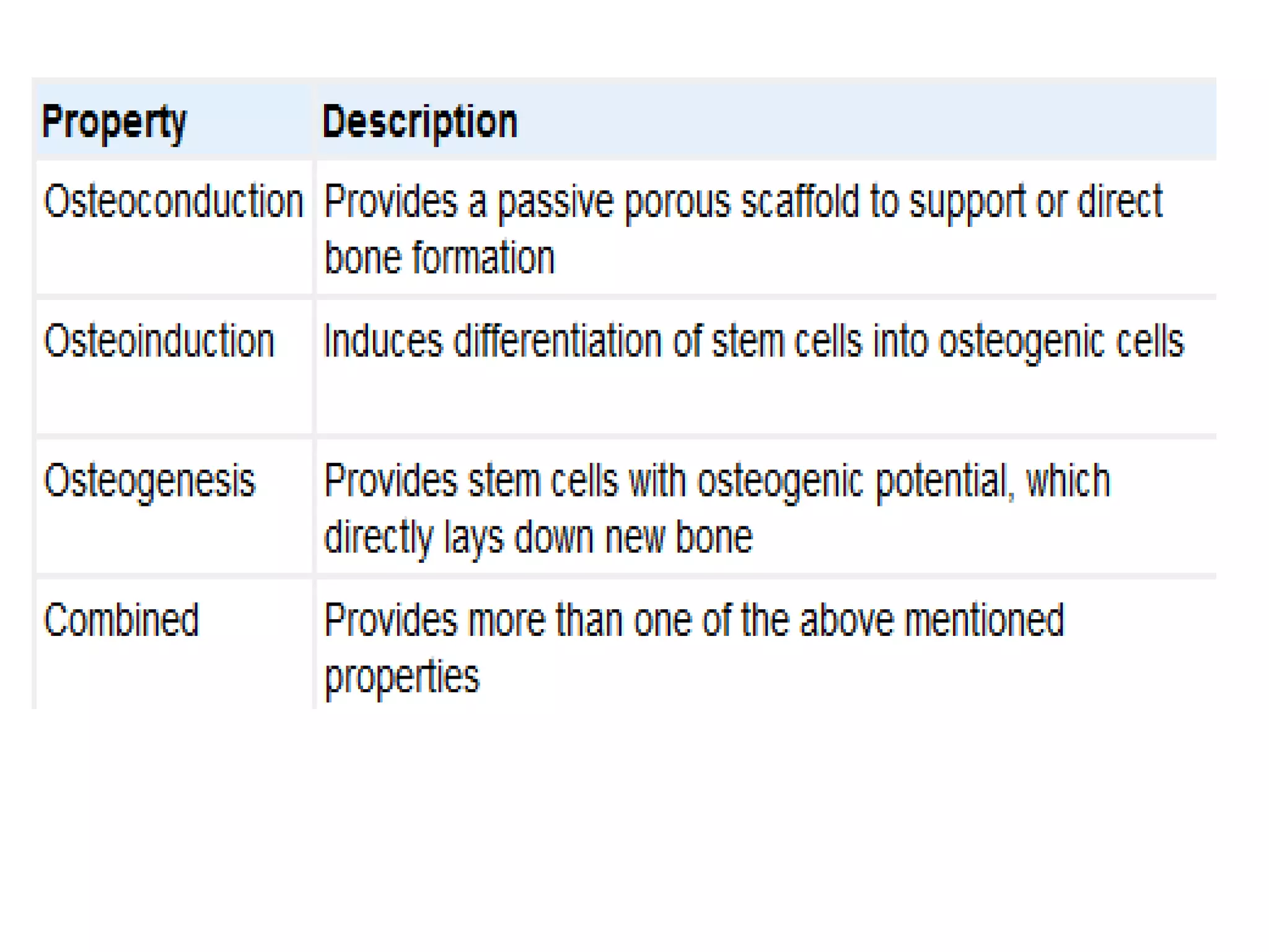
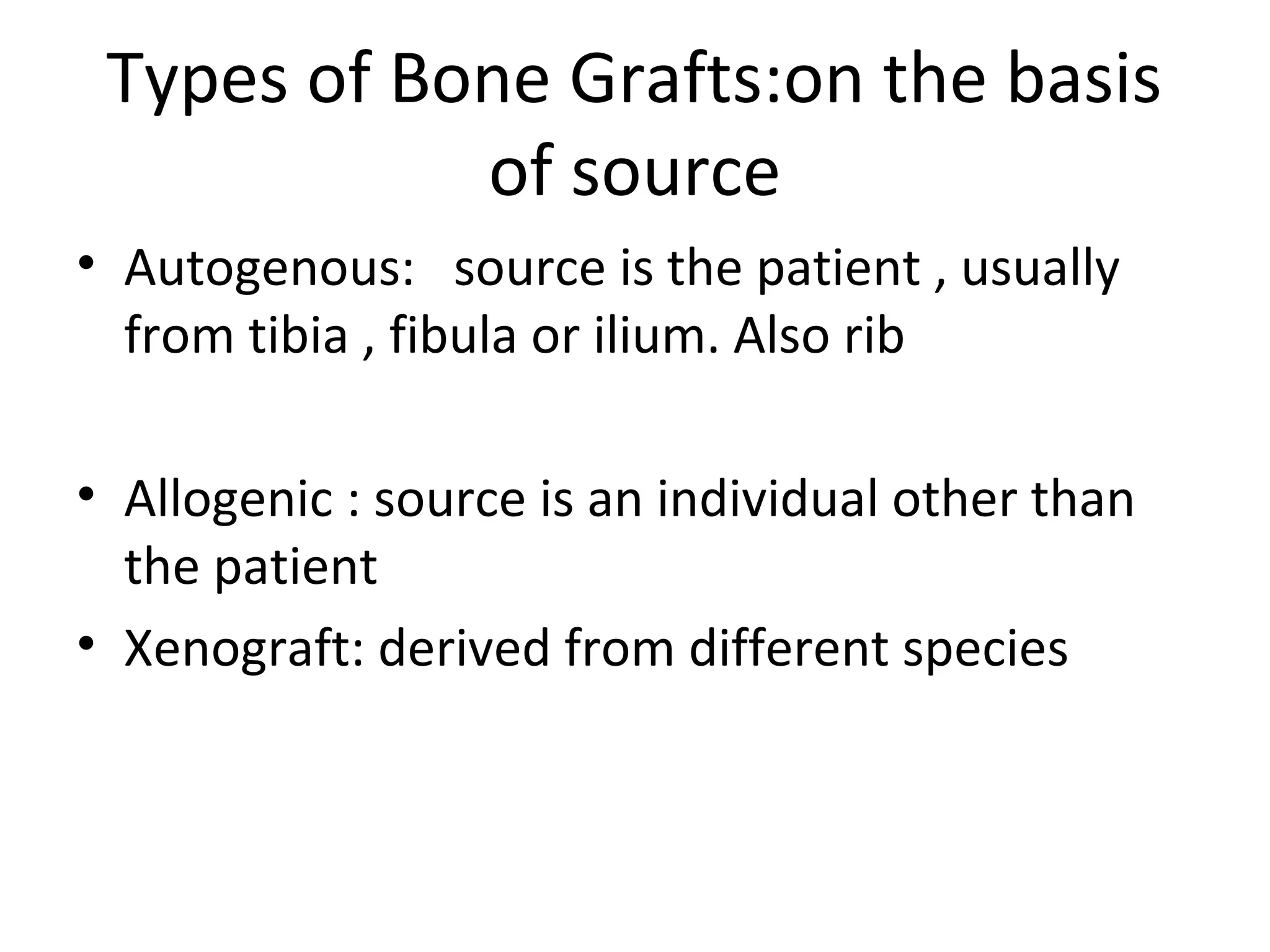
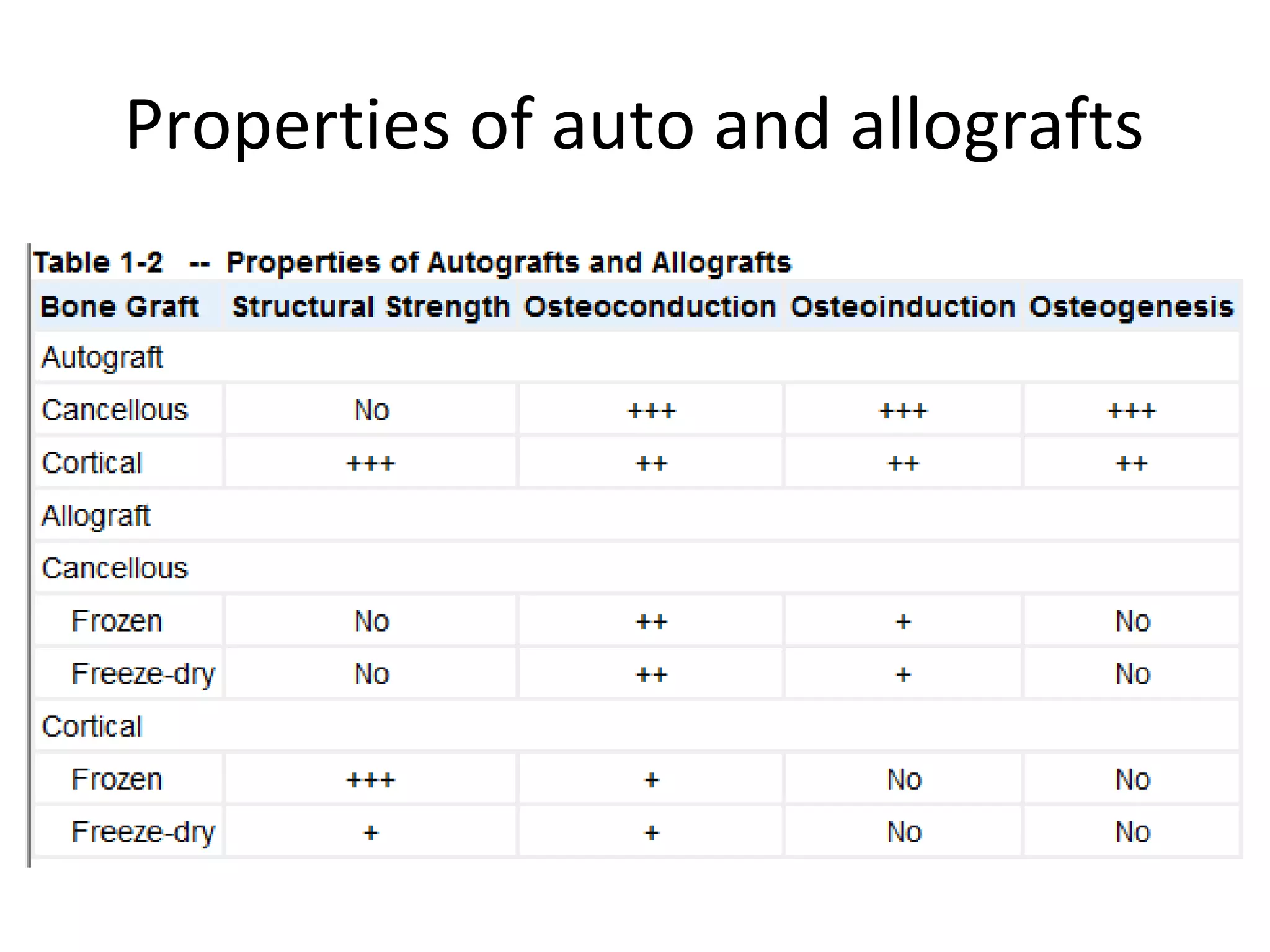
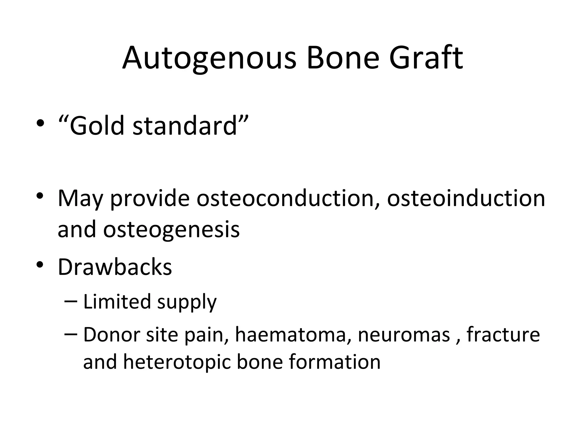
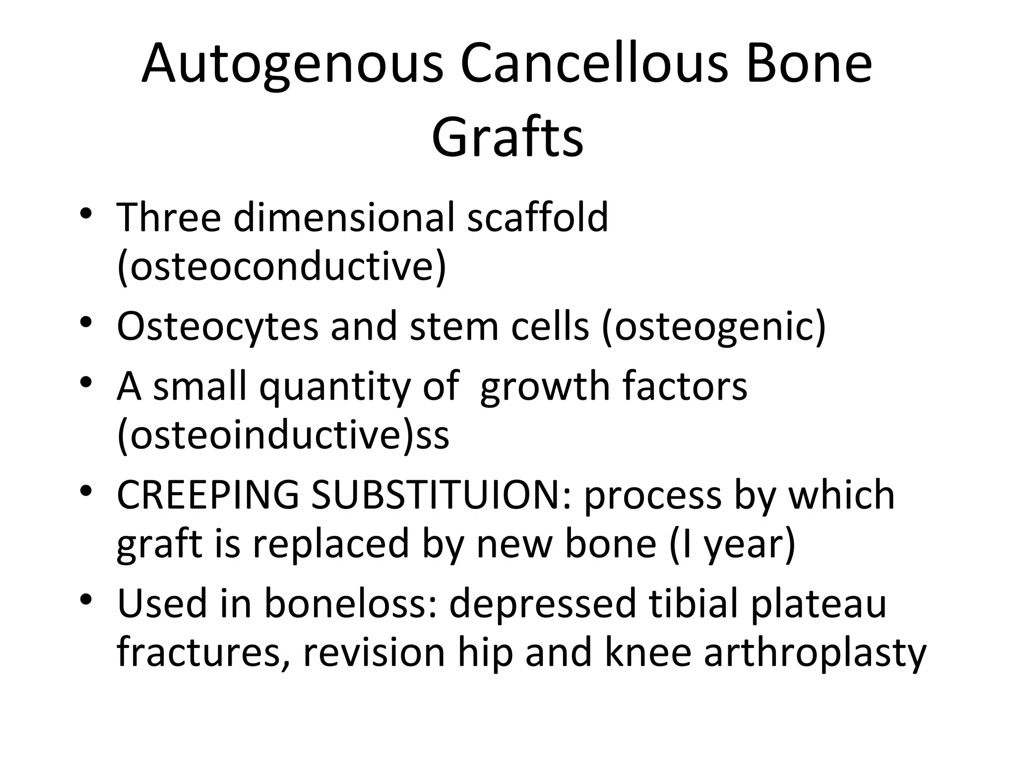
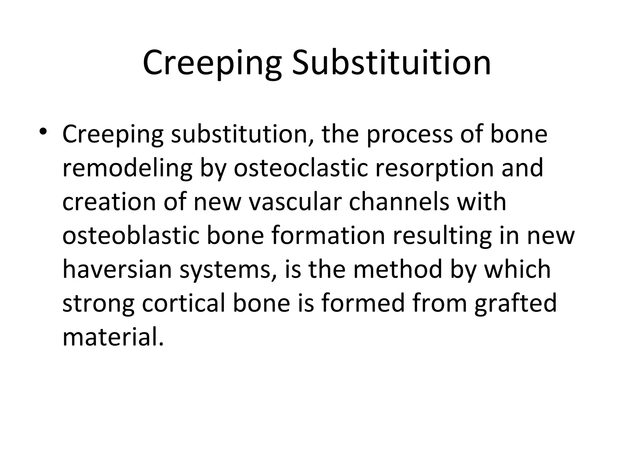
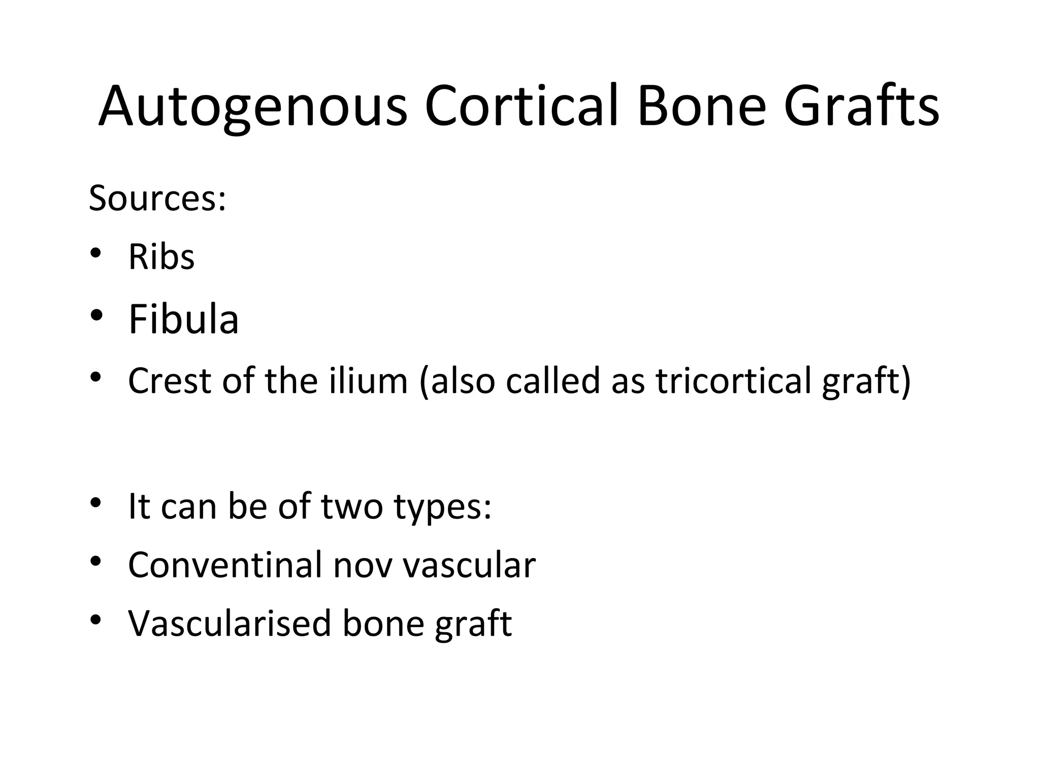
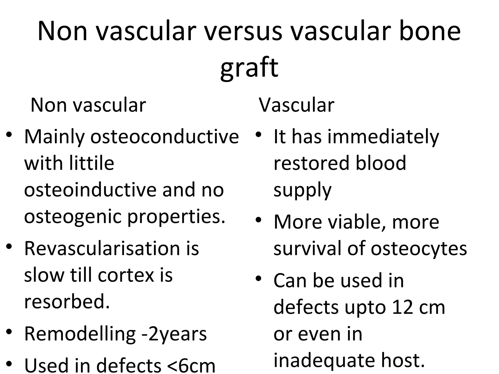
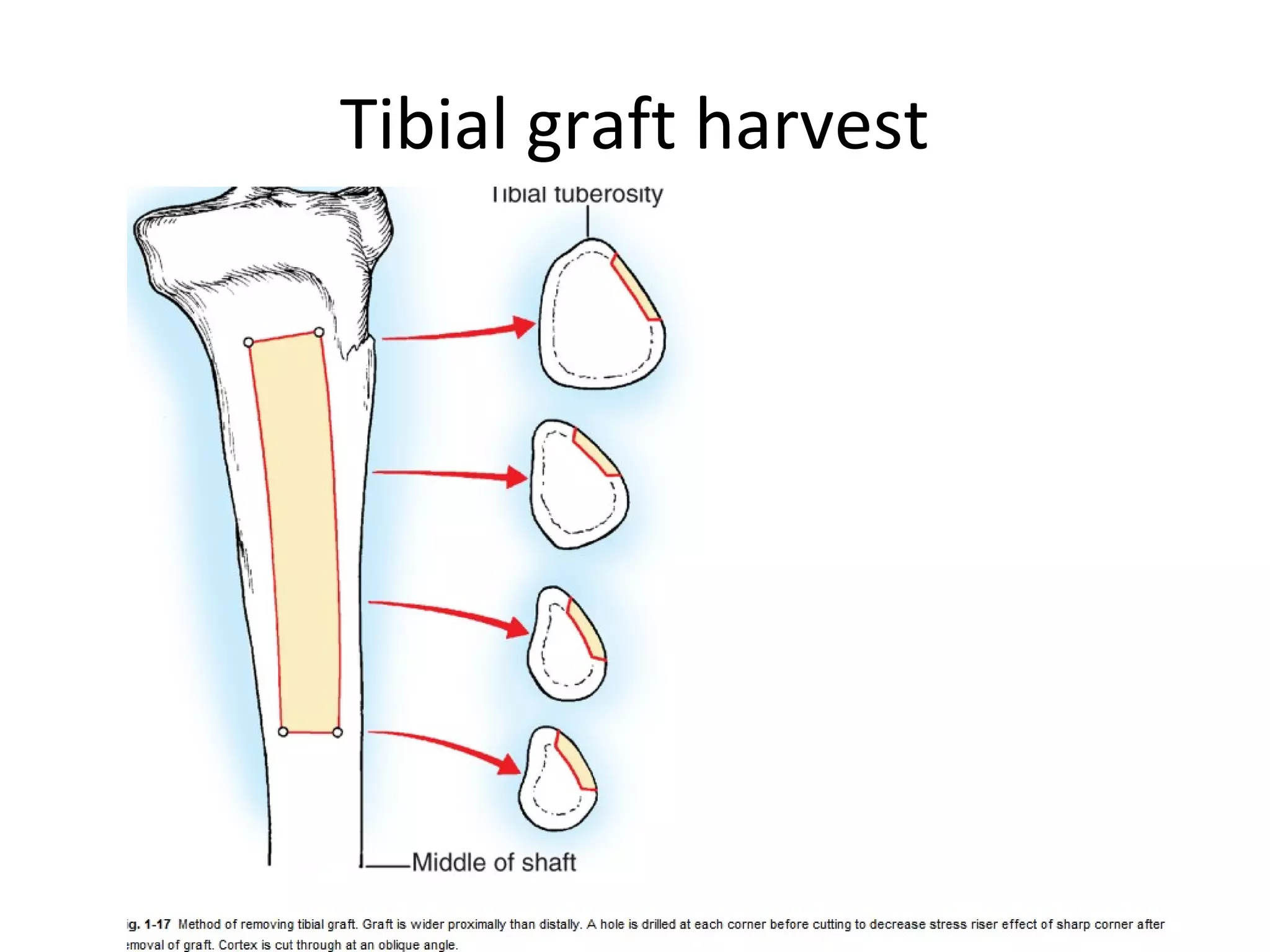
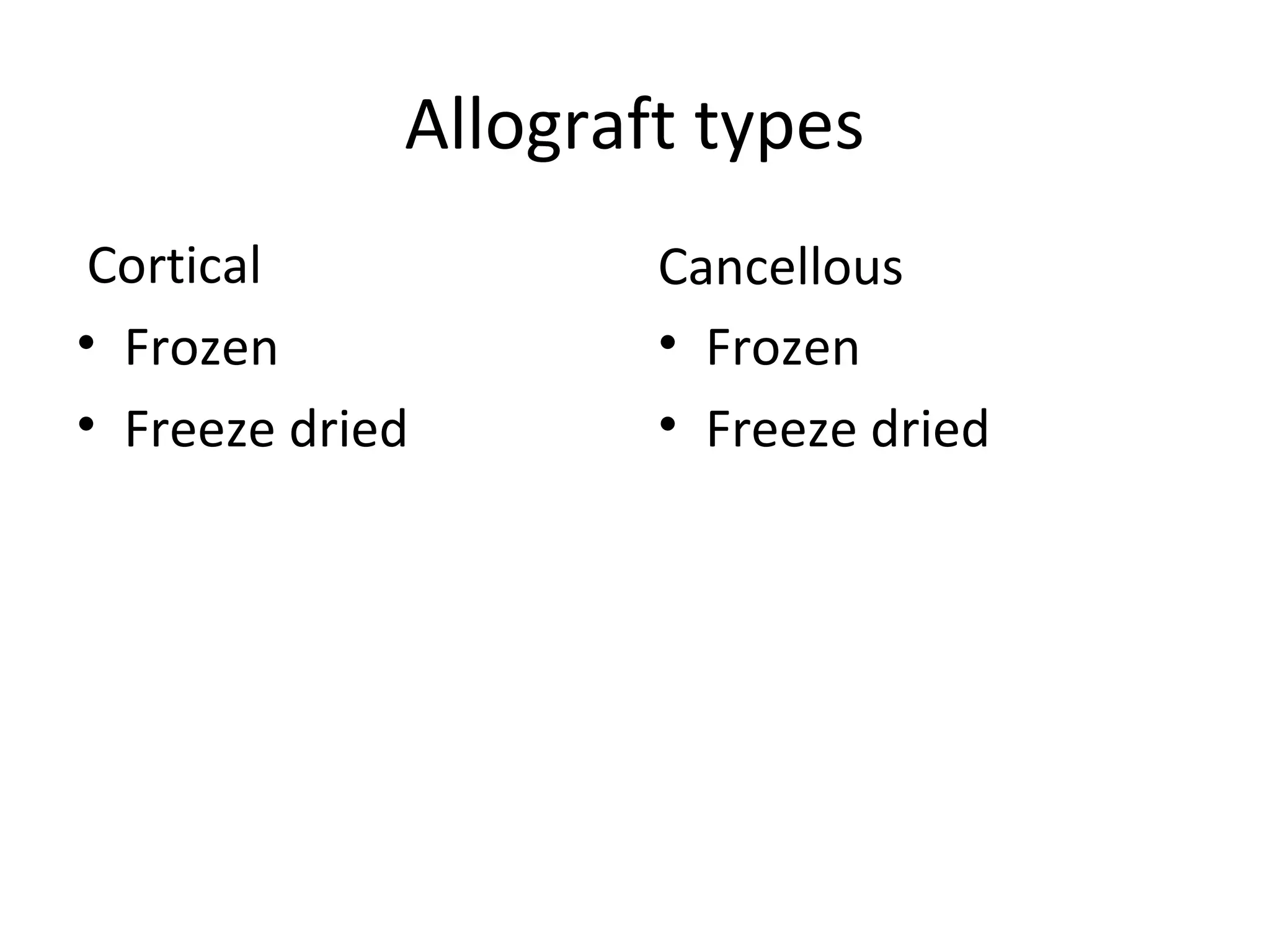
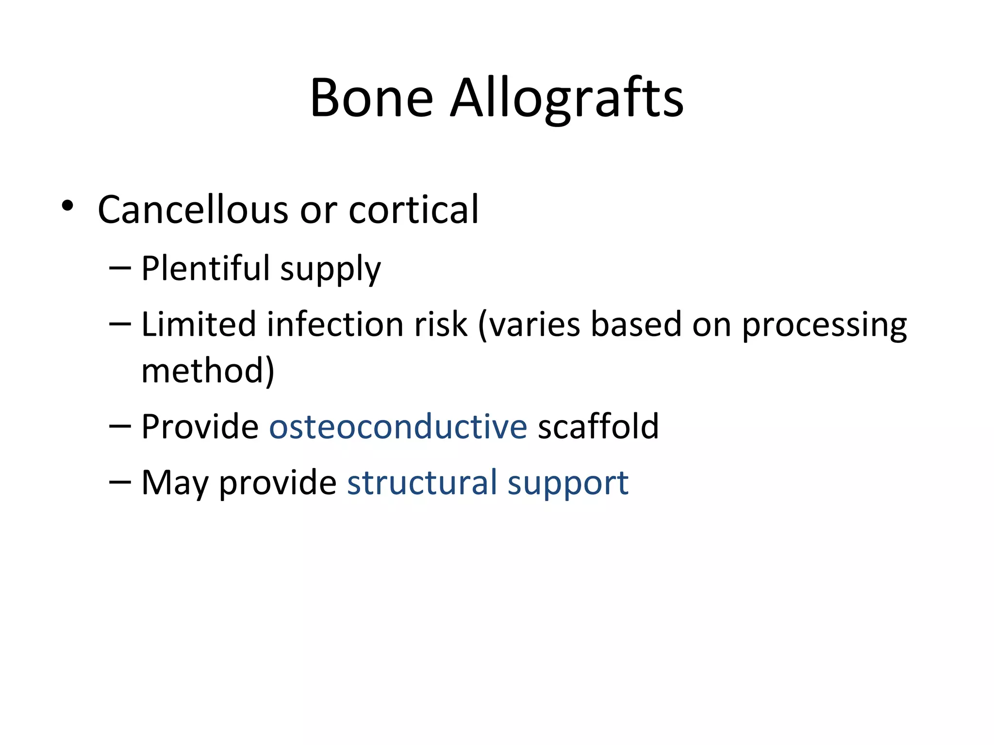
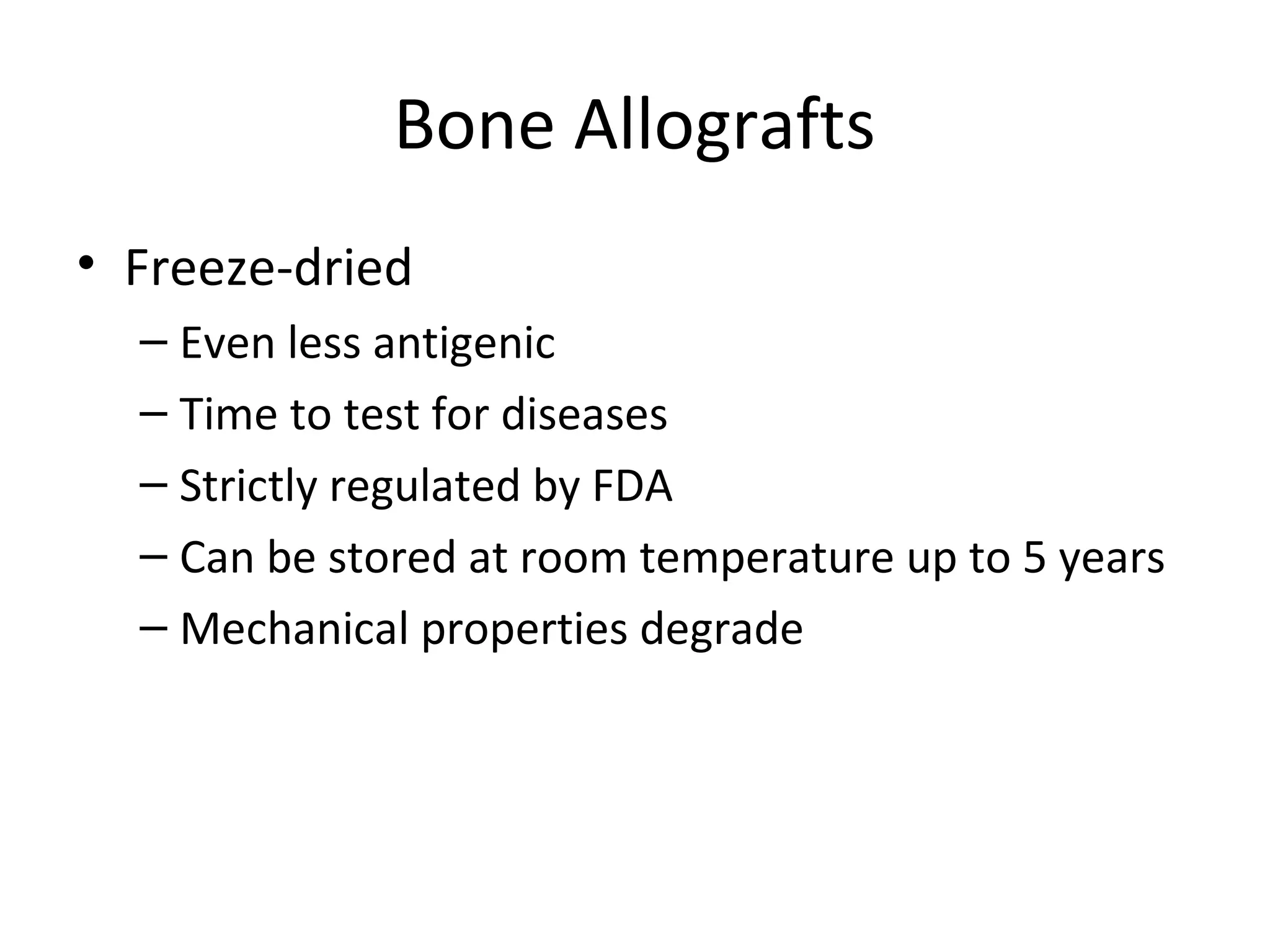
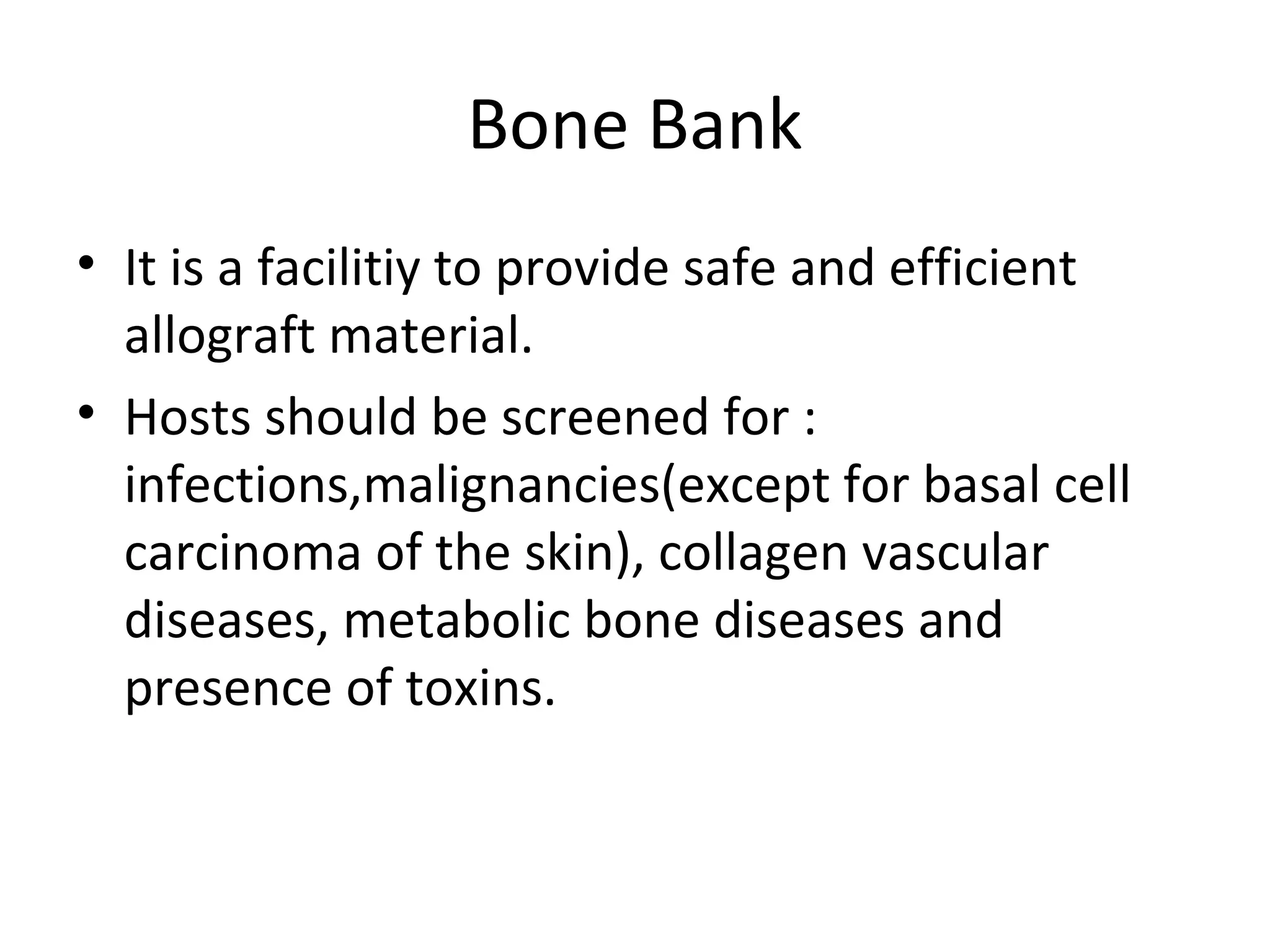
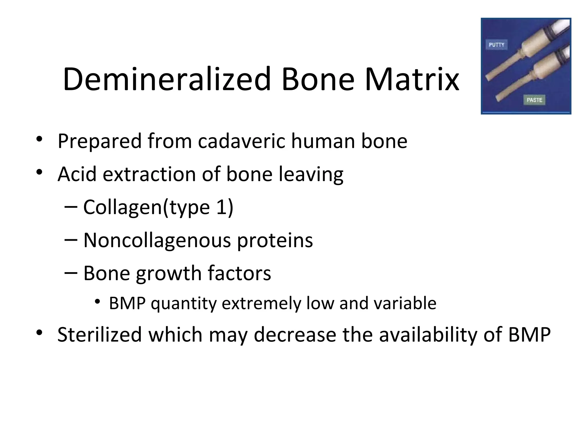
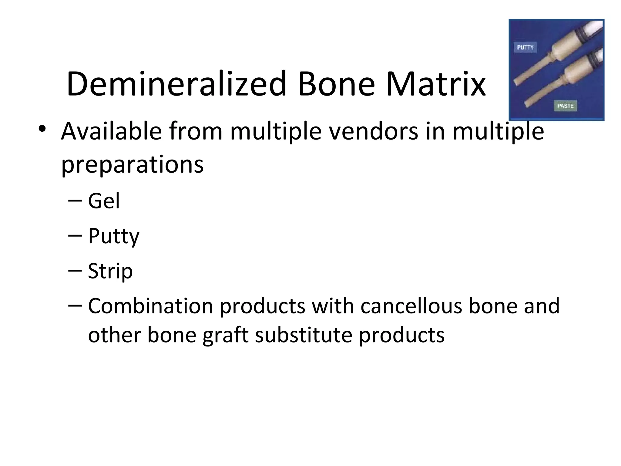
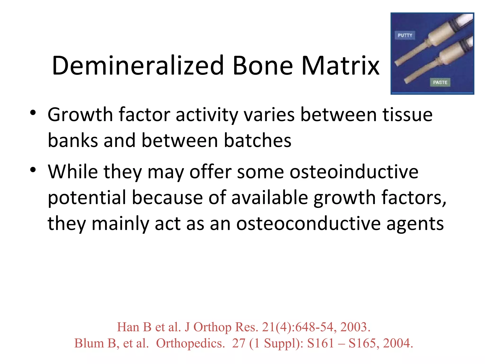
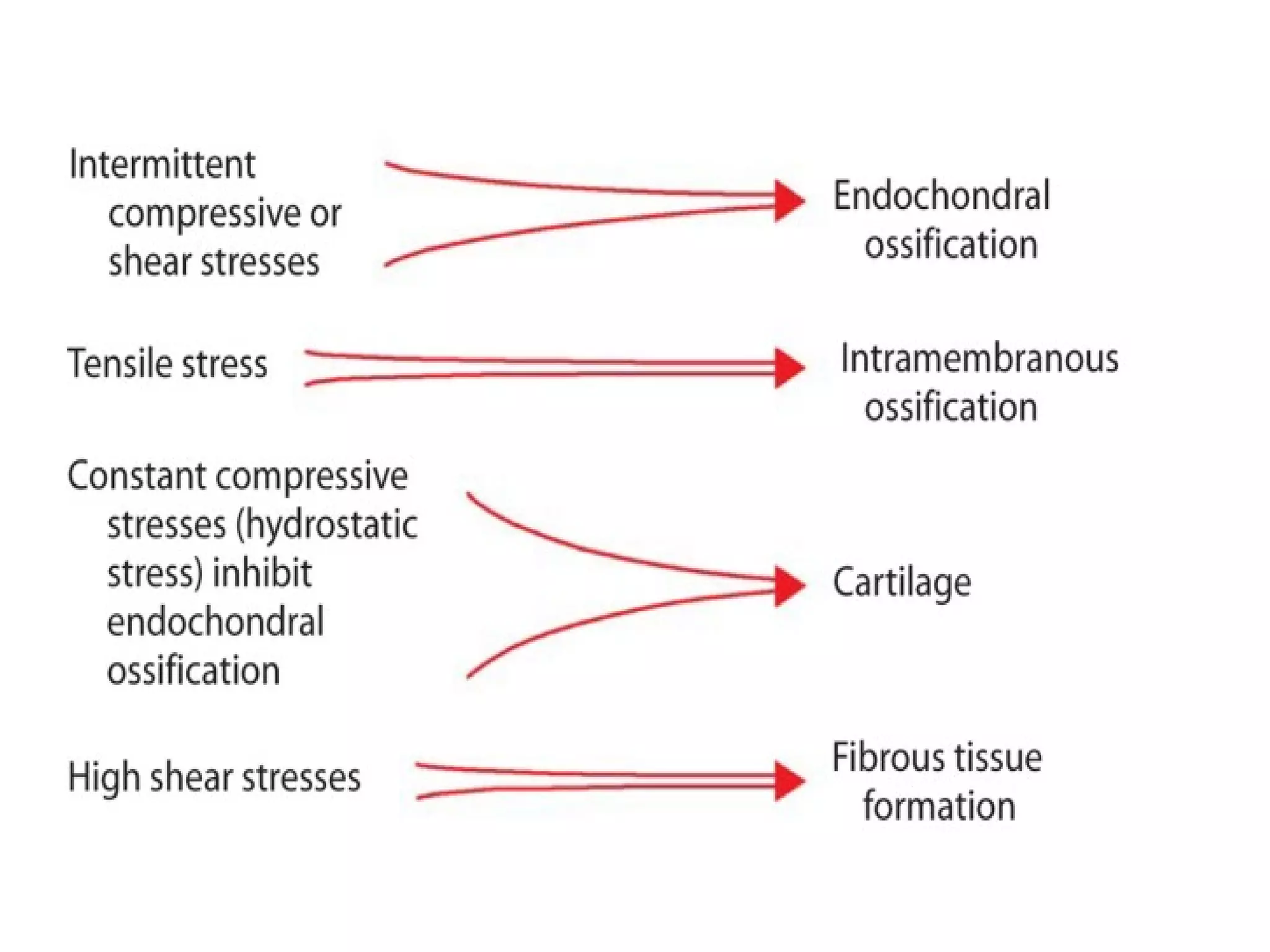
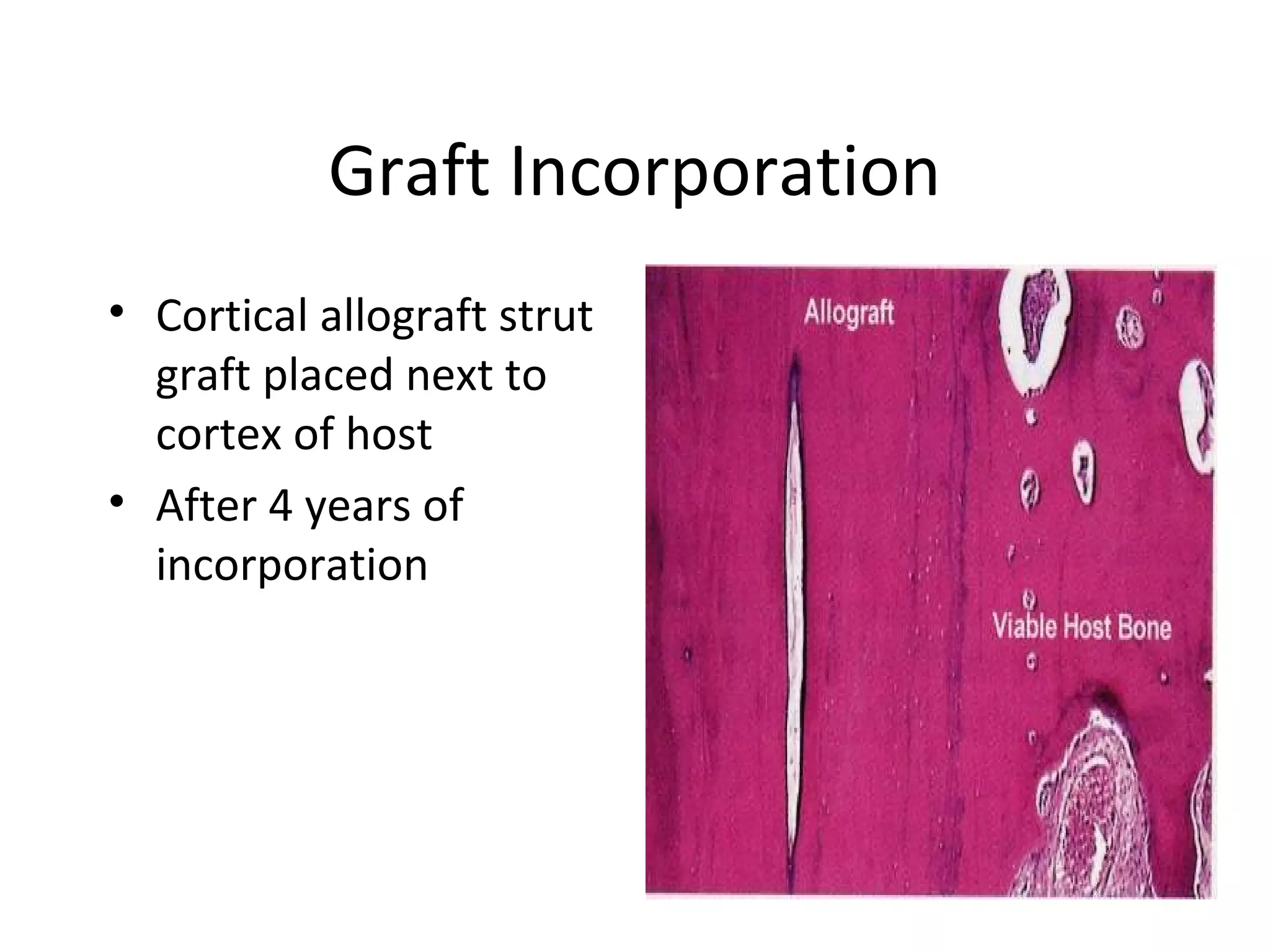
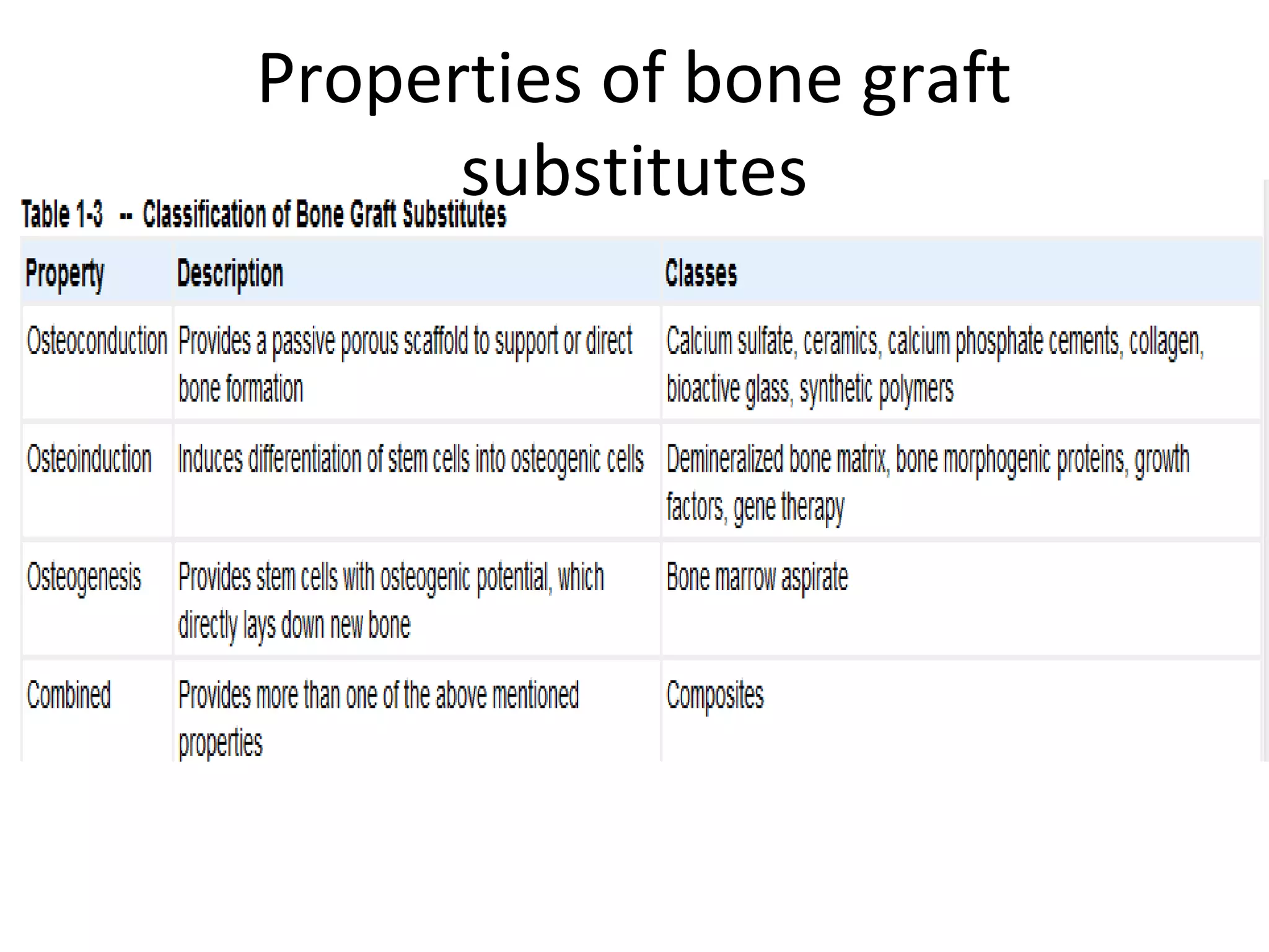
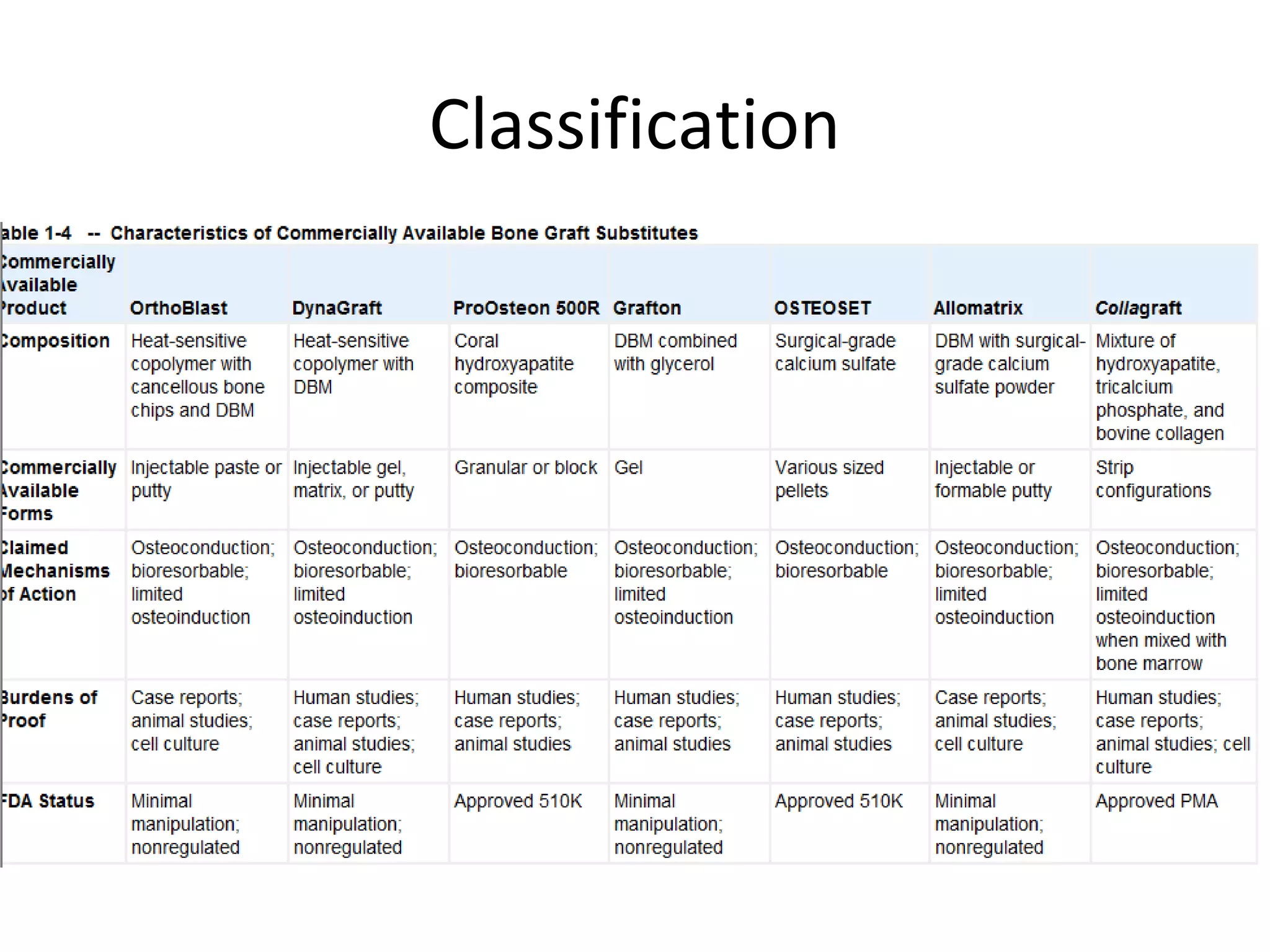
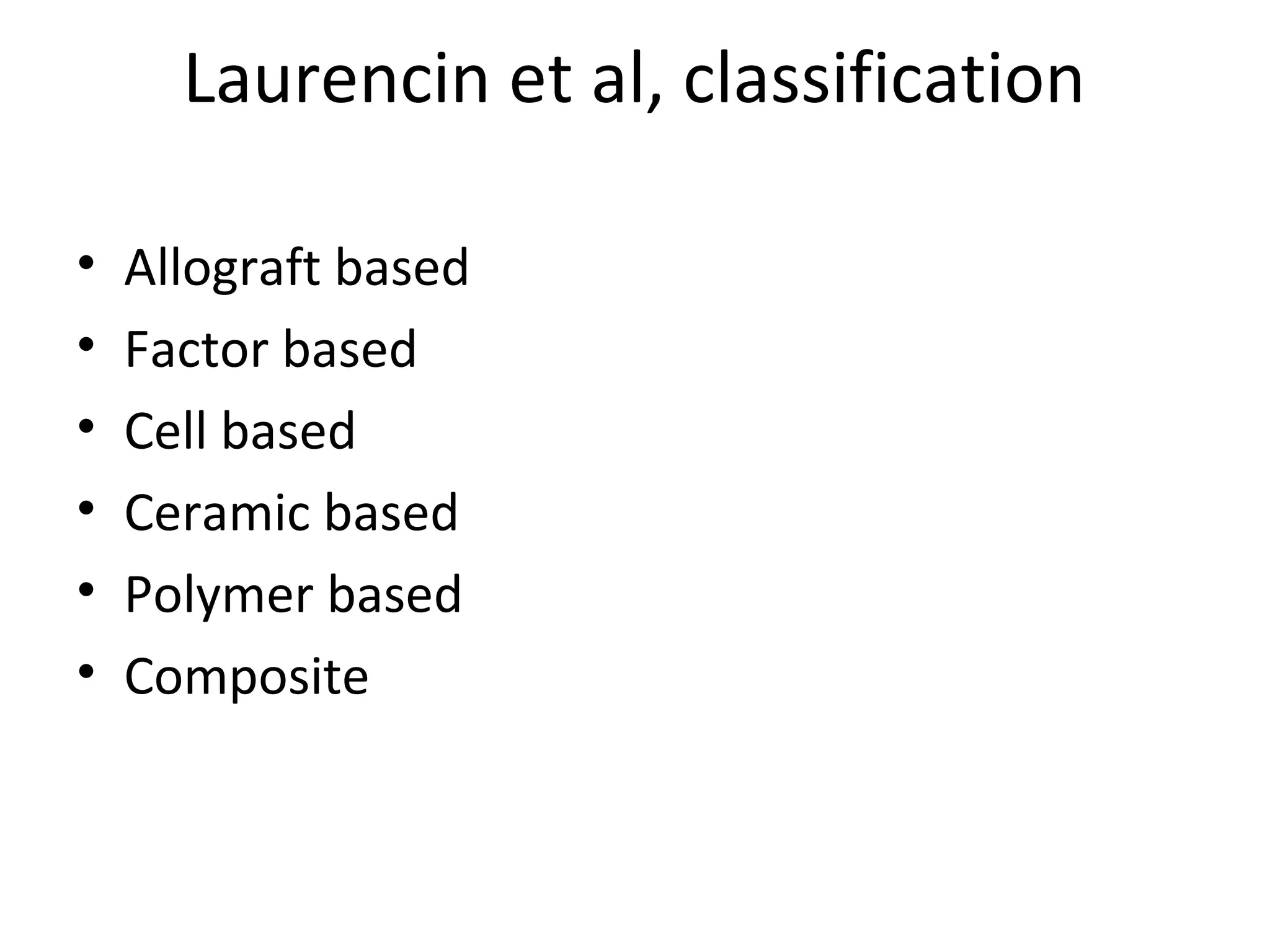
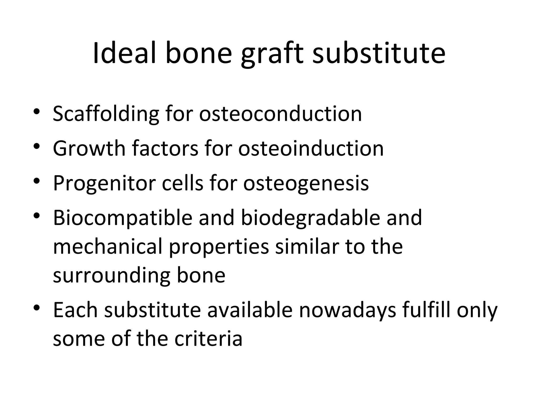
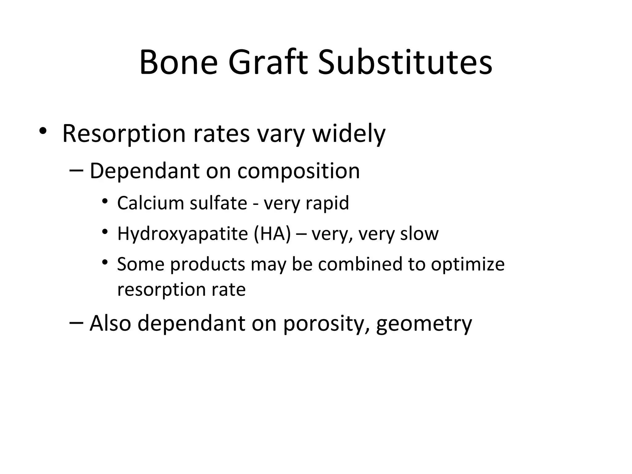
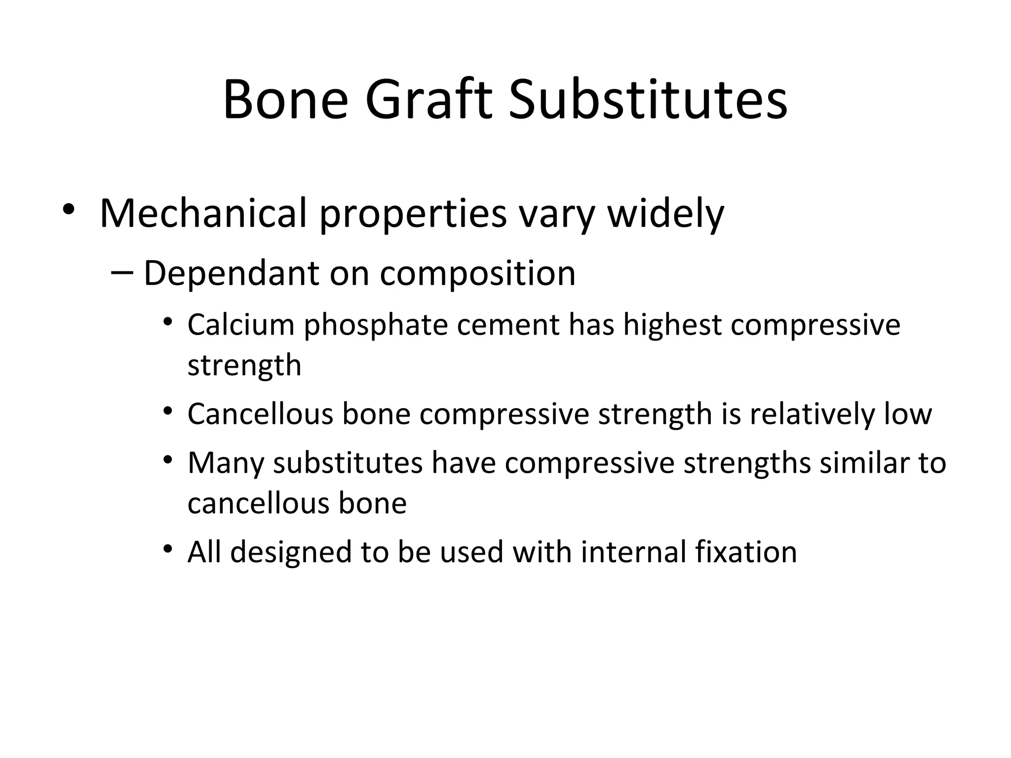
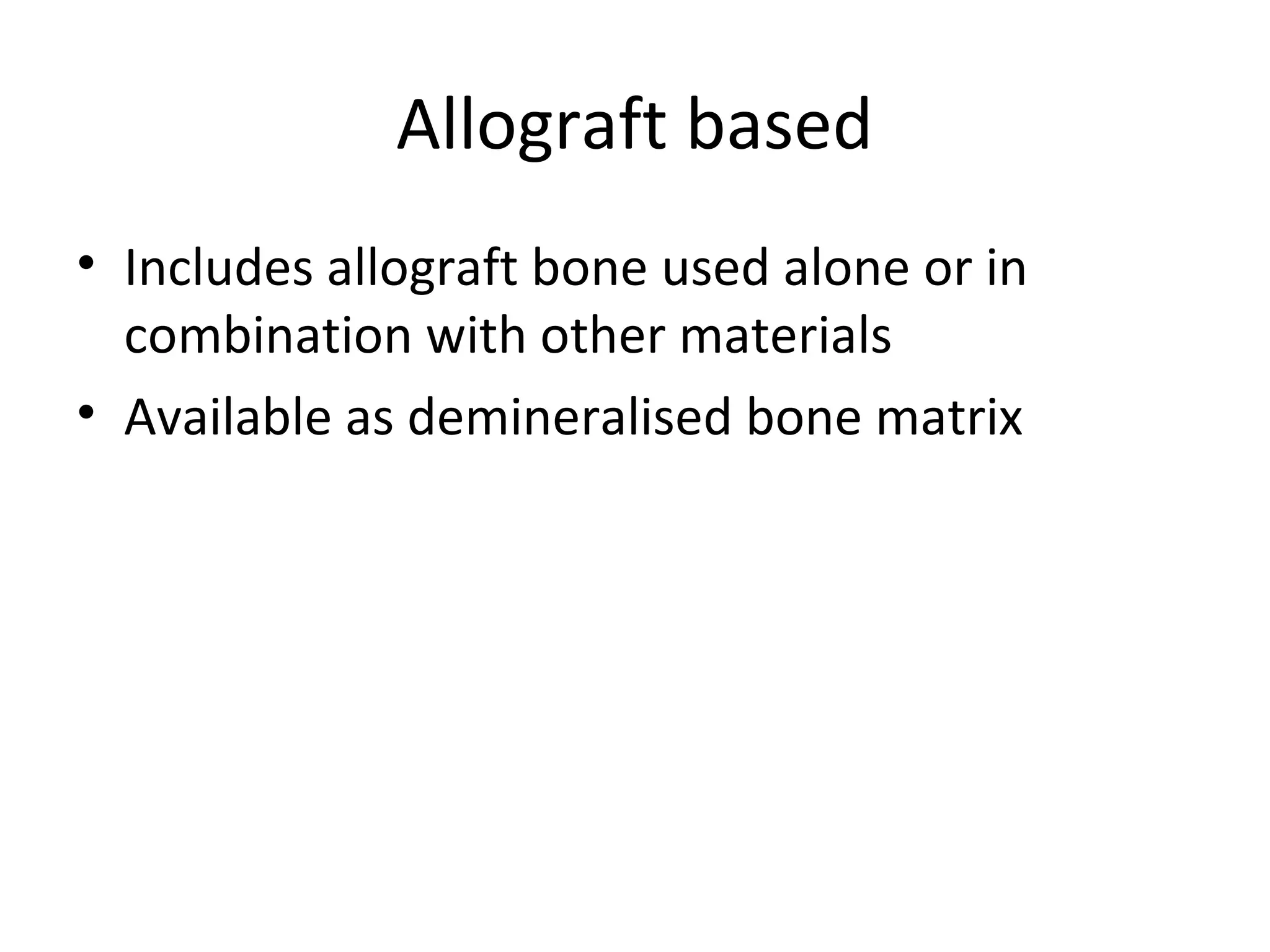
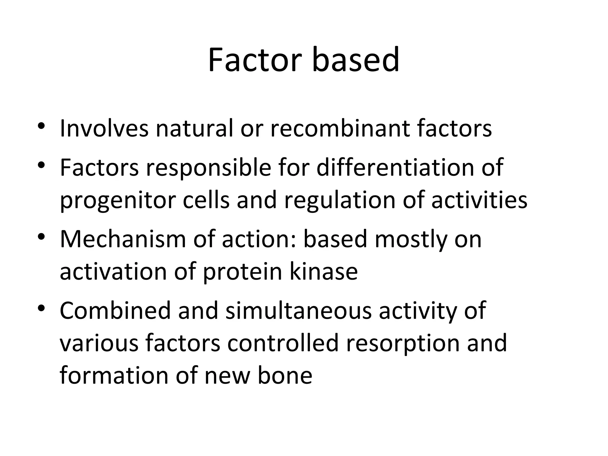
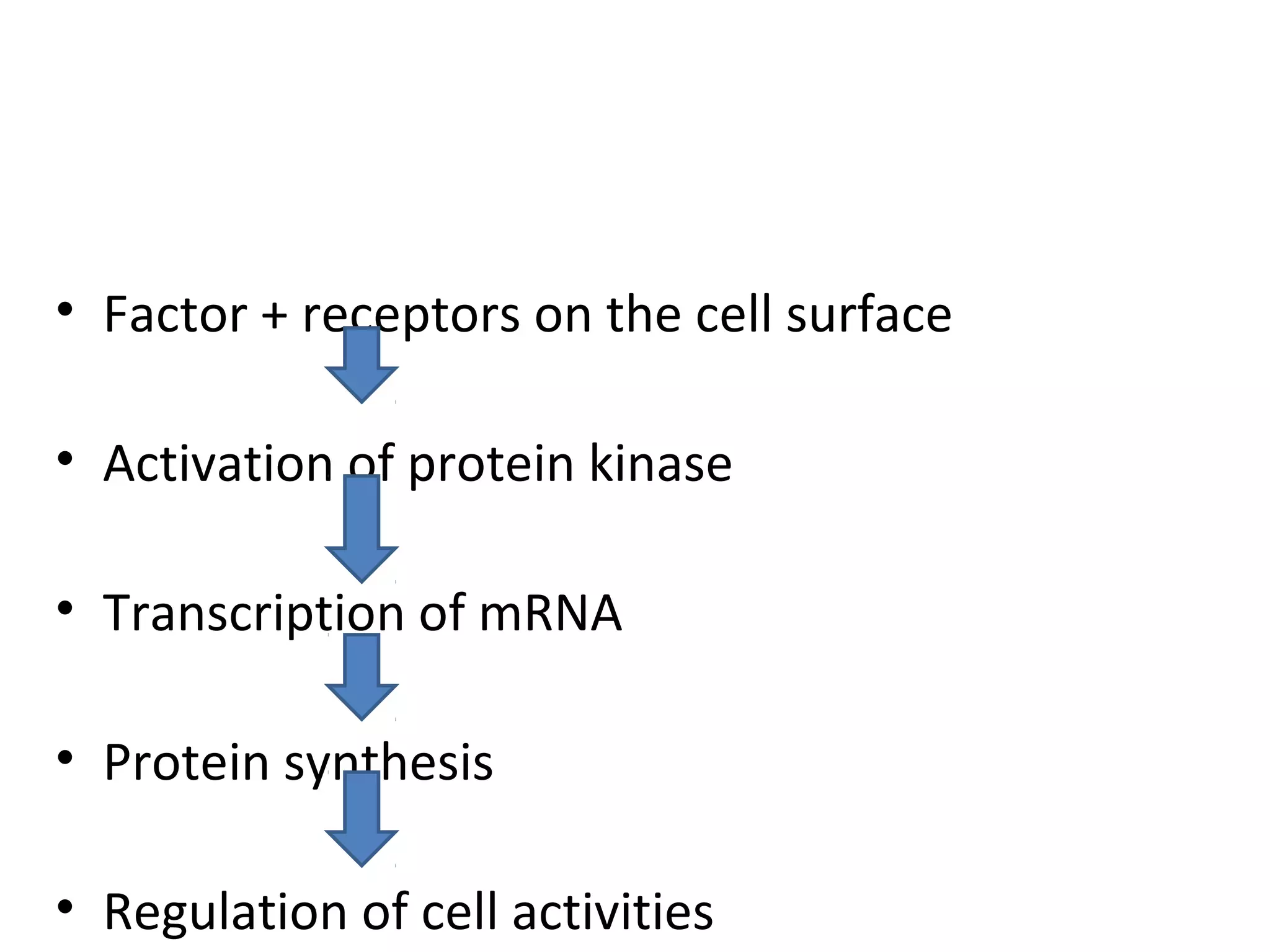
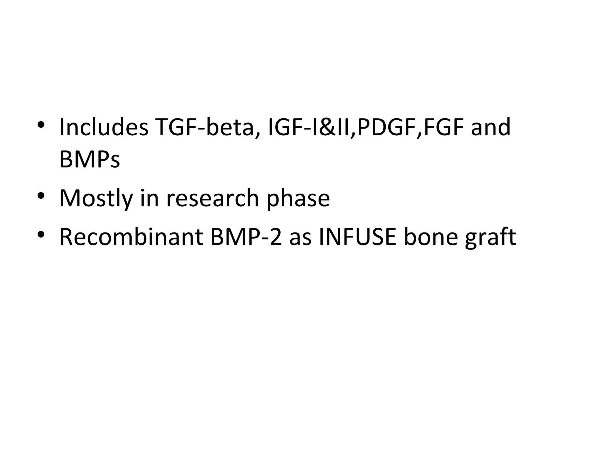
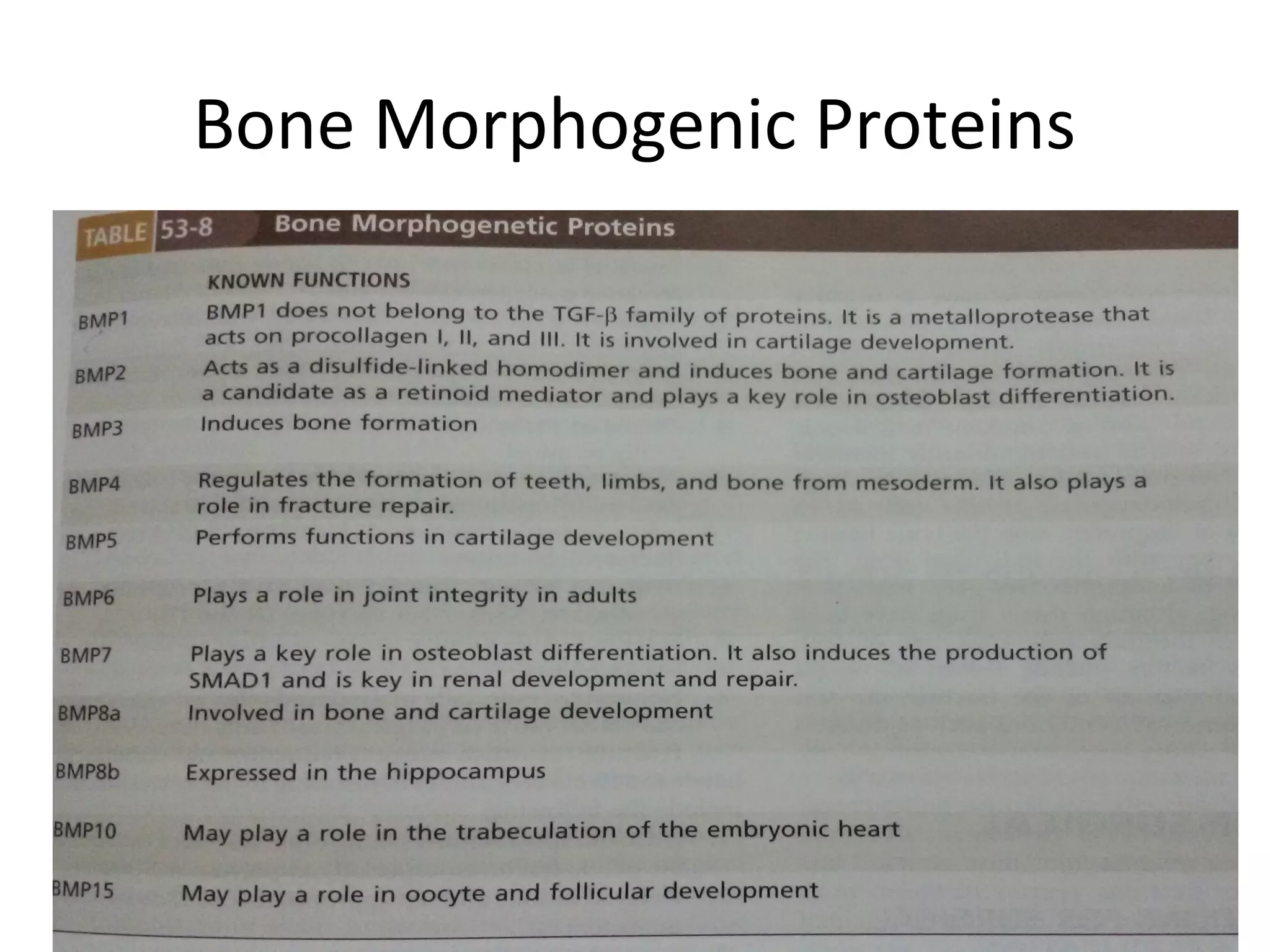
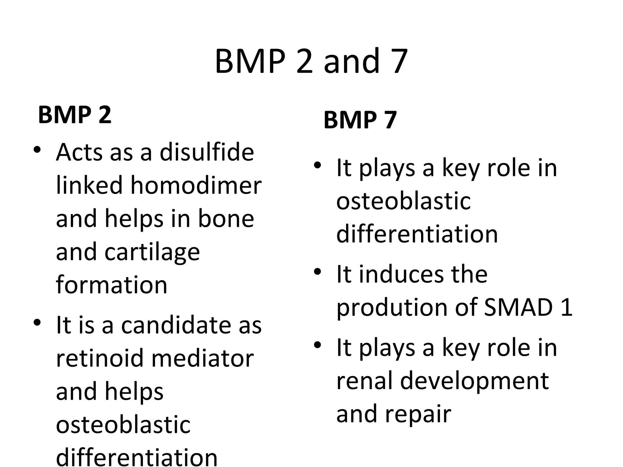
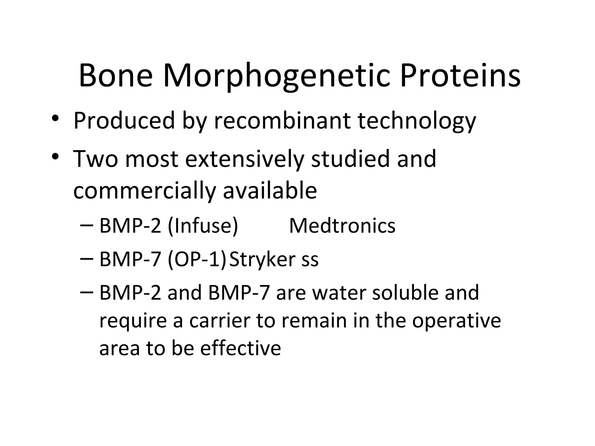
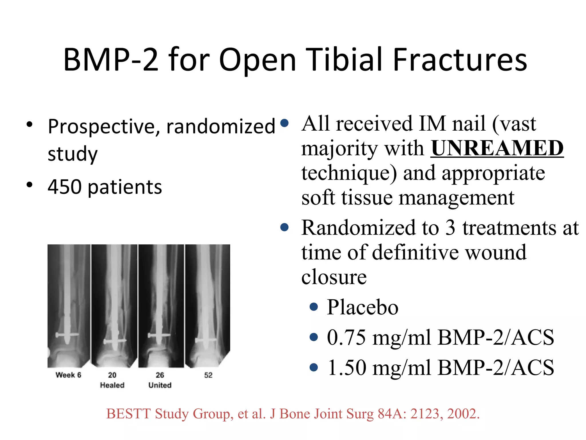
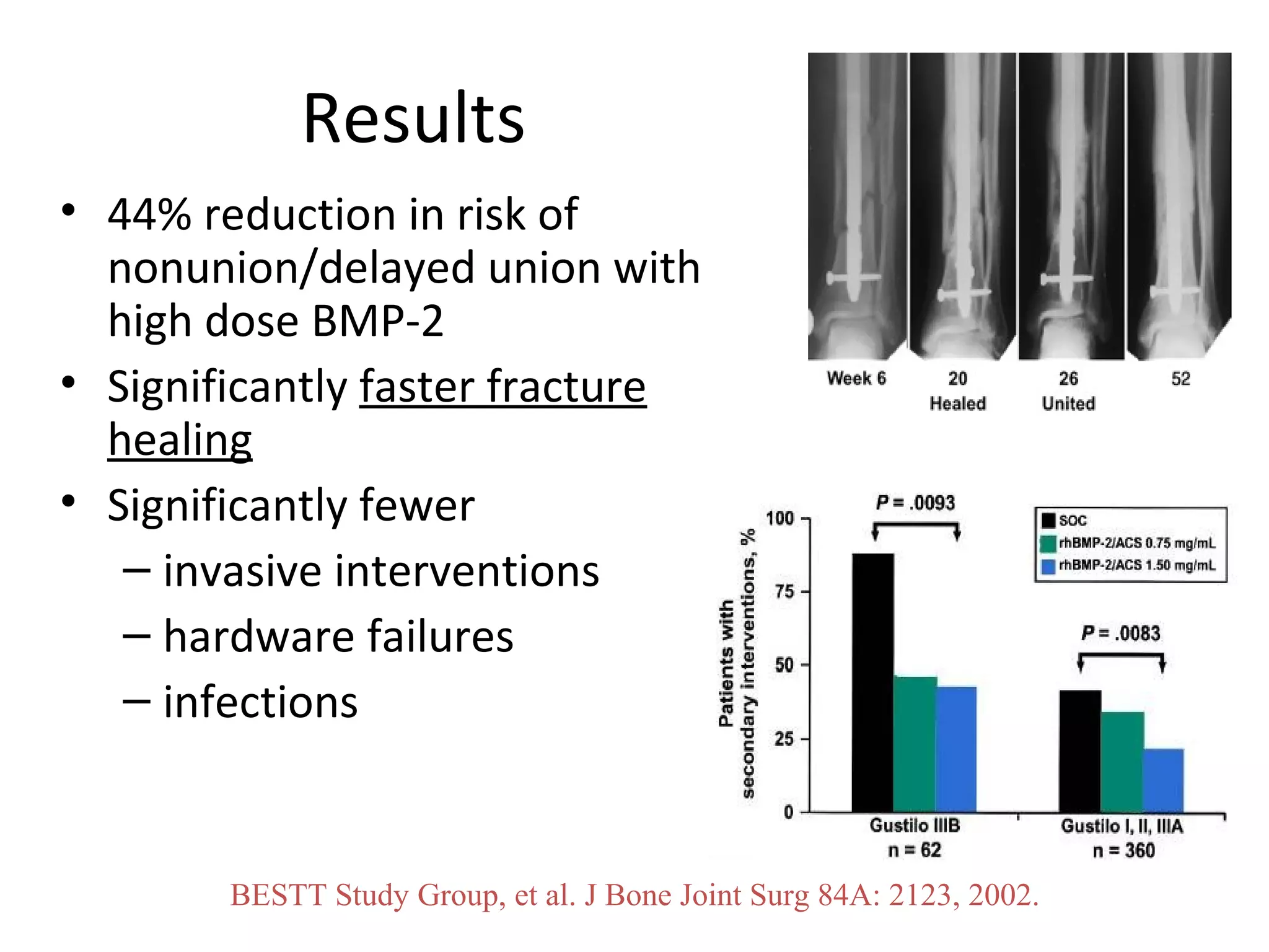
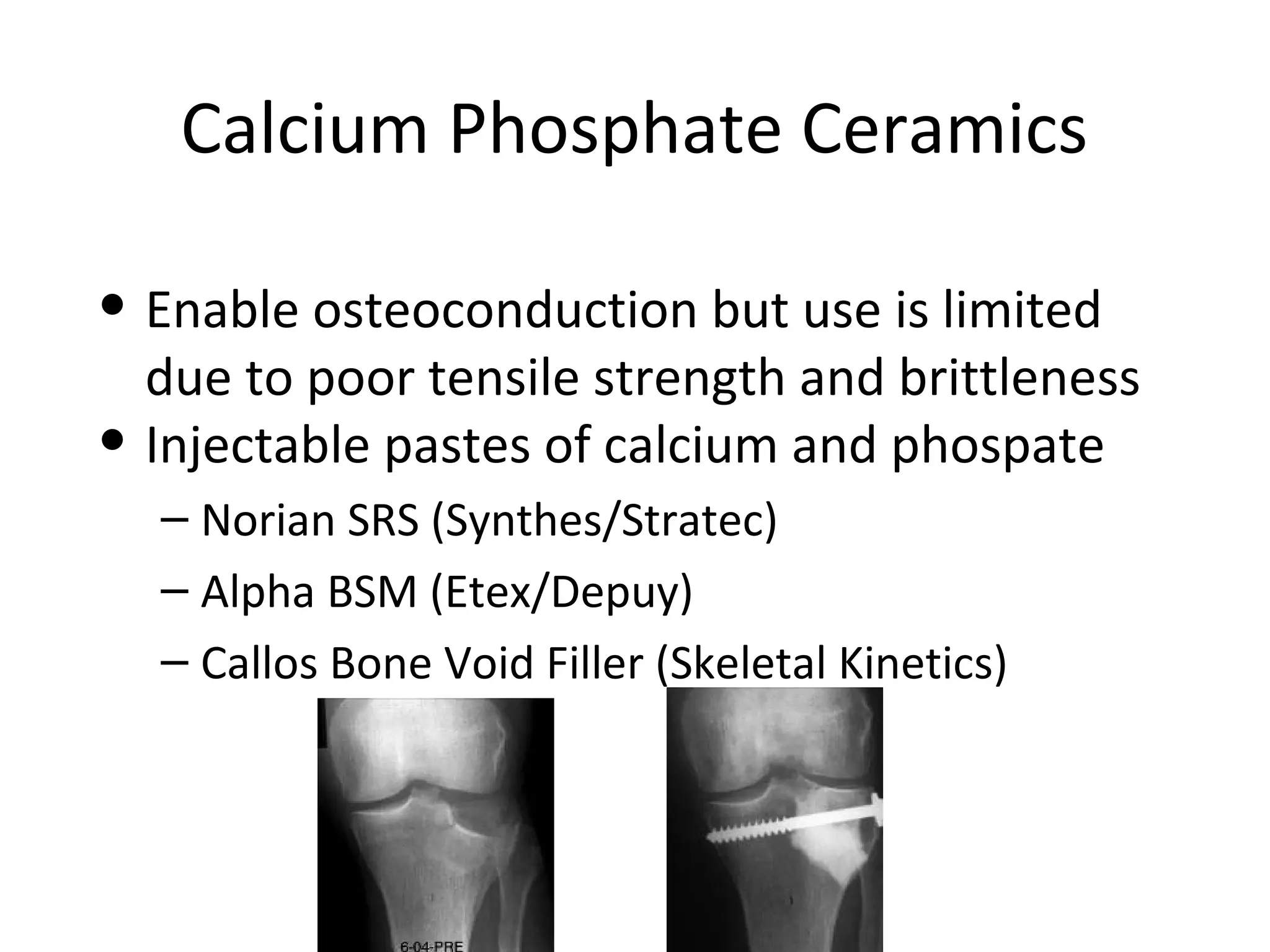
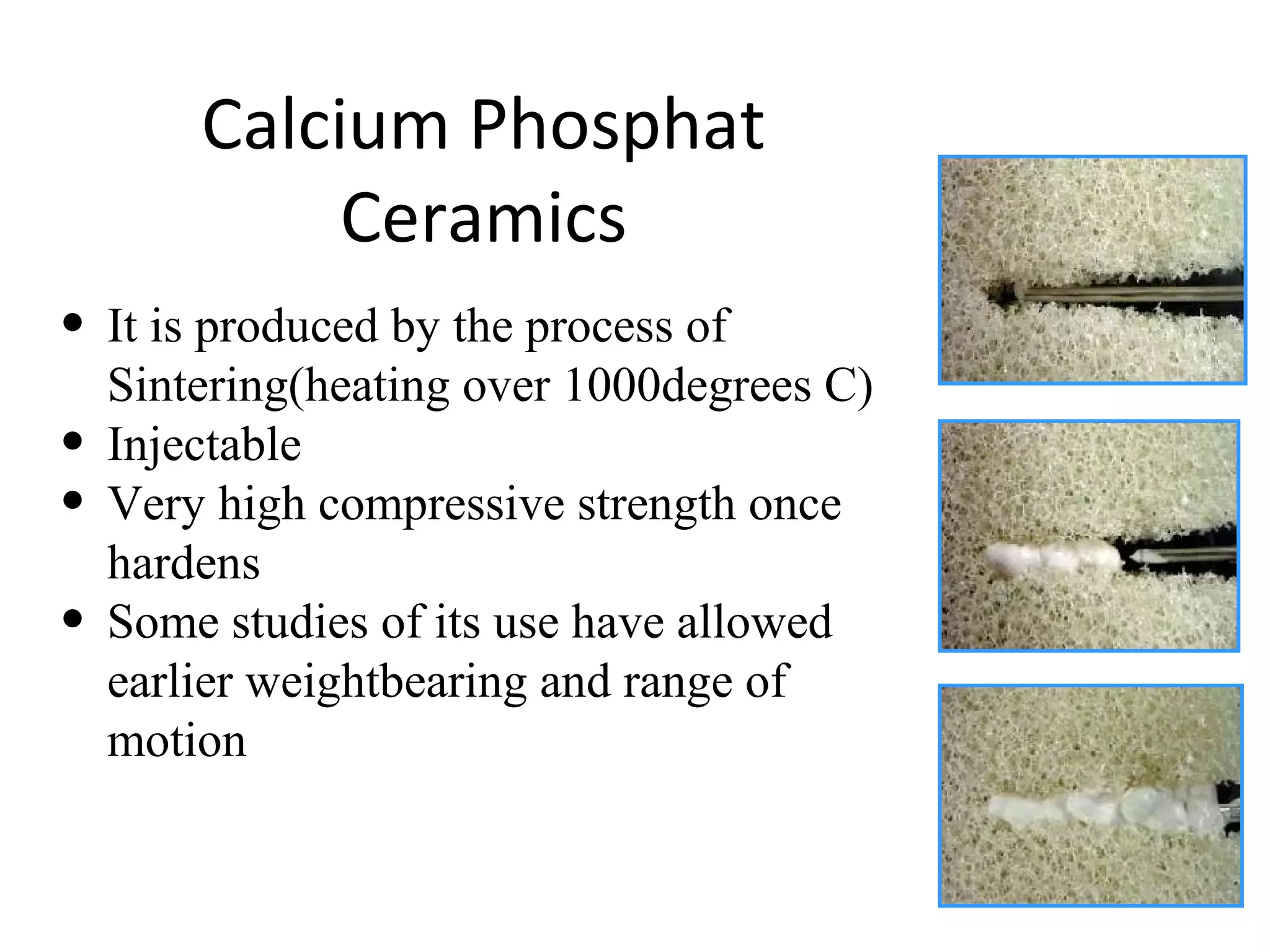
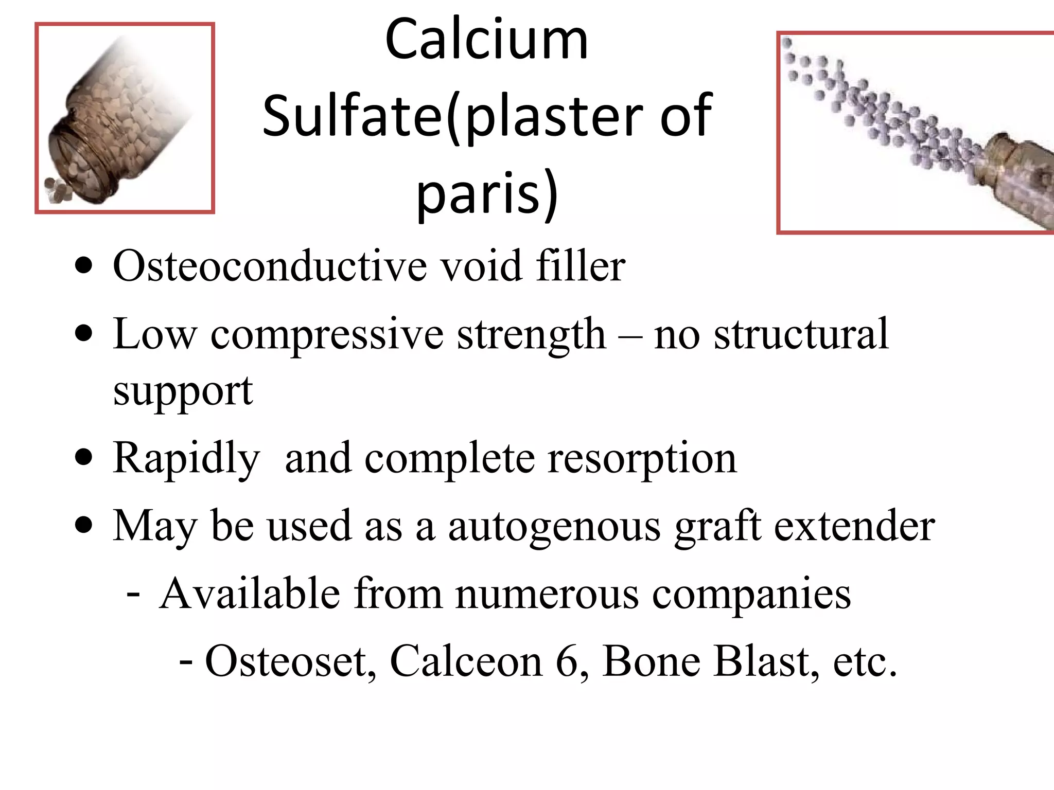
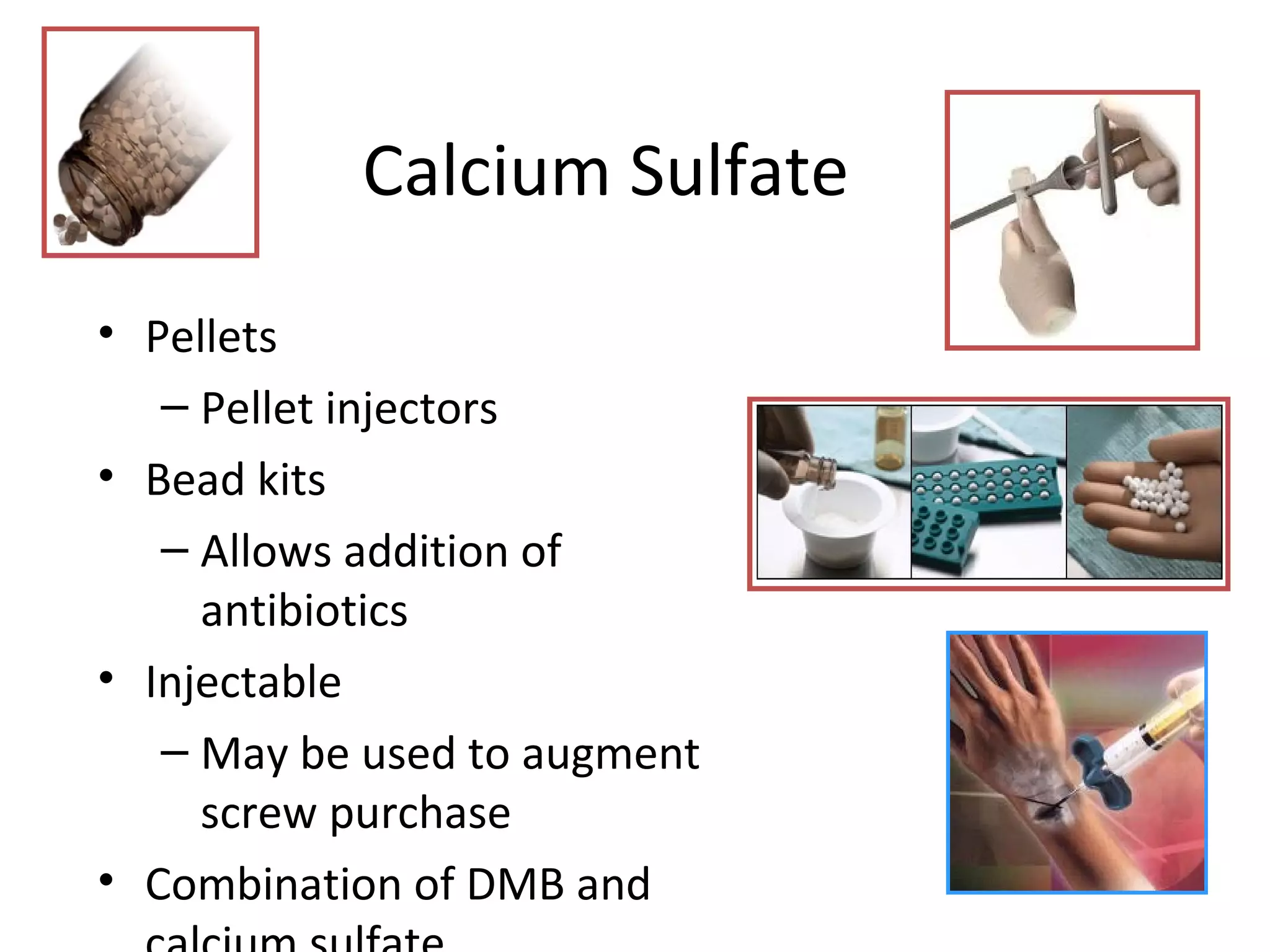
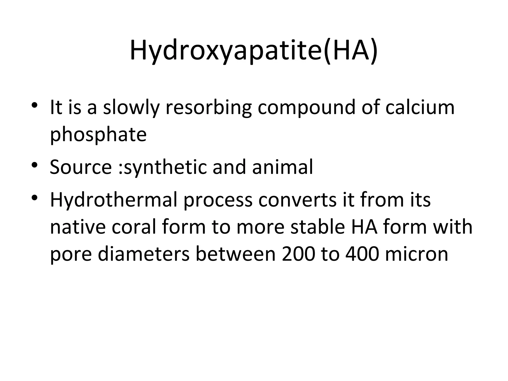
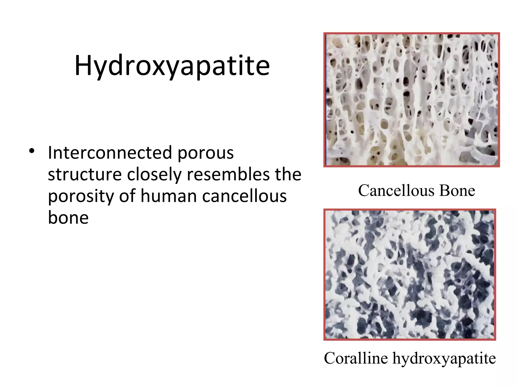
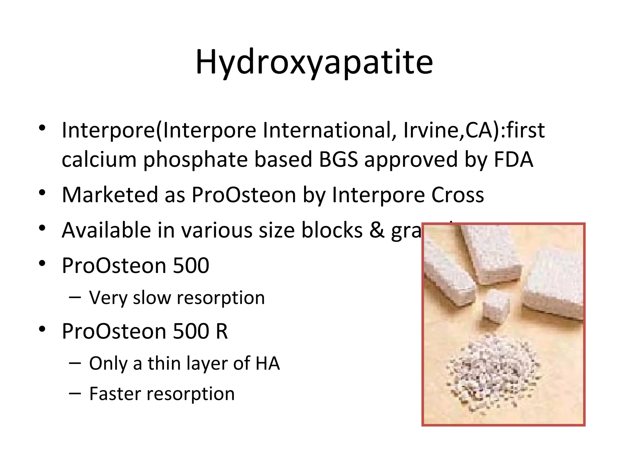
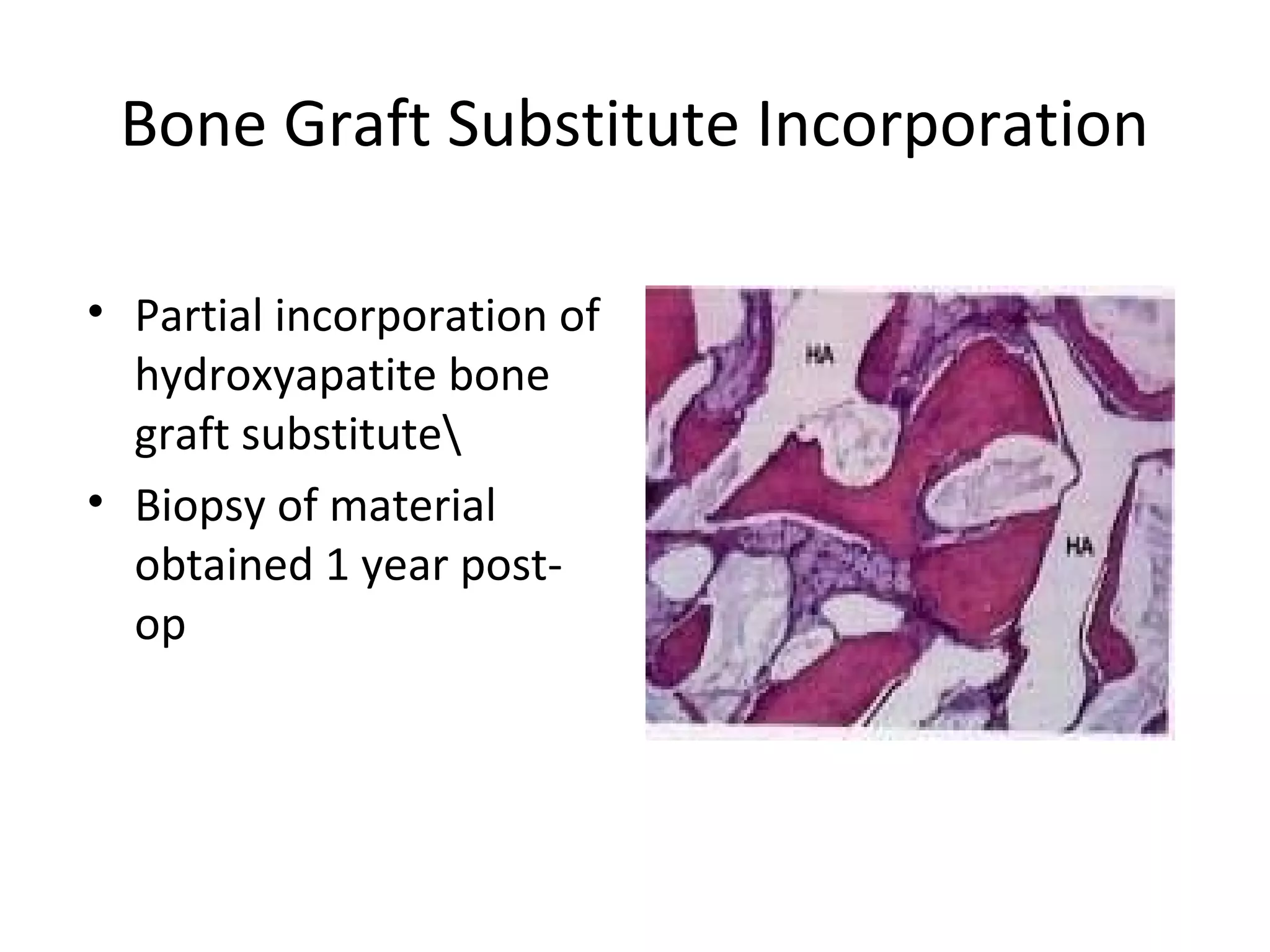
This document summarizes different types of bone grafts and bone graft substitutes. It discusses autogenous bone grafts which are considered the gold standard but have limitations related to donor site morbidity. Allografts from cadaveric donors are also discussed. Bone graft substitutes described include ceramics like calcium sulfate and calcium phosphate, demineralized bone matrix, and growth factors like bone morphogenetic proteins which provide osteoinduction. The properties, advantages, and limitations of each type of graft and substitute are summarized.
Anuman- An inference for helpful in diagnosis and treatment
Bone grafts and bone grafts substitutes
1. Bone Grafting and Bone Graft
Substitutes
Dr. Gurvinder Singh
Dr. Siddharth Gupta
Residents UCMS & GTB, HOSPITAL ,DELHI
2. Bone Graft Uses
• To fill cavities or defects resulting from cysts, tumors,
or other cause
• To bridge joints and provide arthrodesis
• To bridge major defects or establish the continuity
of a long bone
• To provide bone blocks to limit joint motion
(arthroereisis)
• To establish union in a pseudarthrosis
• To promote union or fill defects in delayed union,
malunion, fresh fractures, or osteotomies
4. Mesenchymal Stem cells
• Progenitor cells that provide a source of cells
to differentiate into chondroblasts and
osteoblasts during endochondral and
intramembranous bone formation
• In the elderly the pool of these cells
diminishes
• Bone marrow is the source of adult MSCs
5. Types of Bone Grafts:on the basis
of source
• Autogenous: source is the patient , usually
from tibia , fibula or ilium. Also rib
• Allogenic : source is an individual other than
the patient
• Xenograft: derived from different species
7. Autogenous Bone Graft
• “Gold standard”
• May provide osteoconduction, osteoinduction
and osteogenesis
• Drawbacks
– Limited supply
– Donor site pain, haematoma, neuromas , fracture
and heterotopic bone formation
8. Autogenous Bone Grafts
• Cancellous
• Cortical
• Free vascular transfers
• Muscle pedicle bone graft
• Bone marrow aspirate
9. Autogenous Cancellous Bone
Grafts
• Three dimensional scaffold
(osteoconductive)
• Osteocytes and stem cells (osteogenic)
• A small quantity of growth factors
(osteoinductive)ss
• CREEPING SUBSTITUION: process by which
graft is replaced by new bone (I year)
• Used in boneloss: depressed tibial plateau
fractures, revision hip and knee arthroplasty
10. Creeping Substituition
• Creeping substitution, the process of bone
remodeling by osteoclastic resorption and
creation of new vascular channels with
osteoblastic bone formation resulting in new
haversian systems, is the method by which
strong cortical bone is formed from grafted
material.
11. Autogenous Cortical Bone Grafts
Sources:
• Ribs
• Fibula
• Crest of the ilium (also called as tricortical graft)
• It can be of two types:
• Conventinal nov vascular
• Vascularised bone graft
12. Non vascular versus vascular bone
graft
Non vascular
• Mainly osteoconductive
with littile
osteoinductive and no
osteogenic properties.
• Revascularisation is
slow till cortex is
resorbed.
• Remodelling -2years
• Used in defects <6cm
Vascular
• It has immediately
restored blood
supply
• More viable, more
survival of osteocytes
• Can be used in
defects upto 12 cm
or even in
inadequate host.
13. Bone Marrow Aspirate
• RIA(REAMER IRRIGATOR AND ASPIRATOR)
• It is a technique to harvest sizable amount of
bone marrow,which is particularly rich in
mesenchymal stem cells .
• Growth factors supplied:
Fibroblasts growth factor(FGF)-2
Insulin like growth factor(IGF)-2
Transforming growth factor(TGF) beta
Absence of bone morphogenetic protein-2
14. Advantages of RIA
• Provides enriched osteogenesis
• Decrease intramedullary canal pressure
• Minimal risk of fat embolism
• Potential source of autologous bone,
mesenchymal cells and bone growth factors.
18. Complications of RIA
• Perforation of the meduallary canal which
may require prophylactic intramedullary
fixation.
• Significant blood loss
19. Steps to minimize risks
• Preoperatively donor radigraph should be
used to measure isthmus
• Blood should be arranged for replacement.
• Switch off the aspirator when there is no
reaming
• Postp of ambulation should be protective
• Check haematocrit
• Avoid in metabolic bone diseases
21. Complications of iliac crest graft
• Full thickness iliac crest graft lead to
herniation.
• The lateral femoral cutaneous and ilioinguinal
nerves are at risk during harvest of bone from
the anterior ilium
• Alter the contour of the anterior crest,
producing significant cosmetic deformity
• Arteriovenous fistula, pseudoaneurysm,
ureteral injury, anterior superior iliac spine
avulsion, and pelvic instability
24. Grafts from tibia
• The subcutaneous anteromedial aspect of
tibia is the source of structural autografts.
• The plateau of tibia supplies cancellous bone.
26. Disadvantages of tibial graft
Normal limb is jeopardized
Increased duration of surgery
Protected weight bearing for atleast 6 to 12
months .
27. Bone graft from fibula
• Entire proximal two third of the fibula can be
used for bone graft
• The proximal rounded configuration of the
fibula is covered with hyaline cartilage.
• May replace distal radius or even distal third
of fibula
• The middle third of fibula can serve as the
peroneal artery based vascular graft
28. Points to remember for fibular
bone graft
• The peroneal nerve must not be damaged;
• The distal fourth of the bone must be left to
maintain a stable ankle
• The peroneal muscles should not be cut.
29. Phemister bone graft
• It is subcortical cancellous bone grafting
• A bone graft of cortical bone with cancellous
bone chips to enhance callus formation.
• Bone-grafting without disturbing the pre-existing
callus
• Bone graft is taken by elevating the
osteoperiosteal flaps.
30. Requisites for phemister graft
• Petalling should carried out at the fracture site
• The mobility at the fracture site should be
minimal
• The fracture should have an acceptable
alignment
• The knee joint should have a good range of
motion
31. Urist AAA bone graft
• Composed of bone morphogenic protein and
autolysed , antigen-extracted and autogenic
bone.
• h-BMP/AAA composite implants represent
adjunctive treatment of difficult nonunions
• Composite implants may be implanted in
either partial or complete segmental defects
of long bones
32. Allograft
The morbidity and limited amount of autogenic
bone graft calls for a need of allogenic bone
Graft.
They are indicated in
1.Children 2. Elderly 3.Poor surgical risk
4.Enough graft cannot be harvested
34. Bone Allografts
• Cancellous or cortical
– Plentiful supply
– Limited infection risk (varies based on processing
method)
– Provide osteoconductive scaffold
– May provide structural support
35. Bone Allografts
• Freeze-dried
– Even less antigenic
– Time to test for diseases
– Strictly regulated by FDA
– Can be stored at room temperature up to 5 years
– Mechanical properties degrade
36. Bone Bank
• It is a facilitiy to provide safe and efficient
allograft material.
• Hosts should be screened for :
infections,malignancies(except for basal cell
carcinoma of the skin), collagen vascular
diseases, metabolic bone diseases and
presence of toxins.
37. Technique
• Bone is collected in clean and unsterile
environment.
• It is nibbled to remove the articular cartilage.
• It can be sterilized by irradiation, ethylene
oxide or strong acid( 0.55 % HCl)
• It is subject to deep freeze upto -70 to -80
degrees celcius(frozen)
• Freeze drying involves removal of water and
vacuum packaging of the tissue
38.
39.
40. Freeze dried bone graft
• Decreases expression of MHC 1 complex in
osteoblasts
• Decreased osteoinductive properties
• Reduced mechanical integrity
• Decreased number of viable cells
• Slow revascularisation and delaye remodelling
• Histologically mono nuclear cells surround the
newly developed blood vessels
41. Demineralized Bone Matrix
• Prepared from cadaveric human bone
• Acid extraction of bone leaving
– Collagen(type 1)
– Noncollagenous proteins
– Bone growth factors
• BMP quantity extremely low and variable
• Sterilized which may decrease the availability of BMP
42. Demineralized Bone Matrix
• Available from multiple vendors in multiple
preparations
– Gel
– Putty
– Strip
– Combination products with cancellous bone and
other bone graft substitute products
43. Demineralized Bone Matrix
• Growth factor activity varies between tissue
banks and between batches
• While they may offer some osteoinductive
potential because of available growth factors,
they mainly act as an osteoconductive agents
Han B et al. J Orthop Res. 21(4):648-54, 2003.
Blum B, et al. Orthopedics. 27 (1 Suppl): S161 – S165, 2004.
45. Graft Incorporation
• Hematoma formation
– Release of cytokines and growth factors
• Inflammation
– Development of fibrovascular tissue (18 hours)
46. Graft Incorporation
• Hematoma formation
– Release of cytokines and growth factors
• Inflammation
– Development of fibrovascular tissue
• Vascular ingrowth
– Often extending Haversian canals
47. Graft Incorporation
• Hematoma formation
– Release of cytokines and growth factors
• Inflammation
– Development of fibrovascular tissue(18 hours)
• Vascular ingrowth
– Often extending Haversian canals (10 -12 days)
• Focal osteoclastic resorption of graft
48. Graft Incorporation
• Hematoma formation
– Release of cytokines and growth factors
• Inflammation
– Development of fibrovascular tissue
• Vascular ingrowth
– Often extending Haversian canals
• Focal osteoclastic resorption of graft
• Intramembranous and/or endochondral bone
formation on graft surfaces…….
49.
50. Graft Incorporation
• Cortical allograft strut
graft placed next to
cortex of host
• After 4 years of
incorporation
51. Bone Graft Substitutes
• Need for bone graft alternatives has lead to
development of numerous bone graft substitutes
• Avoid morbidity of autogenous bone graft harvest
• Mechanical properties vary
• Most offer osteoconductive properties
• Some provide osteoinductive properties
52. Bone Graft Substitutes
Potential Roles
• Extender for autogenous bone graft
– Large defects
– Multiple level spinal fusion
• Enhancer
– To improve success of autogenous bone graft
• Substitute
– To replace autogenous bone graft
55. Laurencin et al, classification
• Allograft based
• Factor based
• Cell based
• Ceramic based
• Polymer based
• Composite
56. Ideal bone graft substitute
• Scaffolding for osteoconduction
• Growth factors for osteoinduction
• Progenitor cells for osteogenesis
• Biocompatible and biodegradable and
mechanical properties similar to the
surrounding bone
• Each substitute available nowadays fulfill only
some of the criteria
57. Bone Graft Substitutes
• Resorption rates vary widely
– Dependant on composition
• Calcium sulfate - very rapid
• Hydroxyapatite (HA) – very, very slow
• Some products may be combined to optimize
resorption rate
– Also dependant on porosity, geometry
58. Bone Graft Substitutes
• Mechanical properties vary widely
– Dependant on composition
• Calcium phosphate cement has highest compressive
strength
• Cancellous bone compressive strength is relatively low
• Many substitutes have compressive strengths similar to
cancellous bone
• All designed to be used with internal fixation
59. Allograft based
• Includes allograft bone used alone or in
combination with other materials
• Available as demineralised bone matrix
60. Factor based
• Involves natural or recombinant factors
• Factors responsible for differentiation of
progenitor cells and regulation of activities
• Mechanism of action: based mostly on
activation of protein kinase
• Combined and simultaneous activity of
various factors controlled resorption and
formation of new bone
61. • Factor + receptors on the cell surface
• Activation of protein kinase
• Transcription of mRNA
• Protein synthesis
• Regulation of cell activities
62. • Includes TGF-beta, IGF-I&II,PDGF,FGF and
BMPs
• Mostly in research phase
• Recombinant BMP-2 as INFUSE bone graft
64. BMP 2 and 7
BMP 2
• Acts as a disulfide
linked homodimer
and helps in bone
and cartilage
formation
• It is a candidate as
retinoid mediator
and helps
osteoblastic
differentiation
BMP 7
• It plays a key role in
osteoblastic
differentiation
• It induces the
prodution of SMAD 1
• It plays a key role in
renal development
and repair
65. Bone Morphogenetic Proteins
• Produced by recombinant technology
• Two most extensively studied and
commercially available
– BMP-2 (Infuse) Medtronics
– BMP-7 (OP-1)Stryker ss
– BMP-2 and BMP-7 are water soluble and
require a carrier to remain in the operative
area to be effective
66. BMP-2 for Open Tibial Fractures
• Prospective, randomized
study
• 450 patients
· All received IM nail (vast
majority with UNREAMED
technique) and appropriate
soft tissue management
· Randomized to 3 treatments at
time of definitive wound
closure
· Placebo
· 0.75 mg/ml BMP-2/ACS
· 1.50 mg/ml BMP-2/ACS
BESTT Study Group, et al. J Bone Joint Surg 84A: 2123, 2002.
67. Results
• 44% reduction in risk of
nonunion/delayed union with
high dose BMP-2
• Significantly faster fracture
healing
• Significantly fewer
– invasive interventions
– hardware failures
– infections
BESTT Study Group, et al. J Bone Joint Surg 84A: 2123, 2002.
68. Cell Based
• Based on in vitro differentiation of
mesenchymal stem cells to osteoblastic
lineage
• They have been used along with ceramics
• Proposed to be used in bone repair prosthetic
setting
69. Ceramic based
• About 60% BGS involves ceramics- alone or in
combination
• Eg : calcium sulfate, calcium phosphate,
bioactive glass
• Primary inorganic componet is calcium
hyroxyapatite
• Property of osteointegration, newly formed
mineralised tissue forms intimate bond with
implant materials
70. Calcium Phosphate Ceramics
• Enable osteoconduction but use is limited
due to poor tensile strength and brittleness
• Injectable pastes of calcium and phospate
– Norian SRS (Synthes/Stratec)
– Alpha BSM (Etex/Depuy)
– Callos Bone Void Filler (Skeletal Kinetics)
71. Calcium Phosphat
Ceramics
• It is produced by the process of
Sintering(heating over 1000degrees C)
• Injectable
• Very high compressive strength once
hardens
• Some studies of its use have allowed
earlier weightbearing and range of
motion
72. Calcium
Sulfate(plaster of
paris)
· Osteoconductive void filler
· Low compressive strength – no structural
support
· Rapidly and complete resorption
· May be used as a autogenous graft extender
- Available from numerous companies
- Osteoset, Calceon 6, Bone Blast, etc.
73. Calcium Sulfate
• Pellets
– Pellet injectors
• Bead kits
– Allows addition of
antibiotics
• Injectable
– May be used to augment
screw purchase
• Combination of DMB and
calcium sulfate
74. Hydroxyapatite(HA)
• It is a slowly resorbing compound of calcium
phosphate
• Source :synthetic and animal
• Hydrothermal process converts it from its
native coral form to more stable HA form with
pore diameters between 200 to 400 micron
75. Hydroxyapatite
• Interconnected porous
structure closely resembles the
porosity of human cancellous
bone
Cancellous Bone
Coralline hydroxyapatite
76. Hydroxyapatite
• Interpore(Interpore International, Irvine,CA):first
calcium phosphate based BGS approved by FDA
• Marketed as ProOsteon by Interpore Cross
• Available in various size blocks & granules
• ProOsteon 500
– Very slow resorption
• ProOsteon 500 R
– Only a thin layer of HA
– Faster resorption
77. Hydroxyapatite:indications
• Valgus instability following lateral tibial
plateau fracture
• Varus instability following medial condyle
fracture of tibia
• Articular incongruence of 10 mm or more
• Translation of major condylar fragment of
more than 5mm
78. Tricalcium Phosphate(TCP)
• TCP composition is similar to calcium and
phosphate phase of human bone and has
porous nature
• TCP undergoes partial resorption and some of
it may be converted to HA once implanted in
the human body
• Complete resorption at 6 months
79. Tricalcium Phosphate
• Wet compressive strength slightly less than
cancellous bone
• Available as blocks, wedges, and granules
• Numerous tradenames
– Vitoss (Orthovita)
– ChronOS (Synthes)
– Conduit (DePuy)
– Cellplex TCP (Wright Medical)
– Various Theri__ names (Therics)
80. Calcium Phosphate Cements(CPC)
• CPC is used as void filler in defects
• It consists of inorganic calcium and phosphate
combined to form an injectable paste
81. Polymer based
• Can be divided: natural/synthetic
• Further divided into: biodegradable/non
biodegradable
• eg: Healoss(depuy)-natural
• Eg :Cortoss- injectable resin based product
• Eg : Rhakoss(Orthovita)
82. Collagen Based Matrices
• Highly purified Type 1 bovine
dermal fibrillar collagen
• Bone marrow is added to
provide bone forming cells
• Collagraft (Zimmer)
– Collagen / HA / Tricalcium
phosphate
• Healos (Depuy)
– Collagen / HA
83. Composite graft
• In this two or more type of bone graft
substitutes combined together
• So that osteoconductive and osteoinductive
properties of different BGS, is combined
84. Calcium Phosphate-Collagen
Composite
• Collagen provides binding sites for matrix
proteins
• Type I and III is added to HA,TCP and
autologous bone marrow to form a graft
material
• Although no structural support but augments
frature healing
85. Bone Graft Substitute Incorporation
• Partial incorporation of
hydroxyapatite bone
graft substitute
• Biopsy of material
obtained 1 year post-op
Editor's Notes
Bone grafts, and bone graft substitutes, are used primarily for three main purposes. One, to provide structural support, such as after elevation of a depressed tibial plateau fracture. Second, to fill a void, such as after excision of a bone cyst in an area that does not demand any structural support. Third, to improve fracture healing. Either by speeding healing or increasing the successful union rate.
The most common form of autogenous bone graft is cancellous bone, usually harvested from either anterior or posterior iliac crests, but which also may be harvested locally such as in the proximal tibia or distal radius. Cortical bone strips may be harvested from the inner table of the pelvis. Occasionally a vascularized bone graft may be used, such ass a free fibular transfer. Another option for obtaining osteogenic cells is by bone marrow aspirate.
Two bone morphogenetic proteins are commercially available. These are BMP-2,marketed by Medtronics, and BMP-7, more commonly known of osteogenic protein 1, produced by Stryker Biotech.
These products are produced by recombinant technology in which large quantities of the material are produced in cell culture.
BMPs have been studies in several human investigations including open fractures and nonunions.
BMP-2 was studied in a prospective, randomized study of 450 patients with open tibial shaft fractures treated with fracture debridement and an IM nail. At the time of definitive wound closure patients were randomized to one of three treatments
placebo
0.75 mg/ml BMP-2/ACS (lower BMP dose)
1.50 mg/ml BMP-2/ACS (higher BMP dose)
The primary outcome measure was the proportion of patients requiring secondary intervention because of delayed union or nonunion within twelve months postoperatively.
In the group treated with the higher dose BMP-2 there was a 44% reduction in risk of nonunion/delayed union
Significantly faster fracture healing
Significantly fewer invasive interventions
Significantly fewer hardware failures and fewer infections
The graph from their publication shows the placebo control groups in black representing the standard of care. The Gustilo IIIB&apos;s are on the left and the Gustilo I, II and IIIA&apos;s on the right. There is a statistically significant reduction in the number of secondary interventions in those fractures treated with the higher dose of BMP-2.
Calcium phosphate products are an injectable paste of calcium and phosphate.
Norian SRS was the first such product available and is marketed by Synthes / Stratec.
Two other calcium phosphate type products are also available in the U.S.
Medical grade calcium sulfate is an osteoconductive void filler. It has low compressive strength so doesn’t offer structural support. Another disadvantage of the product is that it resorbs rather rapidly, perhaps more rapidly then it can be replaced by new bone. One way it which it may be used is an extender for autogenous bone graft.
Calcium sulfate products are available both as pellets, bead kits, and injectable preparations. The pellets can be poured into position, but some companies also provide an injector apparatus. The bead kits allow the addition of antibiotics if desired.