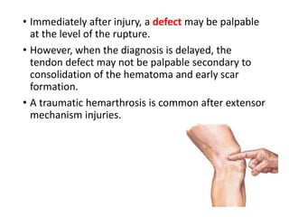
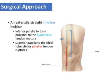
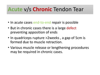
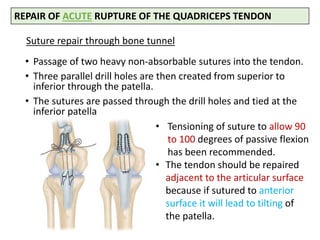
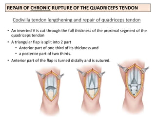
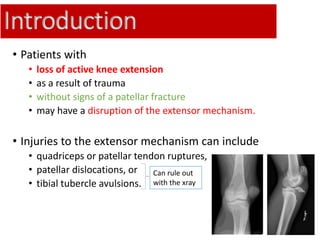
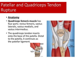
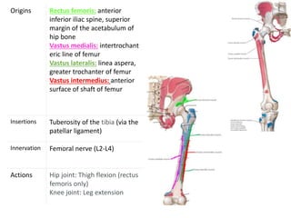
SlideShare a Scribd company logo
Presenter : Dr Yash Oza
Moderator : Dr Ravindra Patil
Introduction
• Patients with
• loss of active knee extension
• as a result of trauma
• without signs of a patellar fracture
• may have a disruption of the extensor mechanism.
• Injuries to the extensor mechanism can include
• quadriceps or patellar tendon ruptures,
• patellar dislocations, or
• tibial tubercle avulsions.
Can rule out
with the xray
Patellar and Quadriceps Tendon
Rupture
• Anatomy
• Quadriceps femoris muscle has
four parts: rectus femoris, vastus
lateralis, vastus medialis, and
vastus intermedius.
• The quadriceps tendon inserts
onto the base of the patella. Distal
to the patella, it continues as
the patellar ligament.
Origins Rectus femoris: anterior
inferior iliac spine, superior
margin of the acetabulum of
hip bone
Vastus medialis: intertrochant
eric line of femur
Vastus lateralis: linea aspera,
greater trochanter of femur
Vastus intermedius: anterior
surface of shaft of femur
Insertions Tuberosity of the tibia (via the
patellar ligament)
Innervation Femoral nerve (L2-L4)
Actions Hip joint: Thigh flexion (rectus
femoris only)
Knee joint: Leg extension
• Patellar Tendon
• Average 4mm thick, but
widens to 5-6mm at
insertion in to tibial
tuberosity
• Merges with Medial &
Lateral retinaculum
• Blood Supply
• Fat pad vessels supply
posterior aspect of tendon
via Med. & Lat. Inferior
Geniculate A.
• Retinacular vessels supply
Anterior portion of tendon
via Lat. IG & Recurrent Tibia
A.
• Proximal & distal insertion
area are relatively avascular
and are a common site of
rupture
Patel
lar
tend
on
Lat.IG &
Rec.TA
Med.IG &
Lat.IG
Relatively avascular
Ant. Post.
• Biomechanics
• Greatest force is applied at 60 degree flexion
• 3-4 times force at @ insertion as compared to
midsubstance
• While climbing stairs forces through patellar tendon is
x3.2 times body weight
Mechanism of Injury
• Quadriceps and patellar tendon ruptures are
typically low-energy injuries.
• Typically, the patient sustains a forceful quadriceps
contraction against a fixed or sudden load of full
body weight with the knee in a flexed position.
• In high-energy injuries it may accompany ACL tear
in 12.5% cases.
Clinical Presentation
• Age Group commonly involved
• Patellar tendon ruptures : <40 years old
• quadriceps tendon ruptures : >40 years of age
• Quadriceps tendon ruptures occur more commonly
in patients with systemic disease or degenerative
changes.
• Bilateral quadriceps tendon rupture may occur in
patients with systemic illness and obesity. Bilateral
rupture of the patellar tendon can occur but is less
frequent.
• History of jumping,squatting or stumbling.
• Pain with an associated tearing or popping
sensation is typical, as is the inability to bear
weight.
• Painful passive knee flexion
• Lack of active knee extension or the inability to
maintain the passively extended knee against
gravity
• Patellar tendon ruptures extend completely through the
retinacular tissue resulting in complete loss of knee
extension.
• In Quadriceps tendon ruptures some extension still may
be possible as whole retinacular tissue is not involved.
• Immediately after injury, a defect may be palpable
at the level of the rupture.
• However, when the diagnosis is delayed, the
tendon defect may not be palpable secondary to
consolidation of the hematoma and early scar
formation.
• A traumatic hemarthrosis is common after extensor
mechanism injuries.
• History of Prodromal symptoms :
fever, malaise, headache
• In general, healthy tendons do not rupture. Tensile
overload of the extensor mechanism usually leads
to # patella which is considered the weakest link.
• Thus Patellar tendon ruptures have been
considered the end stage of long-standing chronic
tendon degeneration.
• So Prodromal Symptoms are frequently associated
with tendon rupture.
• Complaint of chronic pain is also present at site of
the injury.
• Risk factor for Quadriceps/Patellar Tendon Rupture
• Rheumatoid
• SLE
• Diabetes –( Patellar Tendon more association)
• Chronic Renal Failure
• Systemic corticosteroid therapy
• Anabolic steroid use with Heavy Exercise –( Quadriceps
Tendon more association)
• Local steroid injection
• Chronic patellar tendonitis
• Fluoroquinolone antibiotics
Imaging
• Xray
• An AP and lateral plain radiograph
should be obtained in all patients.
• Patellar Tendon Rupture :
• The unopposed pull of the
quadriceps muscle will result in
proximal migration of the patella.
• Patella alta - position of the patella is
considered higher than normal
• position of the patella superior to
Blumensaat's line on the lateral
radiograph
• Insall-Salvati Index - normal value
between 0.8-1.2 . If the ratio is
higher than there is patella alta
Blumensaat's
line should extend to
inferior pole of the
patella at 30 degrees
of knee flexion
Blumensaat line is a line
which corresponds to the
roof of the intercondylar
fossa of the femur as seen
on a lateral radiograph of
the knee joint.
• Xray findings suggestive of a quadriceps tendon
rupture include
• obliteration of the quadriceps tendon shadow,
• a suprapatellar mass,
• suprapatellar calcific densities
• an inferiorly displaced patella.
Degenerative spurring (“tooth sign”), as seen on a lateral
view, may indicate significant changes in the quadriceps
mechanism
•USG
• High-resolution ultrasonography has been
recognized as an effective method of
examining the patellar and quadriceps
tendons in both acute and chronic
injuries.
• Routinely done method to confirm
diagnosis.
• MRI is very effective but expensive, method of
diagnosing.
• It is not recommended in the evaluation of most
suspected extensor mechanism injuries, but may
be helpful in patients with neglected tears or
partial tendon injuries or high-energy injury.
Normal
Quadriceps Rupture Patellar Tendon Rupture
Initial Management
• RICE
• A traumatic hemarthrosis is common after
tendon ruptures.
• Ice, compression, elevation, and anti-
inflammatory medications can be used to treat
local symptoms.
• Role of Knee aspiration
• no studies have shown a benefit for knee aspiration in
these injuries,
• aspiration of a tense hematoma WAS considered to
reduce pain and promote recovery.
Treatment Options :
Conservative & Operative
Indication Relative Contraindication
• Incomplete ruptures
are usually managed
conservatively if full
active extension is
present
• If Medically unfit for
surgery
• Open injury
• Extensor lag
• Loss of knee extensor
function
Conservative Treatment
• The limb is initially immobilized with the knee in full
extension for 4 to 6 weeks,
• after which, protected range of motion and
strengthening are begun.
• Initially, flexion of greater than 90 degrees is
avoided to reduce stress on the tendon.
• Restrictions are removed once the patient achieves
good quadriceps muscle control and is able to
perform a straight-leg raise without discomfort.
• Nonsurgical management of a complete quadriceps
or patellar tendon rupture generally yields poor
results
• long-term disability in gait and weakness.
• Untreated complete ruptures will result in
ambulation with a stiff-knee gait or circumduction
to allow the foot to clear the ground during the
swing phase of gait.
• Patients will also complain of knee buckling and
difficulty in climbing stairs.
• And Loss of extensor mechanism function is an
indication for surgery.
Outcome of conservative treatment
Operative Treatment
• Operative treatment is indicated for all tendon
ruptures in which extensor mechanism function is
compromised.
Patient positioning
• Supine positioning
• A small bump under the hip is helpful for external
rotation of the limb
• Another small bump that can be
• moved below the knee for slight flexion or
• under the ankle for knee extension
Surgical Approach
• An extensile straight midline
incision
• inferior patella to 5 cm
proximal to the quadriceps
tendon rupture
• superior patella to the tibial
tubercle for patellar tendon
ruptures.
5cm
Acute v/s Chronic Tendon Tear
• In acute cases end-to-end repair is possible
• But in chronic cases there is a large defect
preventing apposition of ends
• In quadriceps rupture >2weeks , a gap of 5cm is
formed due to muscle retraction.
• Various muscle release or lengthening procedures
may be required in chronic cases.
Suture repair through bone tunnel
• Passage of two heavy non-absorbable sutures into the tendon.
• Three parallel drill holes are then created from superior to
inferior through the patella.
• The sutures are passed through the drill holes and tied at the
inferior patella
REPAIR OF ACUTE RUPTURE OF THE QUADRICEPS TENDON
• Tensioning of suture to allow 90
to 100 degrees of passive flexion
has been recommended.
• The tendon should be repaired
adjacent to the articular surface
because if sutured to anterior
surface it will lead to tilting of
the patella.
Codivilla tendon lengthening and repair of quadriceps tendon
• An inverted V is cut through the full thickness of the proximal segment of the
quadriceps tendon
• A triangular flap is split into 2 part
• Anterior part of one third of its thickness and
• a posterior part of two thirds.
• Anterior part of the flap is turned distally and is sutured.
REPAIR OF CHRONIC RUPTURE OF THE QUADRICEPS TENDON
• The cylinder cast or knee brace for 6 weeks.
• Weight bearing with crutches is allowed at 3 weeks.
• After Cast removal range of knee motion from 0 to
60 degrees; the range is increased 10 to 15 degrees
each week.
• An aggressive strengthening program is essential
for good functional recovery.
POSTOPERATIVE CARE FOLLOWING
QUADRICEPS TENDON REPAIR :
SUTURE REPAIR OF ACUTE PATELLAR
TENDON RUPTURE
• The site of the tear (proximal, midsubstance,
or distal) will dictate the preferred surgical
repair technique.
• Most patellar tendon ruptures occur at the
insertion on the inferior pole of the patella as
an avulsion.
• It is done by passage of 3 heavy non-
absorbable sutures into the tendon.
• 4 parallel drill holes are then created from
inferior to superior in the patella.
• The sutures are passed through the drill holes
and tied at the superior pole of the patella.
If the patellar tendon is extensively
frayed, 2 running interlocking non-
absorbable sutures can be used to secure
the tendon.
• Use a suture retriever or Beath pin to
thread the suture strands through 3-
mm drill tunnels,
• one horizontally into the tibial tubercle
• two vertically into the patella
*If secure fixation cannot be obtained
with this method, augment the repair
with the semitendinosus or gracilis
tendon.
MERSILENE LOOP TENDON REPAIR
(ACUTE)
RUPTURES THROUGH THE SUBSTANCE
OF THE TENDON
• The tendon is split in 3 bundles. (2 with
long arm directed upwards & 1 downwards)
• Running interlocking sutures placed in
bundles
• 2 parallel vertical holes drilled in the patella
& 1 transverse hole drilled in the tibial
tuberosity.
• Sutures are passed through holes & tied.
• Repair the individual bundles side-to-side
after appropriate tendon length is
determined.
*If needed place a circumferential tension
suture of non-absorbable box wire.
(ACUTE)
SUTURE ANCHOR REPAIR OF PATELLAR
TENDON RUPTURE
• For proximal avulsion of the tendon
from the patella, place three suture
anchors equidistant along the inferior
pole of patella.
• Pull the suture through the anchor eyelet
Pass the one suture arm down and back
of the tendon stump in a locking Krackow
fashion, and pull the another arm to
reduce the tendon & Tie each suture
securely.
• For distal avulsion of the tendon, the
same procedure is used but the suture
anchors are placed in the tibial tubercle
(ACUTE)
POSTOPERATIVE CARE following repair
of acute patellar tendon repair
• Weight bearing is allowed with the knee braced in
full extension.
• Range of Motion is progressed as tolerated, with
the goal of
• 90 degrees of flexion by 4 to 6 weeks and
• full motion by 10 to 12 weeks.
• Isometric quadriceps contractions can be done
immediately after surgery,
• Progressing to straight-leg raises start at 6 weeks.
• Full return to activities after 6 months.
TREATMENT OF CHRONIC
RUPTURE OF PATELLAR TENDON
In chronic cases patella is retracted proximally
and may require extensive surgical release to draw it
distally to the appropriate level.
Before surgery, lateral radiographs of the
uninvolved extremity should be obtained with the
knee flexed to 45 degrees to evaluate patellar height;
• These are compared with radiographs of the
involved knee during surgery to determine the
appropriate tendon length.
• Preoperative traction through a
K-wire placed transversely in the
patella was used
• But, better results can be
obtained with proximal release
of scar tissue and a modified
Thompson quadricepsplasty, if
necessary.
• Modified Thompson
quadricepsplasty is a Z-plasty
lengthening of the rectus
femoris tendon
If sufficient patellar
tendon is left for
repair,
augmentation with
the
semitendinosus or
gracilis tendon
may be indicated.
If the rupture is
several months old,
an allograft can be
used, and an
Achilles tendon
allograft is used.
ACHILLES TENDON ALLOGRAFT FOR
CHRONIC PATELLAR TENDON RUPTURE
• Use an oscillating saw to make a slot in
Tibial tuberosity
• Contour the bone attached to the Achilles
tendon to fit flush in the slot
• Split the Achilles tendon graft into three
branches, the central branch should be 8 to
9 mm in diameter.
• Central arm of graft is placed through 9 mm
longitudinal tunnel and then through
vertical slit in quadriceps tendon.
• Tunnel is placed centrally to avoid
penetration of articular cartilage.
• Graft is secured with multiple sutures.
POSTOPERATIVE CARE.
• Apply cylindrical cast post-OP. At 10 to 14 days, the cast
is removed for wound evaluation.
• A cylinder cast or locked brace is worn for 4 to 6 weeks.
• At 4 to 6 weeks, after removing cast Active and passive
range-of-motion exercises are begun.
• Weight bearing to tolerance with crutches is allowed
until sufficient motion and strength is gained.
• A progressive strengthening and range-of-motion
exercise program is essential to regain function.
HAMSTRING (SEMITENDINOSUS AND GRACILIS)
AUTOGRAFT AUGMENTATION FOR CHRONIC PATELLAR
TENDON RUPTURE
Technique for one-stage delayed reconstruction of patellar tendon.
A, Steinmann pin through transverse hole in patella is used for distal traction.
B, Proximally divided semitendinosus and gracilis tendons are placed through
holes and fixation wire is inserted.
C, With patella in normal position, fixation wire is secured and gracilis and
semitendinosus tendons are sutured to each other.
HAMSTRING AUTOGRAFT AUGMENTATION FOR
CHRONIC PATELLAR TENDON RUPTURE
A, Z-shortening of patellar tendon
and Z-lengthening of quadriceps
tendon.
B, Tack sutures are placed in tendons
after confirming patellar height
C&D, a transverse hole in patella is
made Semitendinosus and gracilis
tendons are harvested with tendon
stripper and sutured together.
E, Tendons are passed through
transverse hole in patella and sutured
together and also to underlying
patellar tendon.
POSTOPERATIVE CARE.
• At 2 weeks, the cast is removed for wound
evaluation and a new cylinder cast or locked brace
is applied.
• At 6 weeks, vigorous straight-leg raising with
weights and active flexion exercises are instituted.
Outcome
ACUTE QUADRICEPS TENDON REPAIR
• good to excellent results in 80% to 100% of
operatively treated quadriceps tendon ruptures
• Many different techniques of primary repair have
been described and no single technique has
demonstrated superiority.
• The only factor that has been associated with
inferior results is delay in timing of surgical repair of
greater than 2 to 3 weeks.
CHRONIC QUADRICEPS TENDON REPAIR
• Overall, less satisfactory than repair of an acute
tear
• Residual functional deficits present in most
patients.
• Some reported good to excellent results in patients
treated with their modification of the Codivilla
lengthening technique.
• More than half delayed repairs lost between 10 and
20 degrees of full active extension at final follow-up
in few studies.
ACUTE PATELLAR TENDON REPAIR
• Most series have reported between 70% and 100% good to
excellent results.
• The majority of patients who undergo early primary repair
achieve a functional range of motion and normal quadriceps
strength.
• Persistent quadriceps muscle atrophy commonly occurs, but
has not been correlated with loss of strength.
• No relationship has been demonstrated between the
configuration of the rupture, the method of repair, and clinical
outcome.
• An early repair within 2 to 3 weeks of injury is the only factor
that has been associated with better outcomes.
• In patients treated within 7 days ,96% had good to excellent
results.
• Patients with multiple injuries may have contributed to the
slightly lower success rate in some series.
CHRONIC OR NEGLECTED PATELLAR TENDON
RUPTURE
• A vey few series recommended preoperative
traction to overcome the contracted quadriceps
muscle so that the tendon ends could be re-
approximated.
• Results have generally been less satisfactory
compared to acute repair.

More Related Content

What's hot

Galleazi fracture dislocation
Galleazi fracture dislocationGalleazi fracture dislocation
Galleazi fracture dislocation
Fawas Muhammad
 
TALUS FRACTURE AND MANAGEMENT.
TALUS FRACTURE AND MANAGEMENT.TALUS FRACTURE AND MANAGEMENT.
TALUS FRACTURE AND MANAGEMENT.
Dr. Anshu Sharma
 
Clavicle fractures
Clavicle fractures Clavicle fractures
Clavicle fractures
Hardik Pawar
 
Monteggia fracture &amp; galeazzi fracture
Monteggia fracture &amp; galeazzi fractureMonteggia fracture &amp; galeazzi fracture
Monteggia fracture &amp; galeazzi fracture
BipulBorthakur
 
Dr. pl srinivas ug class 1
Dr. pl srinivas ug class 1Dr. pl srinivas ug class 1
Dr. pl srinivas ug class 1varuntandra
 
Calcaneal fractures
Calcaneal fracturesCalcaneal fractures
Calcaneal fractures
Rohit Vikas
 
Bennetts Fracture
Bennetts FractureBennetts Fracture
Bennetts Fracturejfreshour
 
Shaft of humerus fracture
Shaft of humerus fractureShaft of humerus fracture
Shaft of humerus fracture
BipulBorthakur
 
Kienbock disease
Kienbock  diseaseKienbock  disease
Kienbock disease
azhanrubeesh
 
Hemiarthroplasty of Hip joint
Hemiarthroplasty  of  Hip joint Hemiarthroplasty  of  Hip joint
Hemiarthroplasty of Hip joint
Dr Thouseef Abdul Majeed
 
Supracondylar fractures humerus
Supracondylar fractures humerusSupracondylar fractures humerus
Supracondylar fractures humerus
M A Roshan Zameer
 
Shoulder dislocation: Types and Management Methods of Reduction
Shoulder dislocation: Types and Management Methods of ReductionShoulder dislocation: Types and Management Methods of Reduction
Shoulder dislocation: Types and Management Methods of Reduction
Uzair Siddiqui
 
Calcaneal fractures
Calcaneal fracturesCalcaneal fractures
Calcaneal fractures
dr.pradeep pathak
 
Galeazzi fracture dislocation
Galeazzi fracture  dislocationGaleazzi fracture  dislocation
Galeazzi fracture dislocation
rashree-singh
 
derangement knee ppt
derangement knee pptderangement knee ppt
derangement knee ppt
dralizameer
 
Supracondylar Fractures
Supracondylar FracturesSupracondylar Fractures
Supracondylar Fractures
Pulasthi Kanchana
 
Blood supply of femoral head at various ages
Blood supply of femoral head at various agesBlood supply of femoral head at various ages
Blood supply of femoral head at various ages
songao
 
Cubitus varus deformity
Cubitus varus deformityCubitus varus deformity
Cubitus varus deformity
ramachandra reddy
 
Humerus fracture
Humerus fractureHumerus fracture
Humerus fracturevaruntandra
 

What's hot (20)

Galleazi fracture dislocation
Galleazi fracture dislocationGalleazi fracture dislocation
Galleazi fracture dislocation
 
TALUS FRACTURE AND MANAGEMENT.
TALUS FRACTURE AND MANAGEMENT.TALUS FRACTURE AND MANAGEMENT.
TALUS FRACTURE AND MANAGEMENT.
 
Clavicle fractures
Clavicle fractures Clavicle fractures
Clavicle fractures
 
Monteggia fracture &amp; galeazzi fracture
Monteggia fracture &amp; galeazzi fractureMonteggia fracture &amp; galeazzi fracture
Monteggia fracture &amp; galeazzi fracture
 
Dr. pl srinivas ug class 1
Dr. pl srinivas ug class 1Dr. pl srinivas ug class 1
Dr. pl srinivas ug class 1
 
Calcaneal fractures
Calcaneal fracturesCalcaneal fractures
Calcaneal fractures
 
Bennetts Fracture
Bennetts FractureBennetts Fracture
Bennetts Fracture
 
Shaft of humerus fracture
Shaft of humerus fractureShaft of humerus fracture
Shaft of humerus fracture
 
Kienbock disease
Kienbock  diseaseKienbock  disease
Kienbock disease
 
Hemiarthroplasty of Hip joint
Hemiarthroplasty  of  Hip joint Hemiarthroplasty  of  Hip joint
Hemiarthroplasty of Hip joint
 
Supracondylar fractures humerus
Supracondylar fractures humerusSupracondylar fractures humerus
Supracondylar fractures humerus
 
Ankle injury
Ankle injuryAnkle injury
Ankle injury
 
Shoulder dislocation: Types and Management Methods of Reduction
Shoulder dislocation: Types and Management Methods of ReductionShoulder dislocation: Types and Management Methods of Reduction
Shoulder dislocation: Types and Management Methods of Reduction
 
Calcaneal fractures
Calcaneal fracturesCalcaneal fractures
Calcaneal fractures
 
Galeazzi fracture dislocation
Galeazzi fracture  dislocationGaleazzi fracture  dislocation
Galeazzi fracture dislocation
 
derangement knee ppt
derangement knee pptderangement knee ppt
derangement knee ppt
 
Supracondylar Fractures
Supracondylar FracturesSupracondylar Fractures
Supracondylar Fractures
 
Blood supply of femoral head at various ages
Blood supply of femoral head at various agesBlood supply of femoral head at various ages
Blood supply of femoral head at various ages
 
Cubitus varus deformity
Cubitus varus deformityCubitus varus deformity
Cubitus varus deformity
 
Humerus fracture
Humerus fractureHumerus fracture
Humerus fracture
 

Similar to Patellar and quadriceps tendon rupture

Patella fx and mechanism injuries
Patella fx and mechanism injuriesPatella fx and mechanism injuries
Patella fx and mechanism injuries
rohit raj
 
patellar fracture.ppt. .
patellar fracture.ppt.                 .patellar fracture.ppt.                 .
patellar fracture.ppt. .
AkshayBadore2
 
Patellofemoral disorders
Patellofemoral disordersPatellofemoral disorders
Patellofemoral disorders
Ponnilavan Ponz
 
Knee dislocation powerpoint for medical students
Knee dislocation powerpoint for medical studentsKnee dislocation powerpoint for medical students
Knee dislocation powerpoint for medical students
CasualityShift
 
Open Reduction of carpal bone fractures
Open Reduction of carpal bone fracturesOpen Reduction of carpal bone fractures
Open Reduction of carpal bone fractures
Hamdard Institute of Medical Sciences and Research
 
Recurrent patellar dislocation
Recurrent patellar dislocationRecurrent patellar dislocation
Recurrent patellar dislocation
boneheallerortho
 
Ankle instability
Ankle instabilityAnkle instability
Ankle instability
RziUllah
 
Acl injury
Acl injuryAcl injury
Acl injury
Sivendu P
 
L07 extensor mechnsm injury
L07 extensor mechnsm injuryL07 extensor mechnsm injury
L07 extensor mechnsm injury
Claudiu Cucu
 
Patellar Instability
Patellar InstabilityPatellar Instability
Patellar Instability
Bijay Mehta
 
Intertrochanteric Fractures of Femur
Intertrochanteric Fractures of FemurIntertrochanteric Fractures of Femur
Intertrochanteric Fractures of Femur
Pulasthi Kanchana
 
INJURIES AROUND HIP [Autosaved].pptx
INJURIES AROUND HIP [Autosaved].pptxINJURIES AROUND HIP [Autosaved].pptx
INJURIES AROUND HIP [Autosaved].pptx
bharti pawar
 
Injuries around the knee
Injuries around the kneeInjuries around the knee
Injuries around the knee
Siddhartha Sinha
 
Musculo skeletal problems in the community
Musculo skeletal problems in the communityMusculo skeletal problems in the community
Musculo skeletal problems in the communityAlampallam Venkatachalam
 
ROTATOR CUFF[4273].pptx
ROTATOR CUFF[4273].pptxROTATOR CUFF[4273].pptx
ROTATOR CUFF[4273].pptx
vedant bansal
 
Radial head and neck fractures
Radial head and neck fracturesRadial head and neck fractures
Radial head and neck fractures
Johny Wilbert
 
L01 hip dislocation, pipkin
L01 hip dislocation, pipkinL01 hip dislocation, pipkin
L01 hip dislocation, pipkin
Claudiu Cucu
 
A lecture on Pelvic Fractures part 1.pptx
A lecture on Pelvic Fractures part 1.pptxA lecture on Pelvic Fractures part 1.pptx
A lecture on Pelvic Fractures part 1.pptx
abotsiebenezer2
 
Hip dislocations and femoral head fractures
Hip dislocations and femoral head fracturesHip dislocations and femoral head fractures
Hip dislocations and femoral head fractures
Ahmed Ashour dr.
 
Patellar fractures & Physiotherapy
Patellar fractures & PhysiotherapyPatellar fractures & Physiotherapy
Patellar fractures & Physiotherapy
Dibyendunarayan Bid
 

Similar to Patellar and quadriceps tendon rupture (20)

Patella fx and mechanism injuries
Patella fx and mechanism injuriesPatella fx and mechanism injuries
Patella fx and mechanism injuries
 
patellar fracture.ppt. .
patellar fracture.ppt.                 .patellar fracture.ppt.                 .
patellar fracture.ppt. .
 
Patellofemoral disorders
Patellofemoral disordersPatellofemoral disorders
Patellofemoral disorders
 
Knee dislocation powerpoint for medical students
Knee dislocation powerpoint for medical studentsKnee dislocation powerpoint for medical students
Knee dislocation powerpoint for medical students
 
Open Reduction of carpal bone fractures
Open Reduction of carpal bone fracturesOpen Reduction of carpal bone fractures
Open Reduction of carpal bone fractures
 
Recurrent patellar dislocation
Recurrent patellar dislocationRecurrent patellar dislocation
Recurrent patellar dislocation
 
Ankle instability
Ankle instabilityAnkle instability
Ankle instability
 
Acl injury
Acl injuryAcl injury
Acl injury
 
L07 extensor mechnsm injury
L07 extensor mechnsm injuryL07 extensor mechnsm injury
L07 extensor mechnsm injury
 
Patellar Instability
Patellar InstabilityPatellar Instability
Patellar Instability
 
Intertrochanteric Fractures of Femur
Intertrochanteric Fractures of FemurIntertrochanteric Fractures of Femur
Intertrochanteric Fractures of Femur
 
INJURIES AROUND HIP [Autosaved].pptx
INJURIES AROUND HIP [Autosaved].pptxINJURIES AROUND HIP [Autosaved].pptx
INJURIES AROUND HIP [Autosaved].pptx
 
Injuries around the knee
Injuries around the kneeInjuries around the knee
Injuries around the knee
 
Musculo skeletal problems in the community
Musculo skeletal problems in the communityMusculo skeletal problems in the community
Musculo skeletal problems in the community
 
ROTATOR CUFF[4273].pptx
ROTATOR CUFF[4273].pptxROTATOR CUFF[4273].pptx
ROTATOR CUFF[4273].pptx
 
Radial head and neck fractures
Radial head and neck fracturesRadial head and neck fractures
Radial head and neck fractures
 
L01 hip dislocation, pipkin
L01 hip dislocation, pipkinL01 hip dislocation, pipkin
L01 hip dislocation, pipkin
 
A lecture on Pelvic Fractures part 1.pptx
A lecture on Pelvic Fractures part 1.pptxA lecture on Pelvic Fractures part 1.pptx
A lecture on Pelvic Fractures part 1.pptx
 
Hip dislocations and femoral head fractures
Hip dislocations and femoral head fracturesHip dislocations and femoral head fractures
Hip dislocations and femoral head fractures
 
Patellar fractures & Physiotherapy
Patellar fractures & PhysiotherapyPatellar fractures & Physiotherapy
Patellar fractures & Physiotherapy
 

More from Yash Oza

Radial nerve entrapment
Radial nerve entrapmentRadial nerve entrapment
Radial nerve entrapment
Yash Oza
 
upper limb congenital anomalies
upper limb congenital anomaliesupper limb congenital anomalies
upper limb congenital anomalies
Yash Oza
 
Amputation and disarticulation
Amputation and disarticulationAmputation and disarticulation
Amputation and disarticulation
Yash Oza
 
Supracondylar humerus fracture & complication for MBBS students
Supracondylar humerus fracture & complication for MBBS studentsSupracondylar humerus fracture & complication for MBBS students
Supracondylar humerus fracture & complication for MBBS students
Yash Oza
 
Osteochondrosis of capitellum
Osteochondrosis of capitellumOsteochondrosis of capitellum
Osteochondrosis of capitellum
Yash Oza
 
Osgood schlatter disease
Osgood schlatter diseaseOsgood schlatter disease
Osgood schlatter disease
Yash Oza
 
Kohler's disease
Kohler's diseaseKohler's disease
Kohler's disease
Yash Oza
 
Freiberg's disease
Freiberg's diseaseFreiberg's disease
Freiberg's disease
Yash Oza
 
Iselin's disease
Iselin's diseaseIselin's disease
Iselin's disease
Yash Oza
 
Common Malignant tumors in orthopedics
Common Malignant tumors in orthopedicsCommon Malignant tumors in orthopedics
Common Malignant tumors in orthopedics
Yash Oza
 
Neck of femur fracture & Trochanteric femur fracture
Neck of femur fracture & Trochanteric femur fractureNeck of femur fracture & Trochanteric femur fracture
Neck of femur fracture & Trochanteric femur fracture
Yash Oza
 
Cerebral Palsy for Undergraduate
Cerebral Palsy for UndergraduateCerebral Palsy for Undergraduate
Cerebral Palsy for Undergraduate
Yash Oza
 

More from Yash Oza (12)

Radial nerve entrapment
Radial nerve entrapmentRadial nerve entrapment
Radial nerve entrapment
 
upper limb congenital anomalies
upper limb congenital anomaliesupper limb congenital anomalies
upper limb congenital anomalies
 
Amputation and disarticulation
Amputation and disarticulationAmputation and disarticulation
Amputation and disarticulation
 
Supracondylar humerus fracture & complication for MBBS students
Supracondylar humerus fracture & complication for MBBS studentsSupracondylar humerus fracture & complication for MBBS students
Supracondylar humerus fracture & complication for MBBS students
 
Osteochondrosis of capitellum
Osteochondrosis of capitellumOsteochondrosis of capitellum
Osteochondrosis of capitellum
 
Osgood schlatter disease
Osgood schlatter diseaseOsgood schlatter disease
Osgood schlatter disease
 
Kohler's disease
Kohler's diseaseKohler's disease
Kohler's disease
 
Freiberg's disease
Freiberg's diseaseFreiberg's disease
Freiberg's disease
 
Iselin's disease
Iselin's diseaseIselin's disease
Iselin's disease
 
Common Malignant tumors in orthopedics
Common Malignant tumors in orthopedicsCommon Malignant tumors in orthopedics
Common Malignant tumors in orthopedics
 
Neck of femur fracture & Trochanteric femur fracture
Neck of femur fracture & Trochanteric femur fractureNeck of femur fracture & Trochanteric femur fracture
Neck of femur fracture & Trochanteric femur fracture
 
Cerebral Palsy for Undergraduate
Cerebral Palsy for UndergraduateCerebral Palsy for Undergraduate
Cerebral Palsy for Undergraduate
 

Recently uploaded

GLOBAL WARMING BY PRIYA BHOJWANI @..pptx
GLOBAL WARMING BY PRIYA BHOJWANI @..pptxGLOBAL WARMING BY PRIYA BHOJWANI @..pptx
GLOBAL WARMING BY PRIYA BHOJWANI @..pptx
priyabhojwani1200
 
Artificial Intelligence to Optimize Cardiovascular Therapy
Artificial Intelligence to Optimize Cardiovascular TherapyArtificial Intelligence to Optimize Cardiovascular Therapy
Artificial Intelligence to Optimize Cardiovascular Therapy
Iris Thiele Isip-Tan
 
GURGAON Call Girls ❤8901183002❤ #ℂALL# #gIRLS# In GURGAON ₹,2500 Cash Payment...
GURGAON Call Girls ❤8901183002❤ #ℂALL# #gIRLS# In GURGAON ₹,2500 Cash Payment...GURGAON Call Girls ❤8901183002❤ #ℂALL# #gIRLS# In GURGAON ₹,2500 Cash Payment...
GURGAON Call Girls ❤8901183002❤ #ℂALL# #gIRLS# In GURGAON ₹,2500 Cash Payment...
ranishasharma67
 
India Clinical Trials Market: Industry Size and Growth Trends [2030] Analyzed...
India Clinical Trials Market: Industry Size and Growth Trends [2030] Analyzed...India Clinical Trials Market: Industry Size and Growth Trends [2030] Analyzed...
India Clinical Trials Market: Industry Size and Growth Trends [2030] Analyzed...
Kumar Satyam
 
GENERAL PHARMACOLOGY - INTRODUCTION DENTAL.ppt
GENERAL PHARMACOLOGY - INTRODUCTION DENTAL.pptGENERAL PHARMACOLOGY - INTRODUCTION DENTAL.ppt
GENERAL PHARMACOLOGY - INTRODUCTION DENTAL.ppt
Mangaiarkkarasi
 
ABDOMINAL COMPARTMENT SYSNDROME
ABDOMINAL COMPARTMENT SYSNDROMEABDOMINAL COMPARTMENT SYSNDROME
ABDOMINAL COMPARTMENT SYSNDROME
Rommel Luis III Israel
 
BOWEL ELIMINATION BY ANUSHRI SRIVASTAVA.pptx
BOWEL ELIMINATION BY ANUSHRI SRIVASTAVA.pptxBOWEL ELIMINATION BY ANUSHRI SRIVASTAVA.pptx
BOWEL ELIMINATION BY ANUSHRI SRIVASTAVA.pptx
AnushriSrivastav
 
VERIFICATION AND VALIDATION TOOLKIT Determining Performance Characteristics o...
VERIFICATION AND VALIDATION TOOLKIT Determining Performance Characteristics o...VERIFICATION AND VALIDATION TOOLKIT Determining Performance Characteristics o...
VERIFICATION AND VALIDATION TOOLKIT Determining Performance Characteristics o...
Nguyễn Thị Vân Anh
 
Leading the Way in Nephrology: Dr. David Greene's Work with Stem Cells for Ki...
Leading the Way in Nephrology: Dr. David Greene's Work with Stem Cells for Ki...Leading the Way in Nephrology: Dr. David Greene's Work with Stem Cells for Ki...
Leading the Way in Nephrology: Dr. David Greene's Work with Stem Cells for Ki...
Dr. David Greene Arizona
 
POLYCYSTIC OVARIAN SYNDROME (PCOS)......
POLYCYSTIC OVARIAN SYNDROME (PCOS)......POLYCYSTIC OVARIAN SYNDROME (PCOS)......
POLYCYSTIC OVARIAN SYNDROME (PCOS)......
Ameena Kadar
 
QA Paediatric dentistry department, Hospital Melaka 2020
QA Paediatric dentistry department, Hospital Melaka 2020QA Paediatric dentistry department, Hospital Melaka 2020
QA Paediatric dentistry department, Hospital Melaka 2020
Azreen Aj
 
Antibiotic Stewardship by Anushri Srivastava.pptx
Antibiotic Stewardship by Anushri Srivastava.pptxAntibiotic Stewardship by Anushri Srivastava.pptx
Antibiotic Stewardship by Anushri Srivastava.pptx
AnushriSrivastav
 
一比一原版纽约大学毕业证(NYU毕业证)成绩单留信认证
一比一原版纽约大学毕业证(NYU毕业证)成绩单留信认证一比一原版纽约大学毕业证(NYU毕业证)成绩单留信认证
一比一原版纽约大学毕业证(NYU毕业证)成绩单留信认证
o6ov5dqmf
 
A Community health , health for prisoners
A Community health  , health for prisonersA Community health  , health for prisoners
A Community health , health for prisoners
Ahmed Elmi
 
Global launch of the Healthy Ageing and Prevention Index 2nd wave – alongside...
Global launch of the Healthy Ageing and Prevention Index 2nd wave – alongside...Global launch of the Healthy Ageing and Prevention Index 2nd wave – alongside...
Global launch of the Healthy Ageing and Prevention Index 2nd wave – alongside...
ILC- UK
 
Introduction to Forensic Pathology course
Introduction to Forensic Pathology courseIntroduction to Forensic Pathology course
Introduction to Forensic Pathology course
fprxsqvnz5
 
Haridwar ❤CALL Girls 🔝 89011★83002 🔝 ❤ℂall Girls IN Haridwar ESCORT SERVICE❤
Haridwar ❤CALL Girls 🔝 89011★83002 🔝 ❤ℂall Girls IN Haridwar ESCORT SERVICE❤Haridwar ❤CALL Girls 🔝 89011★83002 🔝 ❤ℂall Girls IN Haridwar ESCORT SERVICE❤
Haridwar ❤CALL Girls 🔝 89011★83002 🔝 ❤ℂall Girls IN Haridwar ESCORT SERVICE❤
ranishasharma67
 
Dimensions of Healthcare Quality
Dimensions of Healthcare QualityDimensions of Healthcare Quality
Dimensions of Healthcare Quality
Naeemshahzad51
 
💘Ludhiana ℂall Girls 📞]][89011★83002][[ 📱 ❤ESCORTS service in Ludhiana💃💦Ludhi...
💘Ludhiana ℂall Girls 📞]][89011★83002][[ 📱 ❤ESCORTS service in Ludhiana💃💦Ludhi...💘Ludhiana ℂall Girls 📞]][89011★83002][[ 📱 ❤ESCORTS service in Ludhiana💃💦Ludhi...
💘Ludhiana ℂall Girls 📞]][89011★83002][[ 📱 ❤ESCORTS service in Ludhiana💃💦Ludhi...
ranishasharma67
 
Jaipur ❤cALL gIRLS 89O1183002 ❤ℂall Girls IN JaiPuR ESCORT SERVICE
Jaipur ❤cALL gIRLS 89O1183002 ❤ℂall Girls IN JaiPuR ESCORT SERVICEJaipur ❤cALL gIRLS 89O1183002 ❤ℂall Girls IN JaiPuR ESCORT SERVICE
Jaipur ❤cALL gIRLS 89O1183002 ❤ℂall Girls IN JaiPuR ESCORT SERVICE
ranishasharma67
 

Recently uploaded (20)

GLOBAL WARMING BY PRIYA BHOJWANI @..pptx
GLOBAL WARMING BY PRIYA BHOJWANI @..pptxGLOBAL WARMING BY PRIYA BHOJWANI @..pptx
GLOBAL WARMING BY PRIYA BHOJWANI @..pptx
 
Artificial Intelligence to Optimize Cardiovascular Therapy
Artificial Intelligence to Optimize Cardiovascular TherapyArtificial Intelligence to Optimize Cardiovascular Therapy
Artificial Intelligence to Optimize Cardiovascular Therapy
 
GURGAON Call Girls ❤8901183002❤ #ℂALL# #gIRLS# In GURGAON ₹,2500 Cash Payment...
GURGAON Call Girls ❤8901183002❤ #ℂALL# #gIRLS# In GURGAON ₹,2500 Cash Payment...GURGAON Call Girls ❤8901183002❤ #ℂALL# #gIRLS# In GURGAON ₹,2500 Cash Payment...
GURGAON Call Girls ❤8901183002❤ #ℂALL# #gIRLS# In GURGAON ₹,2500 Cash Payment...
 
India Clinical Trials Market: Industry Size and Growth Trends [2030] Analyzed...
India Clinical Trials Market: Industry Size and Growth Trends [2030] Analyzed...India Clinical Trials Market: Industry Size and Growth Trends [2030] Analyzed...
India Clinical Trials Market: Industry Size and Growth Trends [2030] Analyzed...
 
GENERAL PHARMACOLOGY - INTRODUCTION DENTAL.ppt
GENERAL PHARMACOLOGY - INTRODUCTION DENTAL.pptGENERAL PHARMACOLOGY - INTRODUCTION DENTAL.ppt
GENERAL PHARMACOLOGY - INTRODUCTION DENTAL.ppt
 
ABDOMINAL COMPARTMENT SYSNDROME
ABDOMINAL COMPARTMENT SYSNDROMEABDOMINAL COMPARTMENT SYSNDROME
ABDOMINAL COMPARTMENT SYSNDROME
 
BOWEL ELIMINATION BY ANUSHRI SRIVASTAVA.pptx
BOWEL ELIMINATION BY ANUSHRI SRIVASTAVA.pptxBOWEL ELIMINATION BY ANUSHRI SRIVASTAVA.pptx
BOWEL ELIMINATION BY ANUSHRI SRIVASTAVA.pptx
 
VERIFICATION AND VALIDATION TOOLKIT Determining Performance Characteristics o...
VERIFICATION AND VALIDATION TOOLKIT Determining Performance Characteristics o...VERIFICATION AND VALIDATION TOOLKIT Determining Performance Characteristics o...
VERIFICATION AND VALIDATION TOOLKIT Determining Performance Characteristics o...
 
Leading the Way in Nephrology: Dr. David Greene's Work with Stem Cells for Ki...
Leading the Way in Nephrology: Dr. David Greene's Work with Stem Cells for Ki...Leading the Way in Nephrology: Dr. David Greene's Work with Stem Cells for Ki...
Leading the Way in Nephrology: Dr. David Greene's Work with Stem Cells for Ki...
 
POLYCYSTIC OVARIAN SYNDROME (PCOS)......
POLYCYSTIC OVARIAN SYNDROME (PCOS)......POLYCYSTIC OVARIAN SYNDROME (PCOS)......
POLYCYSTIC OVARIAN SYNDROME (PCOS)......
 
QA Paediatric dentistry department, Hospital Melaka 2020
QA Paediatric dentistry department, Hospital Melaka 2020QA Paediatric dentistry department, Hospital Melaka 2020
QA Paediatric dentistry department, Hospital Melaka 2020
 
Antibiotic Stewardship by Anushri Srivastava.pptx
Antibiotic Stewardship by Anushri Srivastava.pptxAntibiotic Stewardship by Anushri Srivastava.pptx
Antibiotic Stewardship by Anushri Srivastava.pptx
 
一比一原版纽约大学毕业证(NYU毕业证)成绩单留信认证
一比一原版纽约大学毕业证(NYU毕业证)成绩单留信认证一比一原版纽约大学毕业证(NYU毕业证)成绩单留信认证
一比一原版纽约大学毕业证(NYU毕业证)成绩单留信认证
 
A Community health , health for prisoners
A Community health  , health for prisonersA Community health  , health for prisoners
A Community health , health for prisoners
 
Global launch of the Healthy Ageing and Prevention Index 2nd wave – alongside...
Global launch of the Healthy Ageing and Prevention Index 2nd wave – alongside...Global launch of the Healthy Ageing and Prevention Index 2nd wave – alongside...
Global launch of the Healthy Ageing and Prevention Index 2nd wave – alongside...
 
Introduction to Forensic Pathology course
Introduction to Forensic Pathology courseIntroduction to Forensic Pathology course
Introduction to Forensic Pathology course
 
Haridwar ❤CALL Girls 🔝 89011★83002 🔝 ❤ℂall Girls IN Haridwar ESCORT SERVICE❤
Haridwar ❤CALL Girls 🔝 89011★83002 🔝 ❤ℂall Girls IN Haridwar ESCORT SERVICE❤Haridwar ❤CALL Girls 🔝 89011★83002 🔝 ❤ℂall Girls IN Haridwar ESCORT SERVICE❤
Haridwar ❤CALL Girls 🔝 89011★83002 🔝 ❤ℂall Girls IN Haridwar ESCORT SERVICE❤
 
Dimensions of Healthcare Quality
Dimensions of Healthcare QualityDimensions of Healthcare Quality
Dimensions of Healthcare Quality
 
💘Ludhiana ℂall Girls 📞]][89011★83002][[ 📱 ❤ESCORTS service in Ludhiana💃💦Ludhi...
💘Ludhiana ℂall Girls 📞]][89011★83002][[ 📱 ❤ESCORTS service in Ludhiana💃💦Ludhi...💘Ludhiana ℂall Girls 📞]][89011★83002][[ 📱 ❤ESCORTS service in Ludhiana💃💦Ludhi...
💘Ludhiana ℂall Girls 📞]][89011★83002][[ 📱 ❤ESCORTS service in Ludhiana💃💦Ludhi...
 
Jaipur ❤cALL gIRLS 89O1183002 ❤ℂall Girls IN JaiPuR ESCORT SERVICE
Jaipur ❤cALL gIRLS 89O1183002 ❤ℂall Girls IN JaiPuR ESCORT SERVICEJaipur ❤cALL gIRLS 89O1183002 ❤ℂall Girls IN JaiPuR ESCORT SERVICE
Jaipur ❤cALL gIRLS 89O1183002 ❤ℂall Girls IN JaiPuR ESCORT SERVICE
 

Patellar and quadriceps tendon rupture

  • 1. Presenter : Dr Yash Oza Moderator : Dr Ravindra Patil
  • 2. Introduction • Patients with • loss of active knee extension • as a result of trauma • without signs of a patellar fracture • may have a disruption of the extensor mechanism. • Injuries to the extensor mechanism can include • quadriceps or patellar tendon ruptures, • patellar dislocations, or • tibial tubercle avulsions. Can rule out with the xray
  • 3. Patellar and Quadriceps Tendon Rupture • Anatomy • Quadriceps femoris muscle has four parts: rectus femoris, vastus lateralis, vastus medialis, and vastus intermedius. • The quadriceps tendon inserts onto the base of the patella. Distal to the patella, it continues as the patellar ligament.
  • 4. Origins Rectus femoris: anterior inferior iliac spine, superior margin of the acetabulum of hip bone Vastus medialis: intertrochant eric line of femur Vastus lateralis: linea aspera, greater trochanter of femur Vastus intermedius: anterior surface of shaft of femur Insertions Tuberosity of the tibia (via the patellar ligament) Innervation Femoral nerve (L2-L4) Actions Hip joint: Thigh flexion (rectus femoris only) Knee joint: Leg extension
  • 5. • Patellar Tendon • Average 4mm thick, but widens to 5-6mm at insertion in to tibial tuberosity • Merges with Medial & Lateral retinaculum • Blood Supply • Fat pad vessels supply posterior aspect of tendon via Med. & Lat. Inferior Geniculate A. • Retinacular vessels supply Anterior portion of tendon via Lat. IG & Recurrent Tibia A. • Proximal & distal insertion area are relatively avascular and are a common site of rupture Patel lar tend on Lat.IG & Rec.TA Med.IG & Lat.IG Relatively avascular Relatively avascular Ant. Post.
  • 6. • Biomechanics • Greatest force is applied at 60 degree flexion • 3-4 times force at @ insertion as compared to midsubstance • While climbing stairs forces through patellar tendon is x3.2 times body weight
  • 7. Mechanism of Injury • Quadriceps and patellar tendon ruptures are typically low-energy injuries. • Typically, the patient sustains a forceful quadriceps contraction against a fixed or sudden load of full body weight with the knee in a flexed position. • In high-energy injuries it may accompany ACL tear in 12.5% cases.
  • 8. Clinical Presentation • Age Group commonly involved • Patellar tendon ruptures : <40 years old • quadriceps tendon ruptures : >40 years of age • Quadriceps tendon ruptures occur more commonly in patients with systemic disease or degenerative changes. • Bilateral quadriceps tendon rupture may occur in patients with systemic illness and obesity. Bilateral rupture of the patellar tendon can occur but is less frequent.
  • 9. • History of jumping,squatting or stumbling. • Pain with an associated tearing or popping sensation is typical, as is the inability to bear weight. • Painful passive knee flexion • Lack of active knee extension or the inability to maintain the passively extended knee against gravity • Patellar tendon ruptures extend completely through the retinacular tissue resulting in complete loss of knee extension. • In Quadriceps tendon ruptures some extension still may be possible as whole retinacular tissue is not involved.
  • 10. • Immediately after injury, a defect may be palpable at the level of the rupture. • However, when the diagnosis is delayed, the tendon defect may not be palpable secondary to consolidation of the hematoma and early scar formation. • A traumatic hemarthrosis is common after extensor mechanism injuries.
  • 11. • History of Prodromal symptoms : fever, malaise, headache • In general, healthy tendons do not rupture. Tensile overload of the extensor mechanism usually leads to # patella which is considered the weakest link. • Thus Patellar tendon ruptures have been considered the end stage of long-standing chronic tendon degeneration. • So Prodromal Symptoms are frequently associated with tendon rupture. • Complaint of chronic pain is also present at site of the injury.
  • 12. • Risk factor for Quadriceps/Patellar Tendon Rupture • Rheumatoid • SLE • Diabetes –( Patellar Tendon more association) • Chronic Renal Failure • Systemic corticosteroid therapy • Anabolic steroid use with Heavy Exercise –( Quadriceps Tendon more association) • Local steroid injection • Chronic patellar tendonitis • Fluoroquinolone antibiotics
  • 13. Imaging • Xray • An AP and lateral plain radiograph should be obtained in all patients. • Patellar Tendon Rupture : • The unopposed pull of the quadriceps muscle will result in proximal migration of the patella. • Patella alta - position of the patella is considered higher than normal • position of the patella superior to Blumensaat's line on the lateral radiograph • Insall-Salvati Index - normal value between 0.8-1.2 . If the ratio is higher than there is patella alta
  • 14. Blumensaat's line should extend to inferior pole of the patella at 30 degrees of knee flexion Blumensaat line is a line which corresponds to the roof of the intercondylar fossa of the femur as seen on a lateral radiograph of the knee joint.
  • 15. • Xray findings suggestive of a quadriceps tendon rupture include • obliteration of the quadriceps tendon shadow, • a suprapatellar mass, • suprapatellar calcific densities • an inferiorly displaced patella.
  • 16. Degenerative spurring (“tooth sign”), as seen on a lateral view, may indicate significant changes in the quadriceps mechanism
  • 17. •USG • High-resolution ultrasonography has been recognized as an effective method of examining the patellar and quadriceps tendons in both acute and chronic injuries. • Routinely done method to confirm diagnosis.
  • 18. • MRI is very effective but expensive, method of diagnosing. • It is not recommended in the evaluation of most suspected extensor mechanism injuries, but may be helpful in patients with neglected tears or partial tendon injuries or high-energy injury. Normal Quadriceps Rupture Patellar Tendon Rupture
  • 19. Initial Management • RICE • A traumatic hemarthrosis is common after tendon ruptures. • Ice, compression, elevation, and anti- inflammatory medications can be used to treat local symptoms. • Role of Knee aspiration • no studies have shown a benefit for knee aspiration in these injuries, • aspiration of a tense hematoma WAS considered to reduce pain and promote recovery.
  • 20. Treatment Options : Conservative & Operative Indication Relative Contraindication • Incomplete ruptures are usually managed conservatively if full active extension is present • If Medically unfit for surgery • Open injury • Extensor lag • Loss of knee extensor function Conservative Treatment
  • 21. Conservative Treatment • The limb is initially immobilized with the knee in full extension for 4 to 6 weeks, • after which, protected range of motion and strengthening are begun. • Initially, flexion of greater than 90 degrees is avoided to reduce stress on the tendon. • Restrictions are removed once the patient achieves good quadriceps muscle control and is able to perform a straight-leg raise without discomfort.
  • 22. • Nonsurgical management of a complete quadriceps or patellar tendon rupture generally yields poor results • long-term disability in gait and weakness. • Untreated complete ruptures will result in ambulation with a stiff-knee gait or circumduction to allow the foot to clear the ground during the swing phase of gait. • Patients will also complain of knee buckling and difficulty in climbing stairs. • And Loss of extensor mechanism function is an indication for surgery. Outcome of conservative treatment
  • 23. Operative Treatment • Operative treatment is indicated for all tendon ruptures in which extensor mechanism function is compromised.
  • 24. Patient positioning • Supine positioning • A small bump under the hip is helpful for external rotation of the limb • Another small bump that can be • moved below the knee for slight flexion or • under the ankle for knee extension
  • 25. Surgical Approach • An extensile straight midline incision • inferior patella to 5 cm proximal to the quadriceps tendon rupture • superior patella to the tibial tubercle for patellar tendon ruptures. 5cm
  • 26. Acute v/s Chronic Tendon Tear • In acute cases end-to-end repair is possible • But in chronic cases there is a large defect preventing apposition of ends • In quadriceps rupture >2weeks , a gap of 5cm is formed due to muscle retraction. • Various muscle release or lengthening procedures may be required in chronic cases.
  • 27. Suture repair through bone tunnel • Passage of two heavy non-absorbable sutures into the tendon. • Three parallel drill holes are then created from superior to inferior through the patella. • The sutures are passed through the drill holes and tied at the inferior patella REPAIR OF ACUTE RUPTURE OF THE QUADRICEPS TENDON • Tensioning of suture to allow 90 to 100 degrees of passive flexion has been recommended. • The tendon should be repaired adjacent to the articular surface because if sutured to anterior surface it will lead to tilting of the patella.
  • 28. Codivilla tendon lengthening and repair of quadriceps tendon • An inverted V is cut through the full thickness of the proximal segment of the quadriceps tendon • A triangular flap is split into 2 part • Anterior part of one third of its thickness and • a posterior part of two thirds. • Anterior part of the flap is turned distally and is sutured. REPAIR OF CHRONIC RUPTURE OF THE QUADRICEPS TENDON
  • 29. • The cylinder cast or knee brace for 6 weeks. • Weight bearing with crutches is allowed at 3 weeks. • After Cast removal range of knee motion from 0 to 60 degrees; the range is increased 10 to 15 degrees each week. • An aggressive strengthening program is essential for good functional recovery. POSTOPERATIVE CARE FOLLOWING QUADRICEPS TENDON REPAIR :
  • 30. SUTURE REPAIR OF ACUTE PATELLAR TENDON RUPTURE • The site of the tear (proximal, midsubstance, or distal) will dictate the preferred surgical repair technique. • Most patellar tendon ruptures occur at the insertion on the inferior pole of the patella as an avulsion. • It is done by passage of 3 heavy non- absorbable sutures into the tendon. • 4 parallel drill holes are then created from inferior to superior in the patella. • The sutures are passed through the drill holes and tied at the superior pole of the patella.
  • 31. If the patellar tendon is extensively frayed, 2 running interlocking non- absorbable sutures can be used to secure the tendon. • Use a suture retriever or Beath pin to thread the suture strands through 3- mm drill tunnels, • one horizontally into the tibial tubercle • two vertically into the patella *If secure fixation cannot be obtained with this method, augment the repair with the semitendinosus or gracilis tendon. MERSILENE LOOP TENDON REPAIR (ACUTE)
  • 32. RUPTURES THROUGH THE SUBSTANCE OF THE TENDON • The tendon is split in 3 bundles. (2 with long arm directed upwards & 1 downwards) • Running interlocking sutures placed in bundles • 2 parallel vertical holes drilled in the patella & 1 transverse hole drilled in the tibial tuberosity. • Sutures are passed through holes & tied. • Repair the individual bundles side-to-side after appropriate tendon length is determined. *If needed place a circumferential tension suture of non-absorbable box wire. (ACUTE)
  • 33. SUTURE ANCHOR REPAIR OF PATELLAR TENDON RUPTURE • For proximal avulsion of the tendon from the patella, place three suture anchors equidistant along the inferior pole of patella. • Pull the suture through the anchor eyelet Pass the one suture arm down and back of the tendon stump in a locking Krackow fashion, and pull the another arm to reduce the tendon & Tie each suture securely. • For distal avulsion of the tendon, the same procedure is used but the suture anchors are placed in the tibial tubercle (ACUTE)
  • 34. POSTOPERATIVE CARE following repair of acute patellar tendon repair • Weight bearing is allowed with the knee braced in full extension. • Range of Motion is progressed as tolerated, with the goal of • 90 degrees of flexion by 4 to 6 weeks and • full motion by 10 to 12 weeks. • Isometric quadriceps contractions can be done immediately after surgery, • Progressing to straight-leg raises start at 6 weeks. • Full return to activities after 6 months.
  • 35. TREATMENT OF CHRONIC RUPTURE OF PATELLAR TENDON In chronic cases patella is retracted proximally and may require extensive surgical release to draw it distally to the appropriate level. Before surgery, lateral radiographs of the uninvolved extremity should be obtained with the knee flexed to 45 degrees to evaluate patellar height; • These are compared with radiographs of the involved knee during surgery to determine the appropriate tendon length.
  • 36. • Preoperative traction through a K-wire placed transversely in the patella was used • But, better results can be obtained with proximal release of scar tissue and a modified Thompson quadricepsplasty, if necessary. • Modified Thompson quadricepsplasty is a Z-plasty lengthening of the rectus femoris tendon
  • 37. If sufficient patellar tendon is left for repair, augmentation with the semitendinosus or gracilis tendon may be indicated. If the rupture is several months old, an allograft can be used, and an Achilles tendon allograft is used.
  • 38. ACHILLES TENDON ALLOGRAFT FOR CHRONIC PATELLAR TENDON RUPTURE • Use an oscillating saw to make a slot in Tibial tuberosity • Contour the bone attached to the Achilles tendon to fit flush in the slot • Split the Achilles tendon graft into three branches, the central branch should be 8 to 9 mm in diameter. • Central arm of graft is placed through 9 mm longitudinal tunnel and then through vertical slit in quadriceps tendon. • Tunnel is placed centrally to avoid penetration of articular cartilage. • Graft is secured with multiple sutures.
  • 39. POSTOPERATIVE CARE. • Apply cylindrical cast post-OP. At 10 to 14 days, the cast is removed for wound evaluation. • A cylinder cast or locked brace is worn for 4 to 6 weeks. • At 4 to 6 weeks, after removing cast Active and passive range-of-motion exercises are begun. • Weight bearing to tolerance with crutches is allowed until sufficient motion and strength is gained. • A progressive strengthening and range-of-motion exercise program is essential to regain function.
  • 40. HAMSTRING (SEMITENDINOSUS AND GRACILIS) AUTOGRAFT AUGMENTATION FOR CHRONIC PATELLAR TENDON RUPTURE Technique for one-stage delayed reconstruction of patellar tendon. A, Steinmann pin through transverse hole in patella is used for distal traction. B, Proximally divided semitendinosus and gracilis tendons are placed through holes and fixation wire is inserted. C, With patella in normal position, fixation wire is secured and gracilis and semitendinosus tendons are sutured to each other.
  • 41. HAMSTRING AUTOGRAFT AUGMENTATION FOR CHRONIC PATELLAR TENDON RUPTURE A, Z-shortening of patellar tendon and Z-lengthening of quadriceps tendon. B, Tack sutures are placed in tendons after confirming patellar height C&D, a transverse hole in patella is made Semitendinosus and gracilis tendons are harvested with tendon stripper and sutured together. E, Tendons are passed through transverse hole in patella and sutured together and also to underlying patellar tendon.
  • 42. POSTOPERATIVE CARE. • At 2 weeks, the cast is removed for wound evaluation and a new cylinder cast or locked brace is applied. • At 6 weeks, vigorous straight-leg raising with weights and active flexion exercises are instituted.
  • 43. Outcome ACUTE QUADRICEPS TENDON REPAIR • good to excellent results in 80% to 100% of operatively treated quadriceps tendon ruptures • Many different techniques of primary repair have been described and no single technique has demonstrated superiority. • The only factor that has been associated with inferior results is delay in timing of surgical repair of greater than 2 to 3 weeks.
  • 44. CHRONIC QUADRICEPS TENDON REPAIR • Overall, less satisfactory than repair of an acute tear • Residual functional deficits present in most patients. • Some reported good to excellent results in patients treated with their modification of the Codivilla lengthening technique. • More than half delayed repairs lost between 10 and 20 degrees of full active extension at final follow-up in few studies.
  • 45. ACUTE PATELLAR TENDON REPAIR • Most series have reported between 70% and 100% good to excellent results. • The majority of patients who undergo early primary repair achieve a functional range of motion and normal quadriceps strength. • Persistent quadriceps muscle atrophy commonly occurs, but has not been correlated with loss of strength. • No relationship has been demonstrated between the configuration of the rupture, the method of repair, and clinical outcome. • An early repair within 2 to 3 weeks of injury is the only factor that has been associated with better outcomes. • In patients treated within 7 days ,96% had good to excellent results. • Patients with multiple injuries may have contributed to the slightly lower success rate in some series.
  • 46. CHRONIC OR NEGLECTED PATELLAR TENDON RUPTURE • A vey few series recommended preoperative traction to overcome the contracted quadriceps muscle so that the tendon ends could be re- approximated. • Results have generally been less satisfactory compared to acute repair.