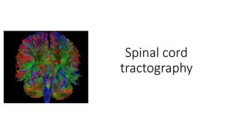
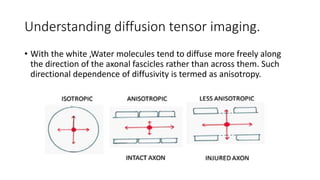
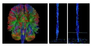
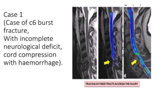
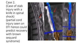
Spinal cord
tractography
Background
• Diffusion tensor imaging with fiber tracking enables a radiologist
/orthopaedic surgeon to non invasively visualize the descending and
ascending tracts of the spinal cord (more commonly used in the
cervical Spine).
• This technique has been recently used in the dorsal spine and lumbar
spine for visualization of tracts and plexus .
• Diffusion tensor imaging of the spinal cord however faces challenges
from physiological motion , susceptibility ,cardiac motion artifact
related image degradation.
Introduction
• It is a non invasive method to study the organisation of white matter
• Diffusion tensor imaging (DTI) :- It is an MRI technique that uses
anisotropic diffusion to estimate the axonal (white matter )
organization of the brain .
• Fiber tractography (FT) :- It is a 3D reconstruction technique to assess
neural tracts using data collected by diffusion tensor imaging.
• Both these tech are combined in diffusion tensor tractography.
Principle
• Based on Brownian motion of water molecules.
• The free water diffusion is termed as “isotropic
”diffusion .If the water diffuses in a medium
with a barrier, the diffusion will be uneven,
which is termed as an isotropic diffusion.
• Relative mobility of the molecules in isotropic
diffusion has a spherical shape, while it
becomes ellipsoid in anisotropic diffusion and
the technique is then called diffusion tensor
imaging
Understanding diffusion tensor imaging.
• With the white ,Water molecules tend to diffuse more freely along
the direction of the axonal fascicles rather than across them. Such
directional dependence of diffusivity is termed as anisotropy.
• Barriers can be many things : cell membranes, axons , myelin etc; but
in white matter the principal barrier is the myelin sheath of axons.
• Bundles of axons provide a barrier to perpendicular diffusion and a
path for parallel diffusion along the orientation of the fibers.
• Anisotropic diffusion is expected to be increased in areas of high
mature axonal order
• Condition where the mylien or the structure of the axon are
disrupted, such as trauma, tumour and inflammation reduce
anisotropy, as the barriers are affected by destruction or
disorganization.
Colour coding
• Basic colours can tell the observer how the fibers are oriented in a 3D
coordinate system, this is termed as an “anisotropic map”. The
software could encode the colours in this way
• Red – indicates direction in the X axis ( right to left or left to right )
• Green – indicates direction in the y axis (anterio to posterior or
posterior to anterior
• Blue – indicates direction in the z axis ( foot to head direction or vice
versa )
• The technique is unable to discriminate the “positive” or “ negative”
direction in the same axis .
Uses in orthopaedics
• Prognosis of Spinal cord Deformity
• Prognosis after spinal Injury
• Prognosis in cases of Myelopathy
• Prognosis in case of tumours in the cord.
Case 1
(Case of c6 burst
fracture,
With incomplete
neurological deficit,
cord compression
with haemorrhage).
Case 2.
(Case of stab
injury with a
knife in spinal
shock)
(partial cord
injury seen on
DTI hence could
predict recovery
with brown
sequard
syndrome)
Limitations
• Where more bundles of fibers coexist or where they cross, approach,
converge or diverge or are non homogeneous the algorithm works poorly.
• These limits in clinical practice could lead to following paths that do not
exist( false positive ) or to not adequately follow paths that exist(false
negative);
• Therefore the interpretation of the tractography reconstruction requires
prior experience and knowledge.
• Cannot identify afferent from efferent projections.
• Cannot determine the functions.
• Presence of synapses along the course of the same neural pathway are not
identified.
Thank you.

Spinal cord tractography

  • 1.
  • 2.
    Background • Diffusion tensorimaging with fiber tracking enables a radiologist /orthopaedic surgeon to non invasively visualize the descending and ascending tracts of the spinal cord (more commonly used in the cervical Spine). • This technique has been recently used in the dorsal spine and lumbar spine for visualization of tracts and plexus . • Diffusion tensor imaging of the spinal cord however faces challenges from physiological motion , susceptibility ,cardiac motion artifact related image degradation.
  • 3.
    Introduction • It isa non invasive method to study the organisation of white matter • Diffusion tensor imaging (DTI) :- It is an MRI technique that uses anisotropic diffusion to estimate the axonal (white matter ) organization of the brain . • Fiber tractography (FT) :- It is a 3D reconstruction technique to assess neural tracts using data collected by diffusion tensor imaging. • Both these tech are combined in diffusion tensor tractography.
  • 4.
    Principle • Based onBrownian motion of water molecules. • The free water diffusion is termed as “isotropic ”diffusion .If the water diffuses in a medium with a barrier, the diffusion will be uneven, which is termed as an isotropic diffusion. • Relative mobility of the molecules in isotropic diffusion has a spherical shape, while it becomes ellipsoid in anisotropic diffusion and the technique is then called diffusion tensor imaging
  • 5.
    Understanding diffusion tensorimaging. • With the white ,Water molecules tend to diffuse more freely along the direction of the axonal fascicles rather than across them. Such directional dependence of diffusivity is termed as anisotropy.
  • 6.
    • Barriers canbe many things : cell membranes, axons , myelin etc; but in white matter the principal barrier is the myelin sheath of axons. • Bundles of axons provide a barrier to perpendicular diffusion and a path for parallel diffusion along the orientation of the fibers. • Anisotropic diffusion is expected to be increased in areas of high mature axonal order • Condition where the mylien or the structure of the axon are disrupted, such as trauma, tumour and inflammation reduce anisotropy, as the barriers are affected by destruction or disorganization.
  • 8.
    Colour coding • Basiccolours can tell the observer how the fibers are oriented in a 3D coordinate system, this is termed as an “anisotropic map”. The software could encode the colours in this way • Red – indicates direction in the X axis ( right to left or left to right ) • Green – indicates direction in the y axis (anterio to posterior or posterior to anterior • Blue – indicates direction in the z axis ( foot to head direction or vice versa ) • The technique is unable to discriminate the “positive” or “ negative” direction in the same axis .
  • 10.
    Uses in orthopaedics •Prognosis of Spinal cord Deformity • Prognosis after spinal Injury • Prognosis in cases of Myelopathy • Prognosis in case of tumours in the cord.
  • 11.
    Case 1 (Case ofc6 burst fracture, With incomplete neurological deficit, cord compression with haemorrhage).
  • 12.
    Case 2. (Case ofstab injury with a knife in spinal shock) (partial cord injury seen on DTI hence could predict recovery with brown sequard syndrome)
  • 13.
    Limitations • Where morebundles of fibers coexist or where they cross, approach, converge or diverge or are non homogeneous the algorithm works poorly. • These limits in clinical practice could lead to following paths that do not exist( false positive ) or to not adequately follow paths that exist(false negative); • Therefore the interpretation of the tractography reconstruction requires prior experience and knowledge. • Cannot identify afferent from efferent projections. • Cannot determine the functions. • Presence of synapses along the course of the same neural pathway are not identified.
  • 14.