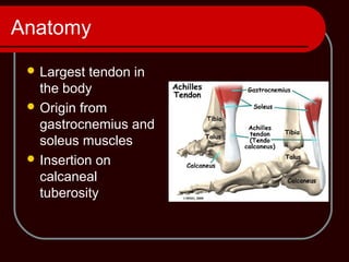
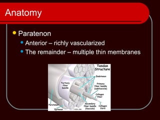
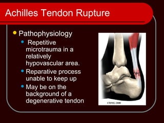
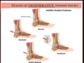
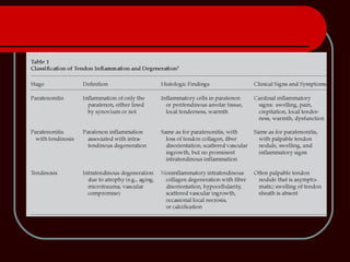
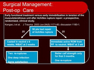
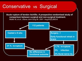
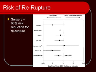
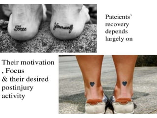
Achilles Tendon Disorders
Dr. Siddharth Gupta
SMO Rajawadi Hospital
Anatomy
 Largest tendon in
the body
 Origin from
gastrocnemius and
soleus muscles
 Insertion on
calcaneal
tuberosity
Anatomy
Lacks a true synovial sheath
Paratenon has visceral and parietal layers
Allows for 1.5cm of tendon glide
Anatomy
Paratenon
Anterior – richly vascularized
The remainder – multiple thin membranes
Anatomy
 Blood supply
1) Musculotendinous junction
2) Osseous insertion on calcaneus
3) Multiple mesotenal vessels on anterior
surface of paratenon (in adipose)
– Transverse vincula
 Fewest @ 2 to 6 cm proximal to osseous insertion
Physiology
Remarkable response to stress
Exercise induces tendon diameter increase
Inactivity or immobilization causes rapid
atrophy
Age-related decreases in cell density,
collagen fibril diameter and density
Older athletes have higher injury
susceptibility
Biomechanics
Gastrocnemius-soleus-Achilles complex
Spans 3 joints
 Flex knee
 Plantar flex tibiotalar joint
 Supinate subtalar joint
Up to 10 times body weight through
tendon when running
Achilles Tendon Rupture
Pathophysiology
 Repetitive
microtrauma in a
relatively
hypovascular area.
Reparative process
unable to keep up
May be on the
background of a
degenerative tendon
Common causes of injury:
•Repetitive or sudden increase of stress
on the tendon.
•Increase in exercise intensity.
•Not allowing recovery time between
exercises.
•Decreased range of motion-tight calf
muscles.
•Bone spur-rubbing against the tendon.
Types of injury
1-Achilles tendonitis Chronic inflammation
Due to
Stresses
Micro-tears
Inflammation and
scar formation
2-Achilles tendon rupture
Achilles tendonitis - presentation
•Pain in the back of the heel
•Occasional swelling due to thickening of
tendon
•Often occurs in runners.
Treatment includes
Anti-inflammatory
Physical therapy.
Massage therapy
Ice therapy
Immobilization
Injections: prolotherapy, platelets or
needling.
Achilles tendon rupture
Patient will feel or hear a “pop” when the
tendon ruptures.
Pain is felt in the back of the ankle.
Foot becomes floppy and weak.
Patient feels as if being kicked in the
back of the ankle where the tendon
snapped.
Achilles Tendon Rupture:
Textbook Facts
Antecedent tendinitis/tendinosis in 15%
75% of sports-related ruptures happen in
patients between 30-40 years of age.
Most ruptures occur in watershed area
4cm proximal to the calcaneal insertion.
Achilles Tendon Rupture
 History
 Feels like being kicked in the leg
 Case reports of fluoroquinolone use, steroid
injections
 Mechanism
 Eccentric loading (running backwards in tennis)
 Sudden unexpected dorsiflexion of ankle
 (Direct blow or laceration)
Physical Exam
 Prone patient with feet over edge of bed
 Palpation of entire length of muscle-tendon
unit during active and passive ROM
 Compare tendon width to other side
 Note tenderness, crepitation, warmth, swelling,
nodularity, palpable defects
Achilles Tendon Rupture
Physical
Partial
 Localized tenderness +/- nodularity
Complete
 Defect
 Cannot heel raise
 Positive Thompson test
Achilles Tendon Rupture
Diagnostic Pitfalls
23% missed by Primary Physician
(Inglis & Sculco)
Tendon defect can be masked by
hematoma
Plantar-flexion power of extrinsic foot
flexors retained
Thompson test can produce a false-
negative if accessory ankle flexors also
squeezed
Imaging
X RAYS
Avulsion fracture at the
insertion , with marked
seperation of fragments.
Imaging
Imaging
Ultrasound
Inexpensive, fast,
reproducable, dynamic
examination possible
Operator dependent
Best to measure
thickness and gap
Good screening test for
complete rupture
Imaging
MRI
Expensive, not
dynamic
Better at detecting
partial ruptures
and staging
degenerative
changes, (monitor
healing)
Management Goals
Restore musculotendinous length and
tension.
Optimize gastro-soleous strength and
function
Avoid ankle stiffness
Conservative Management
Cast in Plantarflexion CAM Walker or cast with
plantarflexion q 2
wks
2 wks
Allow progressive weight-
bearing in removable cast
Remove cast and walk with
shoe lift. Start with 2cm x 1
month, then 1cm x1 month
then D/C
4 weeks
Start physio for ROM
exercises
When WBAT and
foot is plantigrade
Start a strengthening
program
2- 4 weeks
Surgical Management
Preserve anterior paratenon blood
supply
Beware of sural nerve
Debride and approximate tendon ends
Use 2-4 stranded locked suture
technique
May augment with absorbable suture
Close paratenon separately
Surgical Management
 Bunnell Suture
 Modified Kessler
 Many techniques
available
Complications After Open Repair of Achilles Tendon
Rupture
Minor
 􀀀 Wound
 & Superficial infection
 & Wound hematoma
 & Delayed wound healing
 & Adhesion of scar
 & Suture granuloma
 & Skin necrosis
 & General
 & Pain
 & Disturbances in sensibility
 & Suture rupture
Major
 􀀀 Wound
 & Deep infection
 & Chronic fistula
 & General
 & Deep vein thrombosis
 & Tendon lengthening
 & Death
Classification given by - Wong et al.
Surgical Management :
Post– op Care
Assess strength of repair, tension and
ROM intra-op.
Apply cast with ankle in the least amount
of plantarflexion that can be safely
attained.
Patient returns to fracture clinic 2 weeks
post-op.
Variations in Post-op Protocols
Functional Bracing
Post- Op Care
Cast applied in OR Remove sutures, apply a
walking cast with heel lift2 wks
Allow progressive weight-
bearing in removable cast
Remove cast and walk with a
1cm shoe lift x 1 month then
D/C.
2 weeks
Start physio for ROM
exercises. No active
plantarflexion
When WBAT and
foot is plantigrade
Start a strengthening
program
2- 4 weeks
Touch WB
Surgical Management:
Post-op Care
J Trauma. 2003 Jun;54(6):1171-80; discussion 1180-1.Kangas J et al.
Early functional treatment versus early immobilization in tension of the
musculotendinous unit after Achilles rupture repair: a prospective,
randomized, clinical study.
50 pts had repair
of Achilles rupture
Casted in neutral x 6
weeks. WBAT at 3 weeks
Immediate active ROM from
PF to neutral. WBAT at 3 wk
Better calf strength only
for first 3 months.
One re-rupture
Two re-ruptures
One deep infection
Same satisfaction
25 25
Conservative vs Surgical
Acute rupture of tendon Achillis. A prospective randomised study of
comparison between surgical and non-surgical treatment.
Moller M, et al. J Bone Joint Surg Br. 2001 Aug;83(5):863-8
112 patients
Surgery +
Early functional rehab in
brace
Casted x 8 wks
21 % re-rupture 1.7% re-rupture
5% infection
2% Sural nerve inj.
No difference in
functional outcome
Summary of Pooled Outcome Measures
Risk of Re-Rupture
 Surgery =
68% risk
reduction for
re-rupture
Achilles tendon pathology

Achilles tendon pathology

  • 2.
    Achilles Tendon Disorders Dr.Siddharth Gupta SMO Rajawadi Hospital
  • 3.
    Anatomy  Largest tendonin the body  Origin from gastrocnemius and soleus muscles  Insertion on calcaneal tuberosity
  • 4.
    Anatomy Lacks a truesynovial sheath Paratenon has visceral and parietal layers Allows for 1.5cm of tendon glide
  • 5.
    Anatomy Paratenon Anterior – richlyvascularized The remainder – multiple thin membranes
  • 6.
    Anatomy  Blood supply 1)Musculotendinous junction 2) Osseous insertion on calcaneus 3) Multiple mesotenal vessels on anterior surface of paratenon (in adipose) – Transverse vincula  Fewest @ 2 to 6 cm proximal to osseous insertion
  • 7.
    Physiology Remarkable response tostress Exercise induces tendon diameter increase Inactivity or immobilization causes rapid atrophy Age-related decreases in cell density, collagen fibril diameter and density Older athletes have higher injury susceptibility
  • 8.
    Biomechanics Gastrocnemius-soleus-Achilles complex Spans 3joints  Flex knee  Plantar flex tibiotalar joint  Supinate subtalar joint Up to 10 times body weight through tendon when running
  • 9.
    Achilles Tendon Rupture Pathophysiology Repetitive microtrauma in a relatively hypovascular area. Reparative process unable to keep up May be on the background of a degenerative tendon
  • 10.
    Common causes ofinjury: •Repetitive or sudden increase of stress on the tendon. •Increase in exercise intensity. •Not allowing recovery time between exercises. •Decreased range of motion-tight calf muscles. •Bone spur-rubbing against the tendon.
  • 11.
    Types of injury 1-Achillestendonitis Chronic inflammation Due to Stresses Micro-tears Inflammation and scar formation 2-Achilles tendon rupture
  • 12.
    Achilles tendonitis -presentation •Pain in the back of the heel •Occasional swelling due to thickening of tendon •Often occurs in runners.
  • 14.
    Treatment includes Anti-inflammatory Physical therapy. Massagetherapy Ice therapy Immobilization Injections: prolotherapy, platelets or needling.
  • 15.
    Achilles tendon rupture Patientwill feel or hear a “pop” when the tendon ruptures. Pain is felt in the back of the ankle. Foot becomes floppy and weak. Patient feels as if being kicked in the back of the ankle where the tendon snapped.
  • 16.
    Achilles Tendon Rupture: TextbookFacts Antecedent tendinitis/tendinosis in 15% 75% of sports-related ruptures happen in patients between 30-40 years of age. Most ruptures occur in watershed area 4cm proximal to the calcaneal insertion.
  • 18.
    Achilles Tendon Rupture History  Feels like being kicked in the leg  Case reports of fluoroquinolone use, steroid injections  Mechanism  Eccentric loading (running backwards in tennis)  Sudden unexpected dorsiflexion of ankle  (Direct blow or laceration)
  • 19.
    Physical Exam  Pronepatient with feet over edge of bed  Palpation of entire length of muscle-tendon unit during active and passive ROM  Compare tendon width to other side  Note tenderness, crepitation, warmth, swelling, nodularity, palpable defects
  • 20.
    Achilles Tendon Rupture Physical Partial Localized tenderness +/- nodularity Complete  Defect  Cannot heel raise  Positive Thompson test
  • 21.
    Achilles Tendon Rupture DiagnosticPitfalls 23% missed by Primary Physician (Inglis & Sculco) Tendon defect can be masked by hematoma Plantar-flexion power of extrinsic foot flexors retained Thompson test can produce a false- negative if accessory ankle flexors also squeezed
  • 23.
    Imaging X RAYS Avulsion fractureat the insertion , with marked seperation of fragments.
  • 24.
  • 25.
    Imaging Ultrasound Inexpensive, fast, reproducable, dynamic examinationpossible Operator dependent Best to measure thickness and gap Good screening test for complete rupture
  • 26.
    Imaging MRI Expensive, not dynamic Better atdetecting partial ruptures and staging degenerative changes, (monitor healing)
  • 27.
    Management Goals Restore musculotendinouslength and tension. Optimize gastro-soleous strength and function Avoid ankle stiffness
  • 28.
    Conservative Management Cast inPlantarflexion CAM Walker or cast with plantarflexion q 2 wks 2 wks Allow progressive weight- bearing in removable cast Remove cast and walk with shoe lift. Start with 2cm x 1 month, then 1cm x1 month then D/C 4 weeks Start physio for ROM exercises When WBAT and foot is plantigrade Start a strengthening program 2- 4 weeks
  • 30.
    Surgical Management Preserve anteriorparatenon blood supply Beware of sural nerve Debride and approximate tendon ends Use 2-4 stranded locked suture technique May augment with absorbable suture Close paratenon separately
  • 31.
    Surgical Management  BunnellSuture  Modified Kessler  Many techniques available
  • 41.
    Complications After OpenRepair of Achilles Tendon Rupture Minor  􀀀 Wound  & Superficial infection  & Wound hematoma  & Delayed wound healing  & Adhesion of scar  & Suture granuloma  & Skin necrosis  & General  & Pain  & Disturbances in sensibility  & Suture rupture Major  􀀀 Wound  & Deep infection  & Chronic fistula  & General  & Deep vein thrombosis  & Tendon lengthening  & Death Classification given by - Wong et al.
  • 42.
    Surgical Management : Post–op Care Assess strength of repair, tension and ROM intra-op. Apply cast with ankle in the least amount of plantarflexion that can be safely attained. Patient returns to fracture clinic 2 weeks post-op.
  • 43.
  • 44.
  • 45.
    Post- Op Care Castapplied in OR Remove sutures, apply a walking cast with heel lift2 wks Allow progressive weight- bearing in removable cast Remove cast and walk with a 1cm shoe lift x 1 month then D/C. 2 weeks Start physio for ROM exercises. No active plantarflexion When WBAT and foot is plantigrade Start a strengthening program 2- 4 weeks Touch WB
  • 46.
    Surgical Management: Post-op Care JTrauma. 2003 Jun;54(6):1171-80; discussion 1180-1.Kangas J et al. Early functional treatment versus early immobilization in tension of the musculotendinous unit after Achilles rupture repair: a prospective, randomized, clinical study. 50 pts had repair of Achilles rupture Casted in neutral x 6 weeks. WBAT at 3 weeks Immediate active ROM from PF to neutral. WBAT at 3 wk Better calf strength only for first 3 months. One re-rupture Two re-ruptures One deep infection Same satisfaction 25 25
  • 47.
    Conservative vs Surgical Acuterupture of tendon Achillis. A prospective randomised study of comparison between surgical and non-surgical treatment. Moller M, et al. J Bone Joint Surg Br. 2001 Aug;83(5):863-8 112 patients Surgery + Early functional rehab in brace Casted x 8 wks 21 % re-rupture 1.7% re-rupture 5% infection 2% Sural nerve inj. No difference in functional outcome
  • 49.
    Summary of PooledOutcome Measures
  • 50.
    Risk of Re-Rupture Surgery = 68% risk reduction for re-rupture