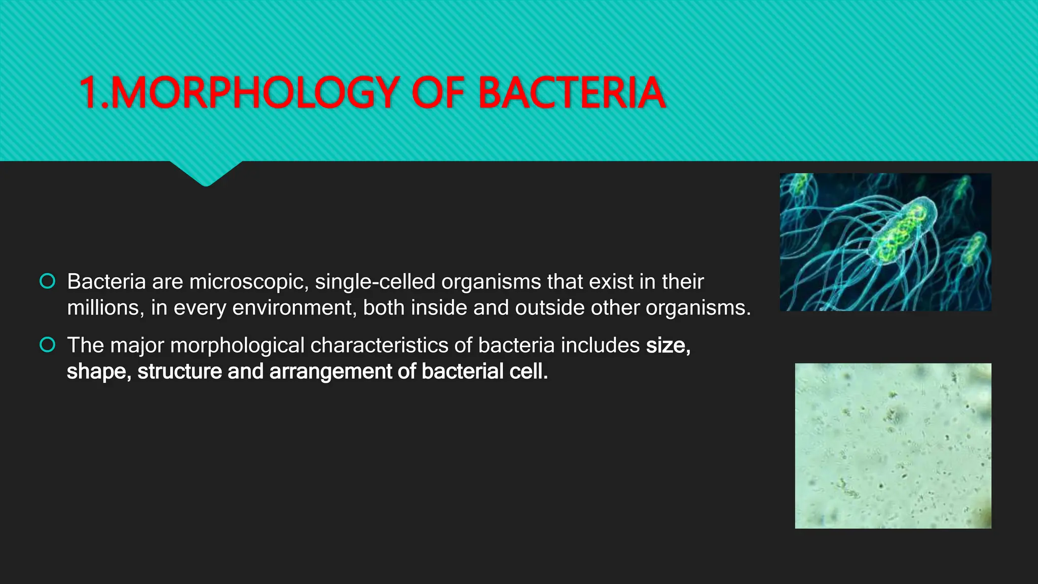
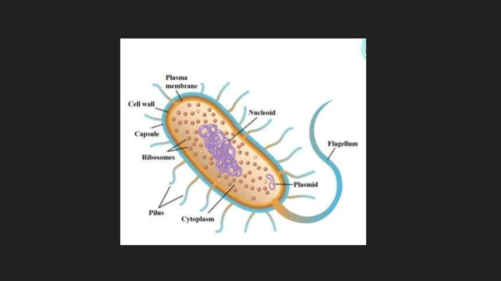
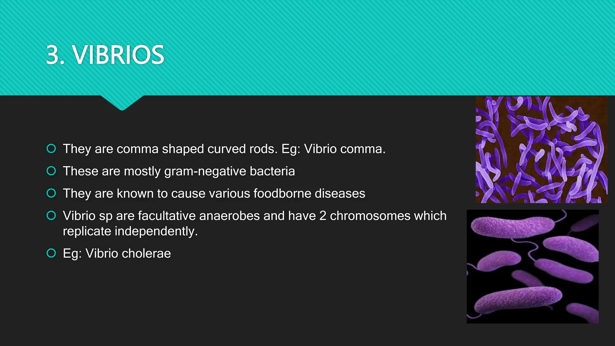
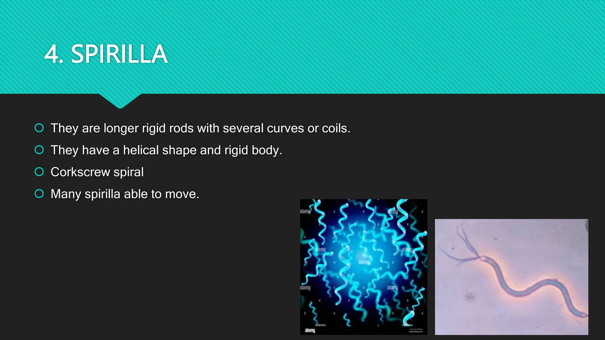
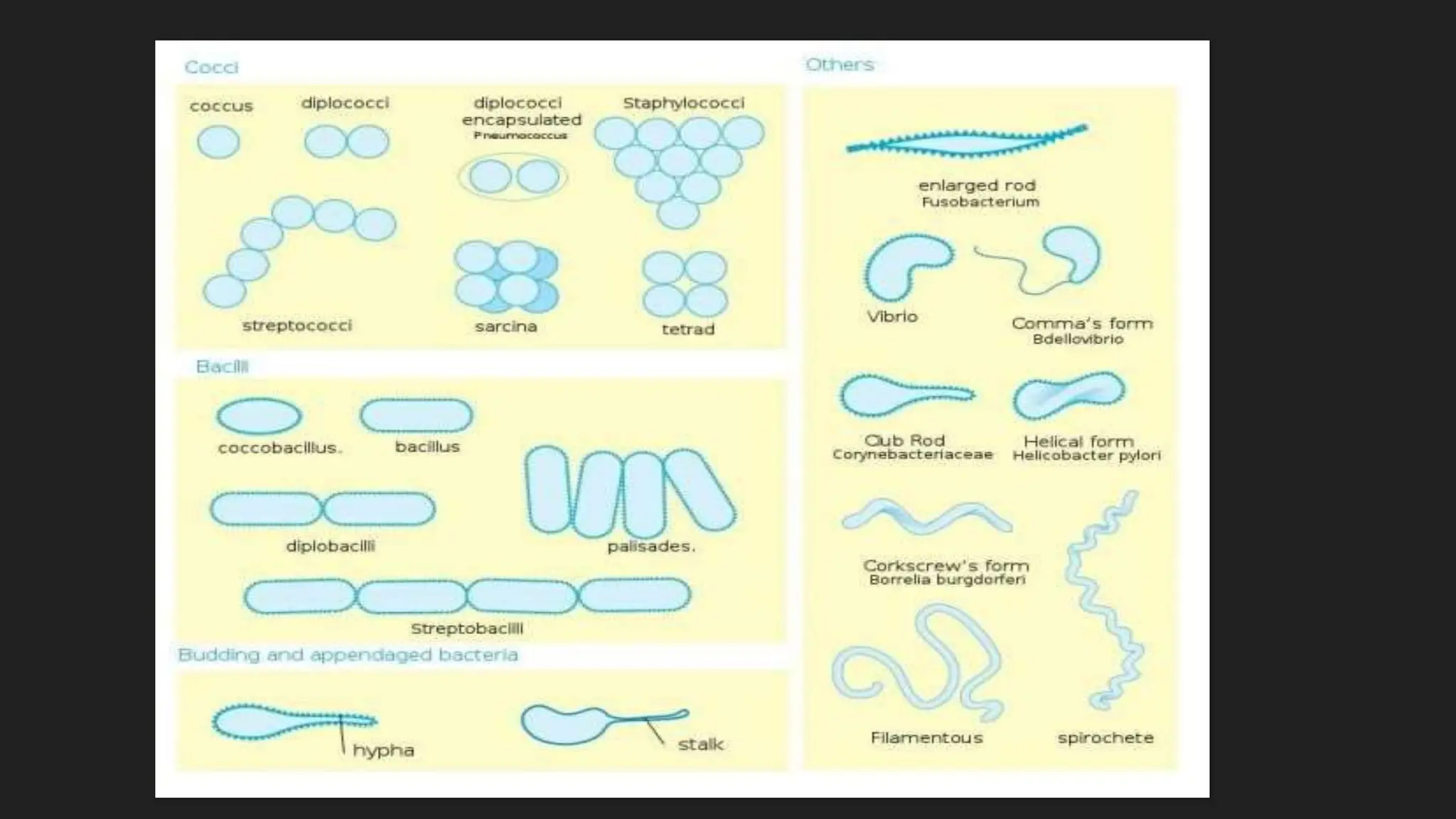
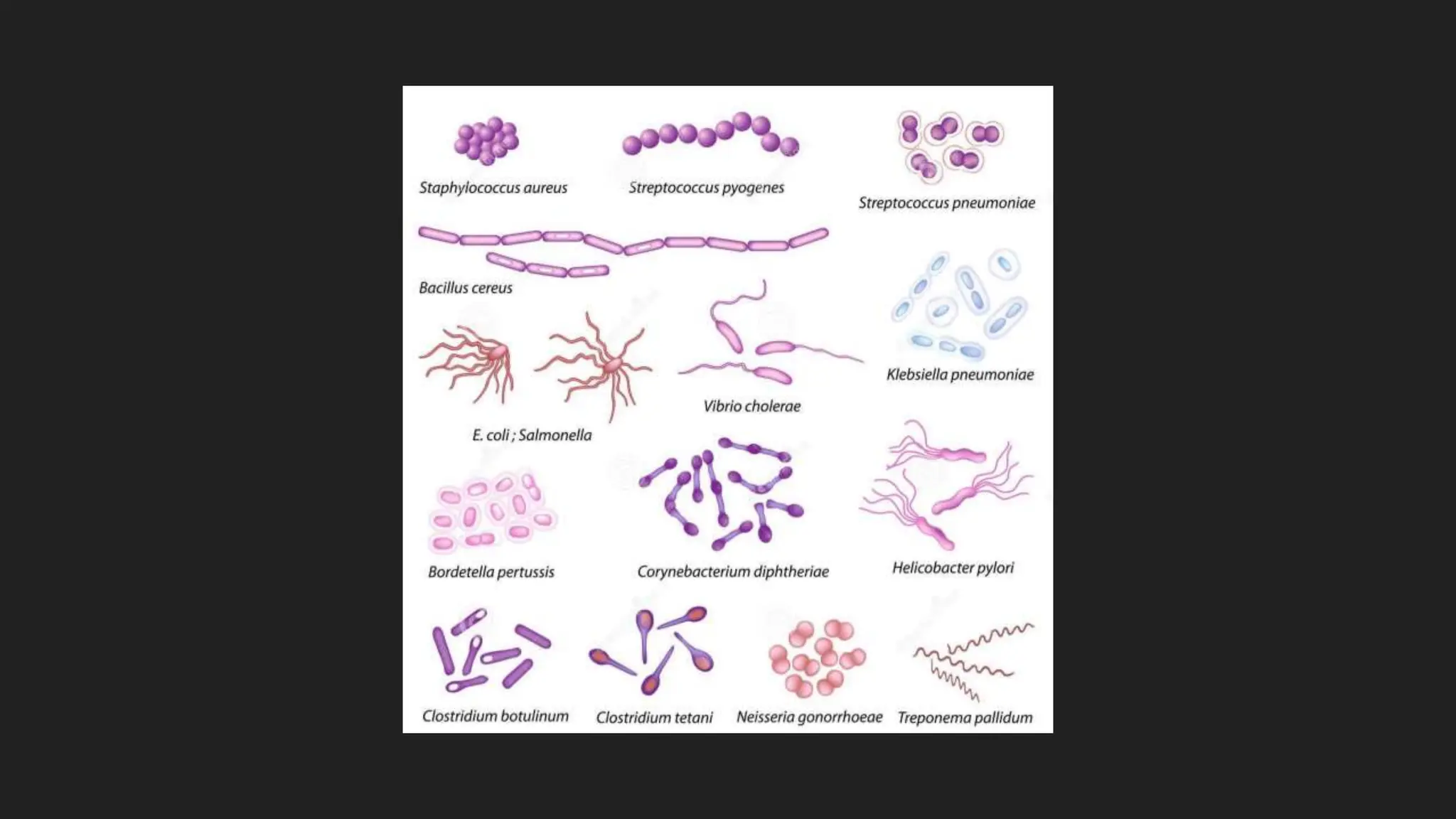
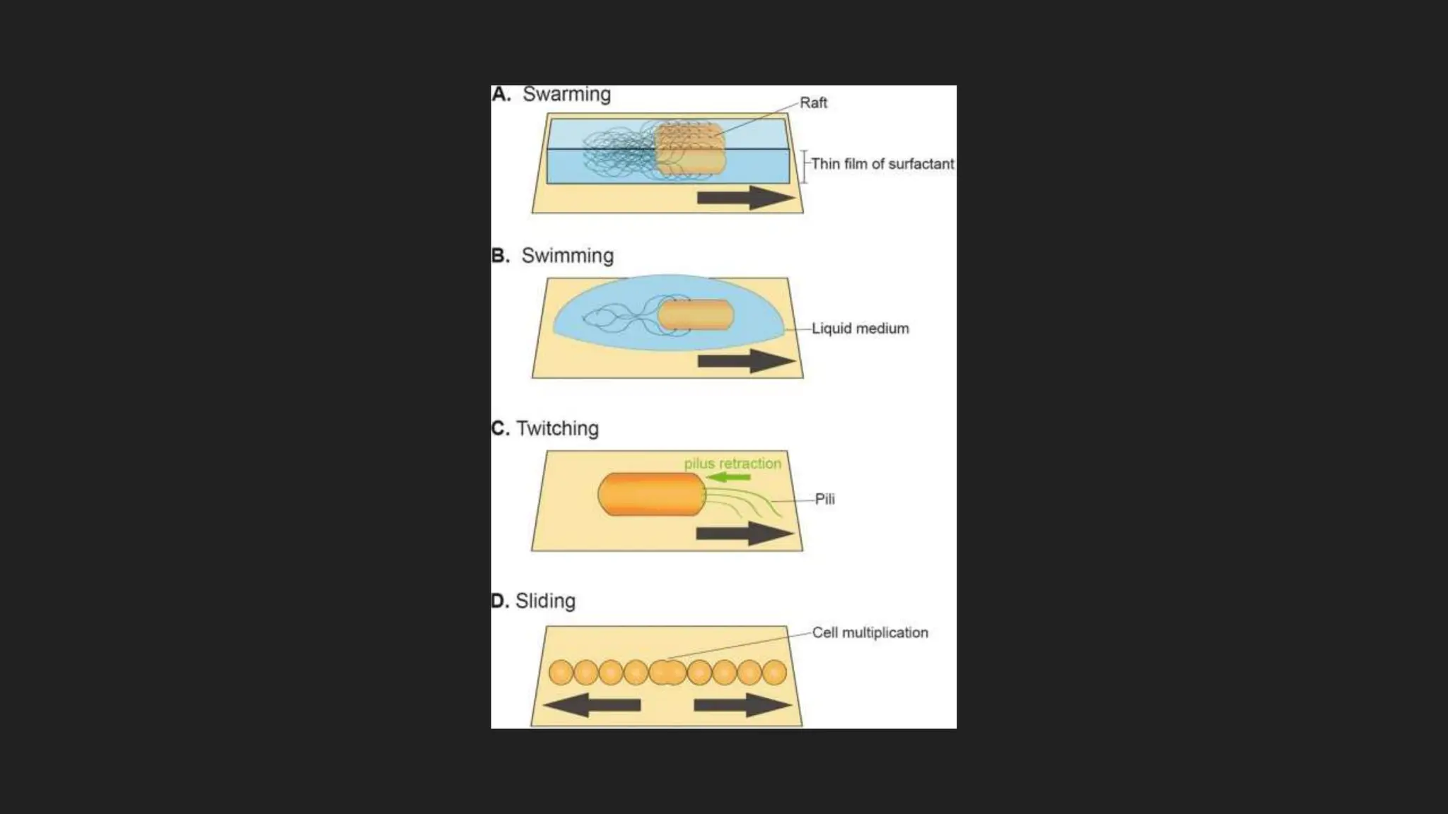
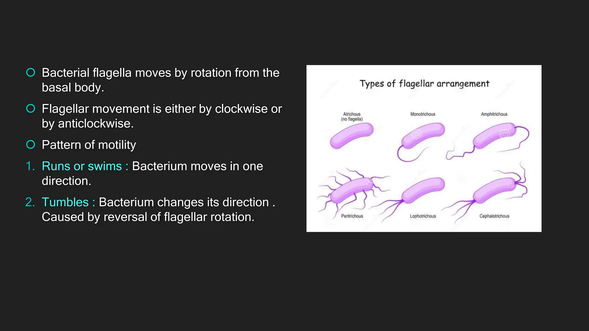
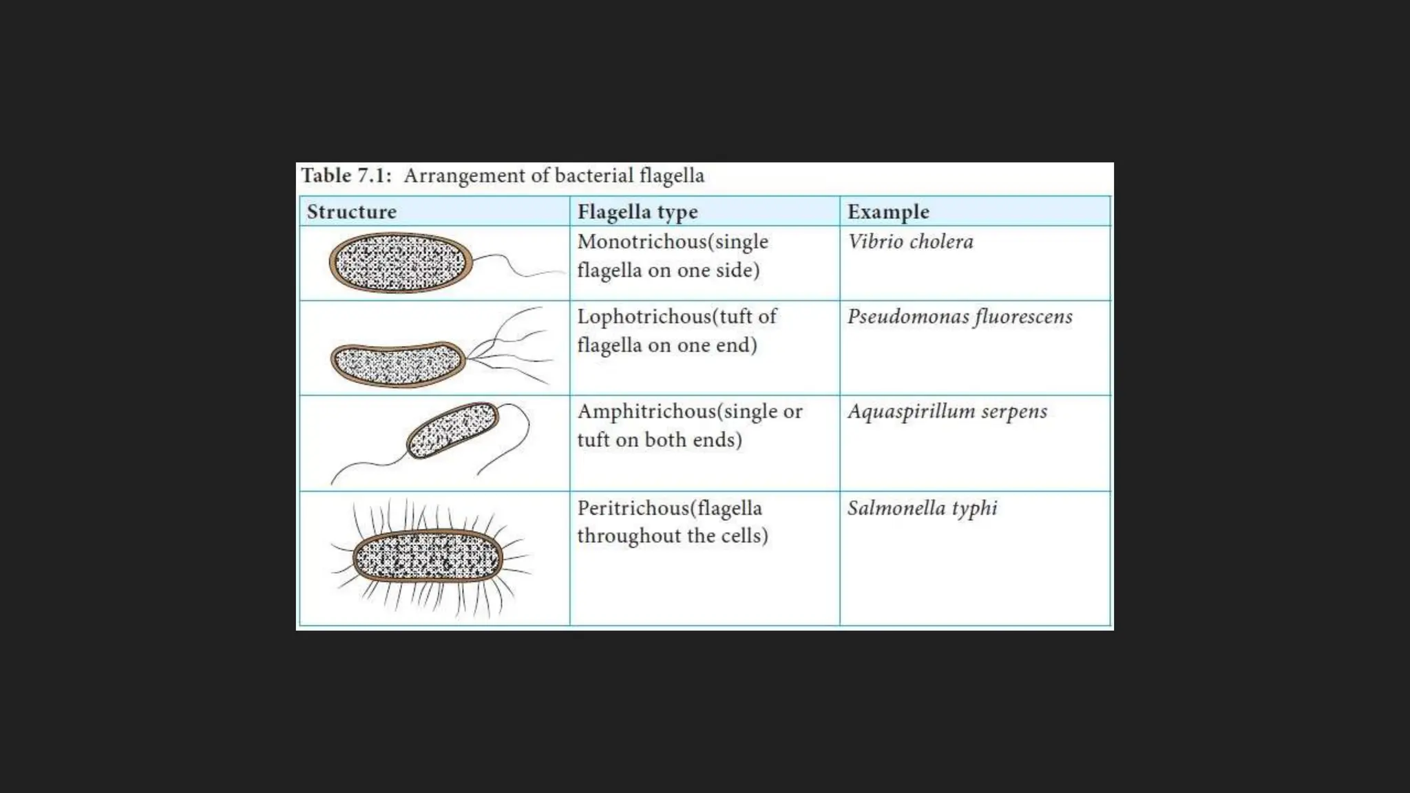
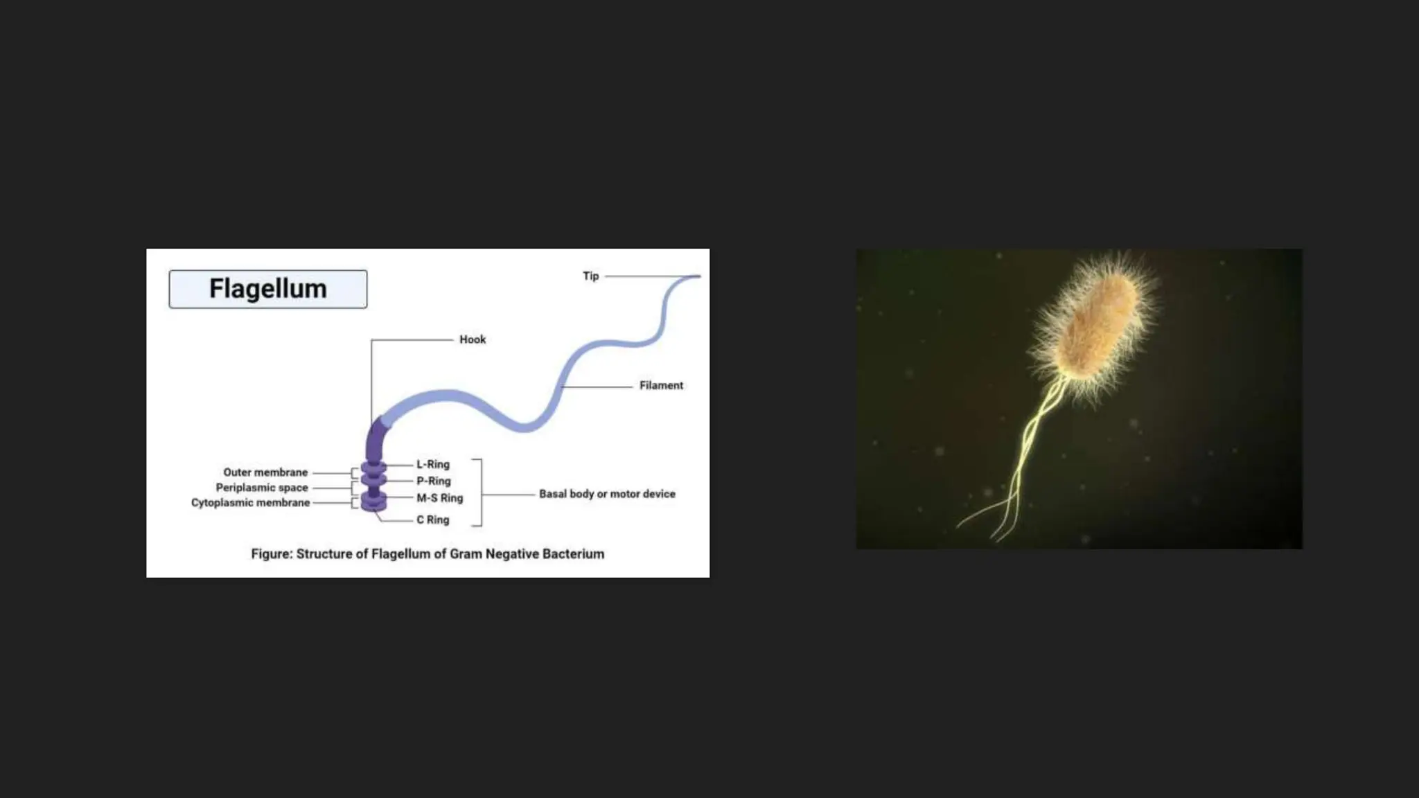
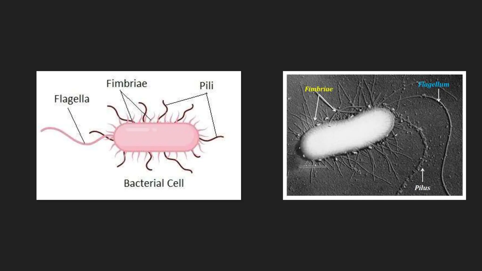
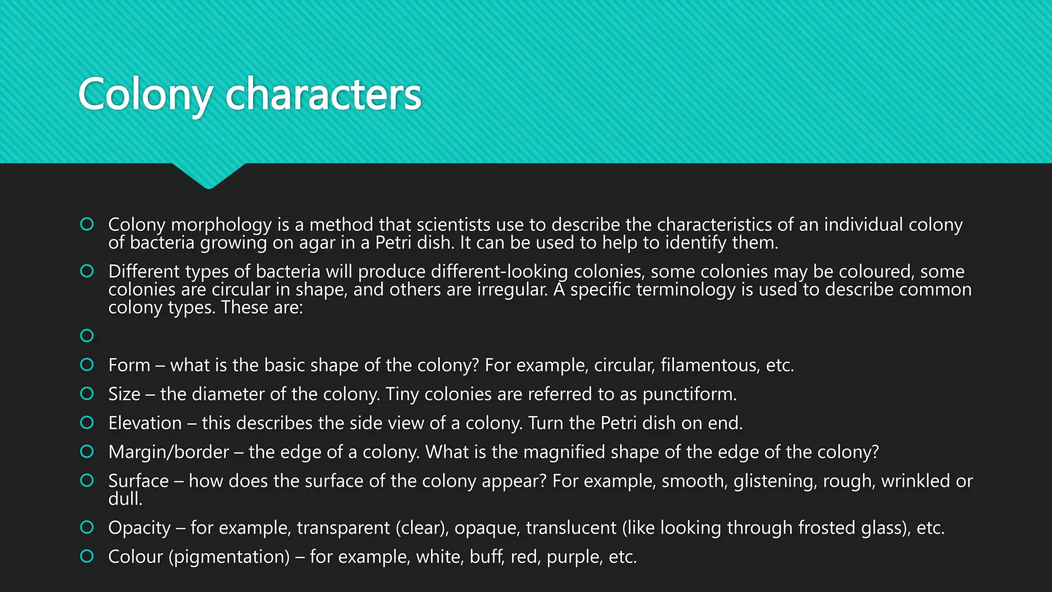
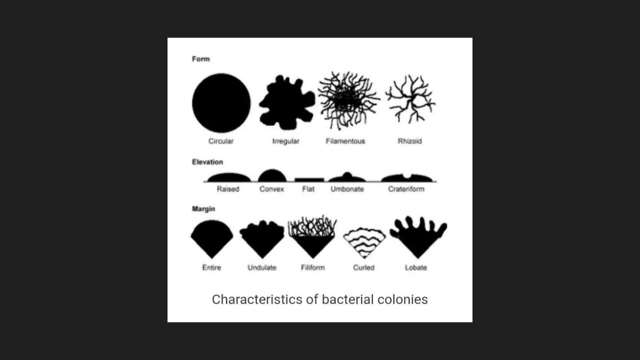
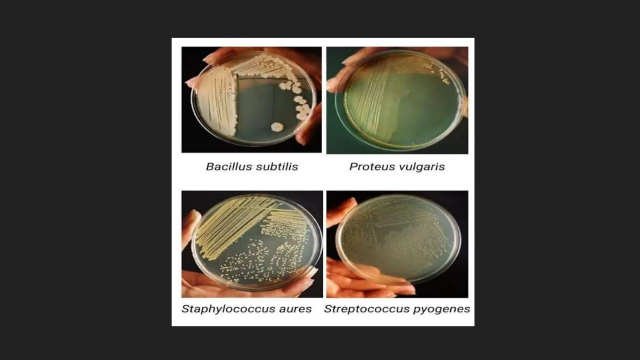
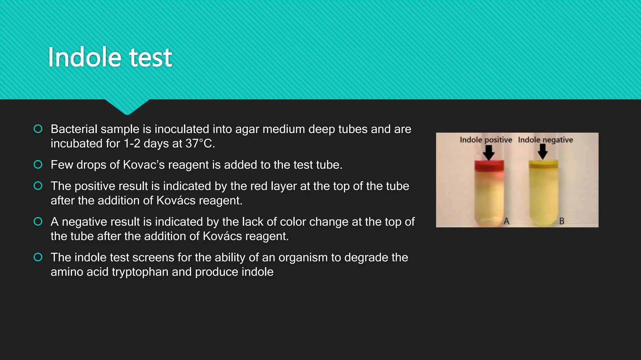
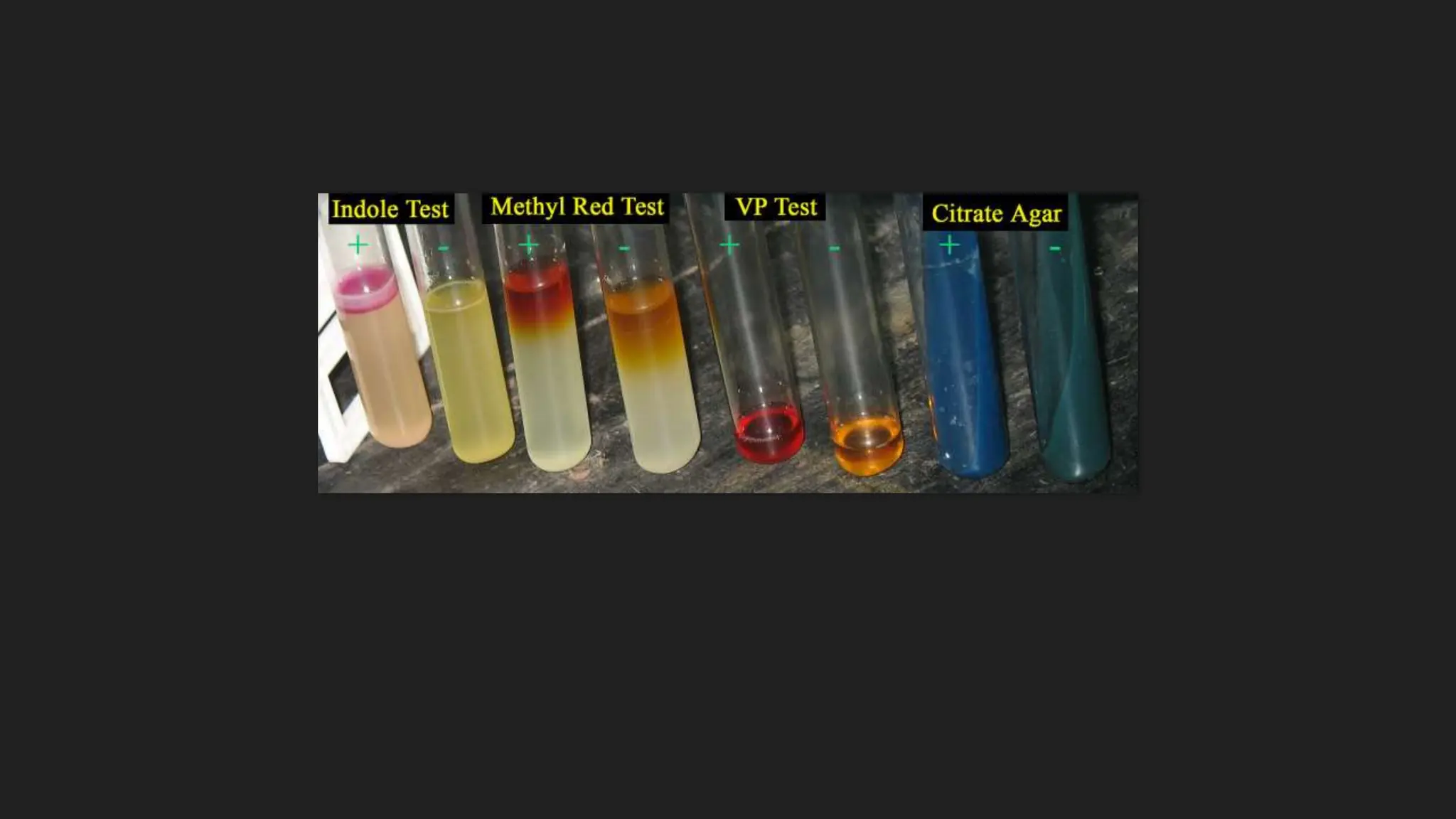
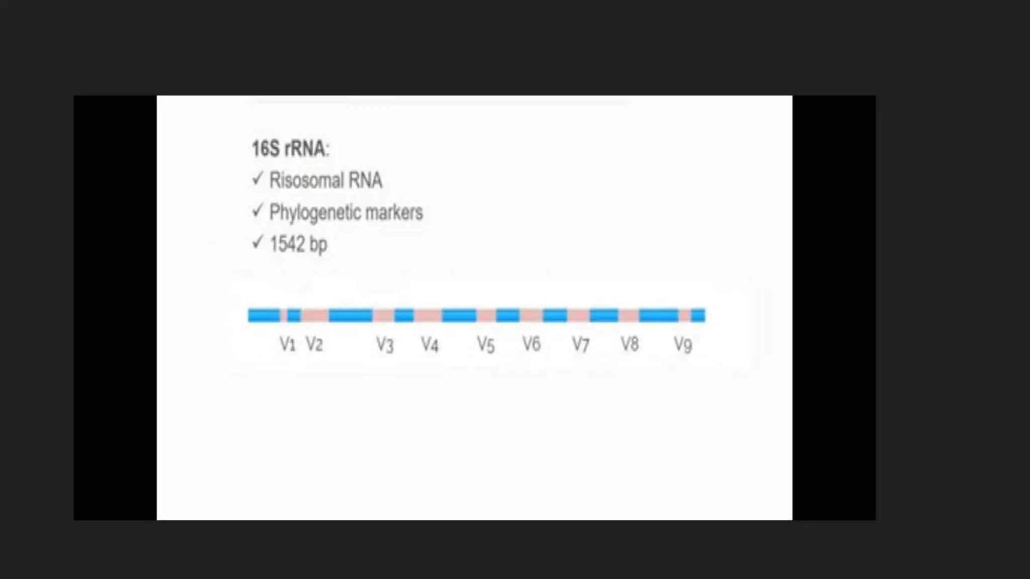
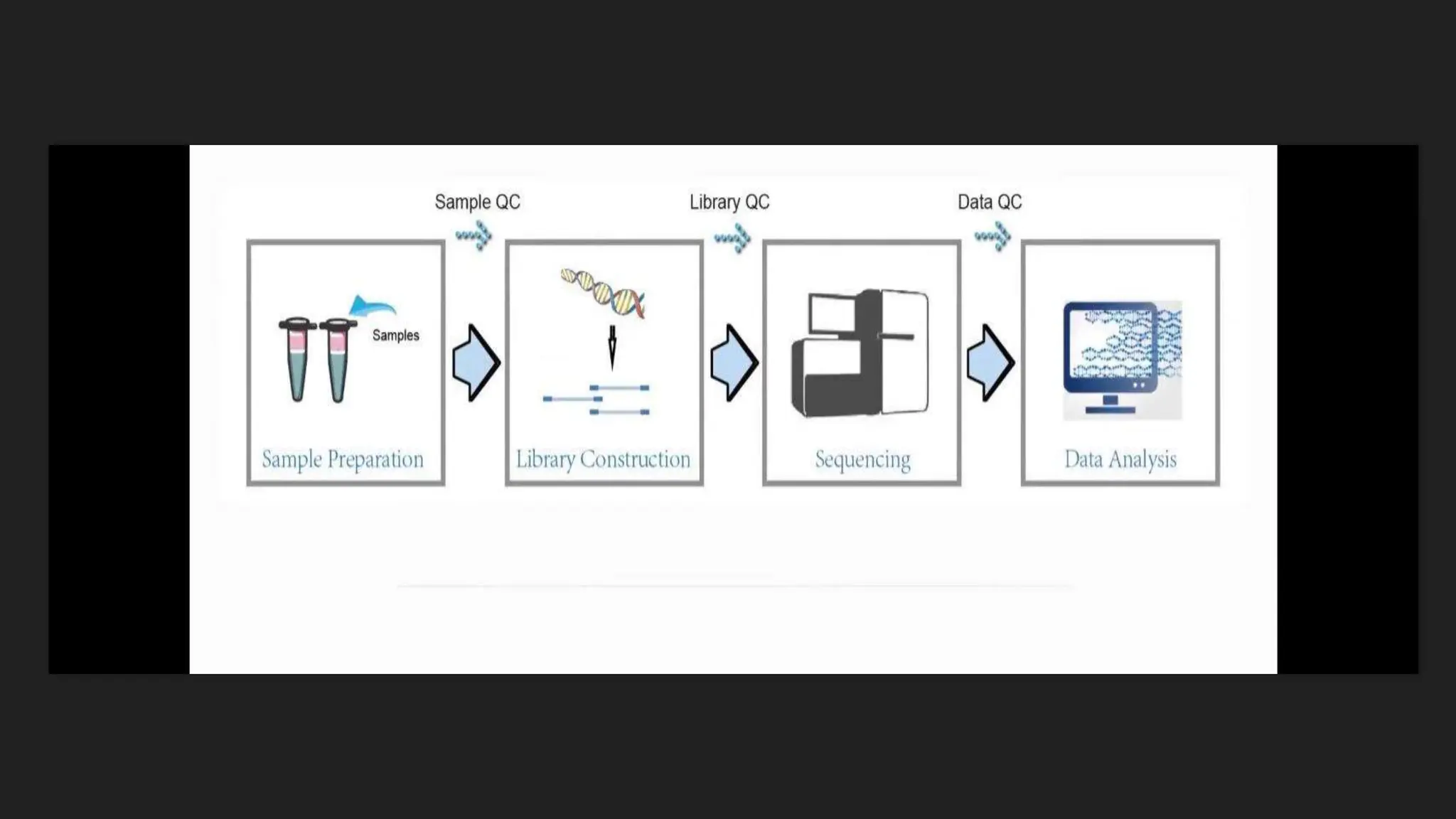
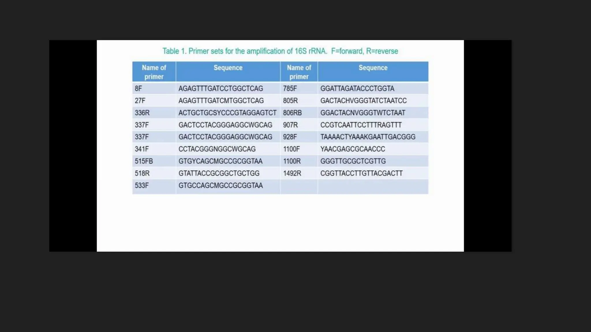
2. Bacterias are usually identified by
morphological and biochemical tests,
3. 1.MORPHOLOGY OF BACTERIA
Bacteria are microscopic, single-celled organisms that exist in their
millions, in every environment, both inside and outside other organisms.
The major morphological characteristics of bacteria includes size,
shape, structure and arrangement of bacterial cell.
4.
5. 1.Shape
On the basis of shape, bacteria are classified as
1. Cocci
2. Bacilli
3. Vibrios
4. Spirilla
5.Spirochetes
6. 1. Cocci
Cocci are small, spherical or oval in shaped.
In Greek ‘kokkos’ means berry.
Cocci can grow in chains, clusters or pairs.
Based on the arrangement of cocci cells, they can be further divided into
1. Streptococci – cocci arranged in chains. E.g., Streptococcus mutans
2. Diplococci – cocci arranged in pairs. Eg., Neisseria gonorrhoeae
3. Monococci – single cocci
4. Staphylococci – clusters of cocci. Eg., Staphylococcus aureus
5. Tetrads – cocci in groups of four. Eg., Pediococcus
6. Sarcina – cocci in groups of eight. E.g., Sarcina ventriculi
7. 2. BACILLI
They are rod shaped cells. Eg: Bacillus anthracis.
It is derived from greek word “ Bacillus” meaning stick.
In some of the bacilli the length of cell may be equal to width. Such
bacillary forms are known as coccobacilli. Eg: Bracella.
1. Diplobacilli: Two bacilli arranged side by side with each other.
2. Streptobacilli: Bacilli arranged in chains.
3. Coccobacillus: Oval and similar to coccus
8. 3. VIBRIOS
They are comma shaped curved rods. Eg: Vibrio comma.
These are mostly gram-negative bacteria
They are known to cause various foodborne diseases
Vibrio sp are facultative anaerobes and have 2 chromosomes which
replicate independently.
Eg: Vibrio cholerae
9. 4. SPIRILLA
They are longer rigid rods with several curves or coils.
They have a helical shape and rigid body.
Corkscrew spiral
Many spirilla able to move.
10. 4.SPIROCHETES
Spirochetes have a spiral shape, a flexible cell wall, and motility mechanisms based on structures
called axial filaments.
Each axial filament is composed of fibrils extending toward each other between two layers of the
cell wall.
11.
12.
13. 2.MOTILITY
Types of movement in bacteria are,
1. Flagellar movement
2. Gliding movement
3. Spirochaetal movement
Bacteria have two different primary mechanisms they use for movement. The flagellum is
used for swimming and swarming, and the pilus (or fimbria) is used for twitching.
14. 1. Flagellar movement : Bacterial flagella are filamentous organelles that drive cell
locomotion. They thrust cells in liquids (swimming) or on surfaces (swarming) so that
cells can move toward favorable environments.
2. Gliding movement : Gliding motility is the ability of certain rod-shaped bacteria to
translocate on surfaces without the aid of external appendages such as flagella, cilia, or
pili.
3. Spirochaetial movement : the spirochetes use long, helical appendages known as
flagella to move; however, the spirochetes enclose their flagella in the periplasm, the
narrow space between the inner and outer membranes. Rotation of the flagella in the
periplasm causes the entire cell body to rotate and/or undulate
15.
16. MOTILITY
FLAGELLA
Flagella are long, slender, thin hair-like cytoplasmic appendages, which are responsible
for the motility of bacteria.
Flagella are made up of a protein- flagellin.
The flagellum has three basic parts ,
1. Filament
2. Hook
3. Basal body
17. Bacterial flagella moves by rotation from the
basal body.
Flagellar movement is either by clockwise or
by anticlockwise.
Pattern of motility
1. Runs or swims : Bacterium moves in one
direction.
2. Tumbles : Bacterium changes its direction .
Caused by reversal of flagellar rotation.
18.
19.
20. PILI OR FIMBRIAE
Fimbriae and pili are extensions of the cytoplasmic membrane and are made up of an
oligomeric protein known as pilin.
They are hair-like appendages present on the bacterial cell wall similar to flagella.
Fimbriae are found in gram negative as well as gram positive bacteria but are shorter in length
as compared to pili. Pili are longer than fimbriae and there are only a few per cell.
21.
22. Colony characters
Colony morphology is a method that scientists use to describe the characteristics of an individual colony
of bacteria growing on agar in a Petri dish. It can be used to help to identify them.
Different types of bacteria will produce different-looking colonies, some colonies may be coloured, some
colonies are circular in shape, and others are irregular. A specific terminology is used to describe common
colony types. These are:
Form – what is the basic shape of the colony? For example, circular, filamentous, etc.
Size – the diameter of the colony. Tiny colonies are referred to as punctiform.
Elevation – this describes the side view of a colony. Turn the Petri dish on end.
Margin/border – the edge of a colony. What is the magnified shape of the edge of the colony?
Surface – how does the surface of the colony appear? For example, smooth, glistening, rough, wrinkled or
dull.
Opacity – for example, transparent (clear), opaque, translucent (like looking through frosted glass), etc.
Colour (pigmentation) – for example, white, buff, red, purple, etc.
23.
24.
25. 2. BIOCHEMICAL TESTS ( IMVIC )
IMVIC series is a group of four individual tests that are commonly used to identify bacterial species,
especially coliforms.
A coliform is a gram negative , aerobic anaerobic rod which produces gas from lactose within 48
hours. The presence of some califorms indicates fecal contamination.
Each of the letter of IMVIC stands for one of these tests.
‘ I’ for Indole
‘M’ is for methyl red
'V’ for Voges proskauer test
'C’ for citrate utilization test
The letter “i” is only for rhyming purpose.
26. Cultures of any members of Enterobacteriaceae have to grow for 24 to 48 hours at 37°C, and the
respective tests can be performed
27. Indole test
Bacterial sample is inoculated into agar medium deep tubes and are
incubated for 1-2 days at 37°C.
Few drops of Kovac’s reagent is added to the test tube.
The positive result is indicated by the red layer at the top of the tube
after the addition of Kovács reagent.
A negative result is indicated by the lack of color change at the top of
the tube after the addition of Kovács reagent.
The indole test screens for the ability of an organism to degrade the
amino acid tryptophan and produce indole
28. Methyl red test
Bacteria which produces acid through fermentation of glucose can be
identified using this test. Bacterial sample is asceptically inoculated into
MR-VP-medium.
The culture is incubated for 1-2 days
Methyl-red indicator is added to the test tubes.
Red coloration indicates the positive results.
29. VP test
Some microorganisms have the capability to produce neutral end products, such as acetyl methyl
carbinol from pyruvic acid. Such organisms can be identified by using this test.
Bacterial sample is inoculated into MR-VP media.
It is then incubated for 1-2 days.
Barrit’s reagent is added to the culture
Deep rose color indicates the presence of VP-positive organisms.
30. Citrate utilization test
Some microbes have the presence of citrate permease enzyme which allows them to
obtain energy from citrate when no other carbon sources are present. Citrase
converts citrate into oxaloacetic acid and acetate which are further converted into
pyruvic acid and carbon dioxide. Carbon dioxide so produces reacts with sodium and
water to form sodium carbonate which is an alkaline product. This changes the color
of bromothymol blue indicator from green to deep Prussian blue.
Bacterial sample is carefully inoculated into Koser’s citrate medium which is also
having presence of bromothymol blue.
A slant is prepared and incubated for 1-2 days.
Presence of deep blue coloration indicates the positive results, while, green color
indicates negative results.
31.
32. 16SrRNA
16S and Internal Transcribed Spacer (ITS) ribosomal RNA (rRNA) sequencing are common
amplicon sequencing methods used to identify and compare bacteria or fungi present within a
given sample.
16SrRNA gene
-1500 bp
Common to all bacteria
Present in 1 or more copies
Critical for cell functioning