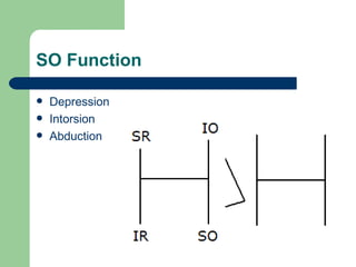
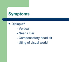
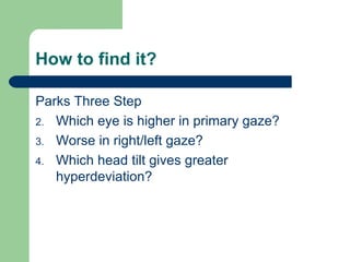
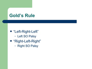
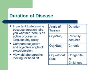
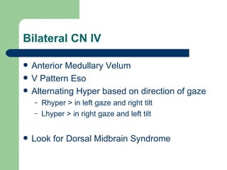
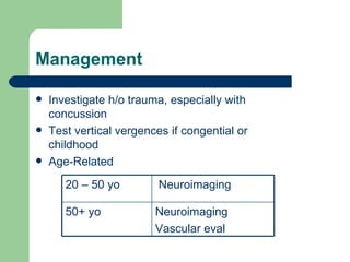
Superior Oblique Palsy  Jeff Guthrie
Anatomy Trochlear nuclei are located in the midbrain at the level of the midbrain Nerve decussates in roof of cerebral aqueduct at the anterior medullary vellum Emerges from the dorsal aspect of the midbrain Courses forward between posterior cerebral and superior cerebellar arteries then enters the cavernous sinus Enters orbit via SOF outside the annulus of zinn
 
 
SO Function Depression Intorsion Abduction
Symptoms Diplopia? - Vertical - Near > Far - Compensatory head tilt - tilting of visual world
How to find it? Parks Three Step Which eye is higher in primary gaze? Worse in right/left gaze? Which head tilt gives greater hyperdeviation?
Gold’s Rule “Left-Right-Left” Left SO Palsy “Right-Left-Right” Right SO Palsy
4 th  Question? Is there excyclotorsion? By defenition, superior oblique palsy should manifest excyclotorsion of the affected eye.
Unilateral vs. Bilateral? How can you determine? Measure subjective excyclotorsion! Using bilateral Maddox rod or Bagolini lenses in trial frame while pt views point source Ask pt to rotate one lens until the lines seen are lined up parallel. This is the amount of subjective torsion If >10 degrees = Bilateral CN IV Palsy
Duration of Disease Important to determine because duration tells you whether there is an active process vs. longstanding palsy. Compare subjective and objective angle of excyclotorsion View old photographs looking for head tilt Congenital or Childhood Obj without Subj Chronic Obj>Subj Recently acquired Obj=Subj Duration Angle of Torsion
Objective Excyclotorsion Found by looking at fundus photo Normal eye; fovea seen just inferior to the midline Objective = difference between where the fovea should be and where it is located on the photo BIO will show the fovea substantially superior to the expected location
Etiology of Isolated CN IV Children Trauma: may be unilateral or bilateral Congenital: only unilateral No subjective excyclotorsion High vertical vergence ranges Amblyogenic!!  But only 12% develop b/c children adopt head tilt and achieve fusion at least while head is tilted.
Etiology of Isolated CN IV Adults 30% Indeterminate  20% Ischemic 10% Aneurysm  40% Traumatic CN IV is the longest and thinnest cranial nerve Passes over the tentorium cerebelli making susceptible to increased ICP Generally regarded as the cranial nerve most susceptible to traumatic injury.
What to look out for Contralateral Horner’s Contralateral INO Dorsal Midbrain Syndrome Bilateral CN IV Palsy Trunctal Ataxia w Ipsilateral Dysmetria Ipsilateral CN III, VI Palsy and Oculosympathetics
Contralateral Horner’s Locus Ceruleus Suggestive of lesion affecting trochlear and Edinger-Westphal nuclei
Contralateral INO “ One & One-Half Syndrome” Lesion affecting trochlear nucleus and MLF tract contralateral to SO palsy Complete gaze palsy to side of lesion Adduction defecit on gaze away from lesion
Dorsal Midbrain Syndrome Anterior Medullary Vellum Light-Near Dissociation Eyelid retraction Headaches Etiology Pinealoma (most common), midbrain tumors, infarcts, syphillis, MS, herniation of temporal lobe, etc.
Bilateral CN IV Anterior Medullary Velum V Pattern Eso Alternating Hyper based on direction of gaze Rhyper > in left gaze and right tilt Lhyper > in right gaze and left tilt Look for Dorsal Midbrain Syndrome
Ipsilateral CN III, VI and Oculosympathetics Cavernous Sinus Syndrome Consider aneurysm (pain?), neoplasm, thrombosis, inflammatory conditions
Management Watch for resolution over 12 months Most will resolve in weeks-months, usually by 6mo Microvascular most likely to resolve spontaneously Traumatic less likely to resolve, but do tend to improve 65% of unilateral resolve, 25% of bilateral Aneurysmal and neoplastic least likely to resolve
Management  Investigate h/o trauma, especially with concussion  Test vertical vergences if congential or childhood Age-Related Neuroimaging Vascular eval 50+ yo Neuroimaging 20 – 50 yo
But Dr, what about my double vision? Prism, BD over the paretic eye Fresnel press on prism if still observing pt for resolution/improvement  2 Pairs!  SV distance and near, deviation greatest at near point Surgical correction after 12 months of observation

Superior Oblique Palsy

  • 1.
  • 2.
    Anatomy Trochlear nucleiare located in the midbrain at the level of the midbrain Nerve decussates in roof of cerebral aqueduct at the anterior medullary vellum Emerges from the dorsal aspect of the midbrain Courses forward between posterior cerebral and superior cerebellar arteries then enters the cavernous sinus Enters orbit via SOF outside the annulus of zinn
  • 3.
  • 4.
  • 5.
    SO Function DepressionIntorsion Abduction
  • 6.
    Symptoms Diplopia? -Vertical - Near > Far - Compensatory head tilt - tilting of visual world
  • 7.
    How to findit? Parks Three Step Which eye is higher in primary gaze? Worse in right/left gaze? Which head tilt gives greater hyperdeviation?
  • 8.
    Gold’s Rule “Left-Right-Left”Left SO Palsy “Right-Left-Right” Right SO Palsy
  • 9.
    4 th Question? Is there excyclotorsion? By defenition, superior oblique palsy should manifest excyclotorsion of the affected eye.
  • 10.
    Unilateral vs. Bilateral?How can you determine? Measure subjective excyclotorsion! Using bilateral Maddox rod or Bagolini lenses in trial frame while pt views point source Ask pt to rotate one lens until the lines seen are lined up parallel. This is the amount of subjective torsion If >10 degrees = Bilateral CN IV Palsy
  • 11.
    Duration of DiseaseImportant to determine because duration tells you whether there is an active process vs. longstanding palsy. Compare subjective and objective angle of excyclotorsion View old photographs looking for head tilt Congenital or Childhood Obj without Subj Chronic Obj>Subj Recently acquired Obj=Subj Duration Angle of Torsion
  • 12.
    Objective Excyclotorsion Foundby looking at fundus photo Normal eye; fovea seen just inferior to the midline Objective = difference between where the fovea should be and where it is located on the photo BIO will show the fovea substantially superior to the expected location
  • 13.
    Etiology of IsolatedCN IV Children Trauma: may be unilateral or bilateral Congenital: only unilateral No subjective excyclotorsion High vertical vergence ranges Amblyogenic!! But only 12% develop b/c children adopt head tilt and achieve fusion at least while head is tilted.
  • 14.
    Etiology of IsolatedCN IV Adults 30% Indeterminate 20% Ischemic 10% Aneurysm 40% Traumatic CN IV is the longest and thinnest cranial nerve Passes over the tentorium cerebelli making susceptible to increased ICP Generally regarded as the cranial nerve most susceptible to traumatic injury.
  • 15.
    What to lookout for Contralateral Horner’s Contralateral INO Dorsal Midbrain Syndrome Bilateral CN IV Palsy Trunctal Ataxia w Ipsilateral Dysmetria Ipsilateral CN III, VI Palsy and Oculosympathetics
  • 16.
    Contralateral Horner’s LocusCeruleus Suggestive of lesion affecting trochlear and Edinger-Westphal nuclei
  • 17.
    Contralateral INO “One & One-Half Syndrome” Lesion affecting trochlear nucleus and MLF tract contralateral to SO palsy Complete gaze palsy to side of lesion Adduction defecit on gaze away from lesion
  • 18.
    Dorsal Midbrain SyndromeAnterior Medullary Vellum Light-Near Dissociation Eyelid retraction Headaches Etiology Pinealoma (most common), midbrain tumors, infarcts, syphillis, MS, herniation of temporal lobe, etc.
  • 19.
    Bilateral CN IVAnterior Medullary Velum V Pattern Eso Alternating Hyper based on direction of gaze Rhyper > in left gaze and right tilt Lhyper > in right gaze and left tilt Look for Dorsal Midbrain Syndrome
  • 20.
    Ipsilateral CN III,VI and Oculosympathetics Cavernous Sinus Syndrome Consider aneurysm (pain?), neoplasm, thrombosis, inflammatory conditions
  • 21.
    Management Watch forresolution over 12 months Most will resolve in weeks-months, usually by 6mo Microvascular most likely to resolve spontaneously Traumatic less likely to resolve, but do tend to improve 65% of unilateral resolve, 25% of bilateral Aneurysmal and neoplastic least likely to resolve
  • 22.
    Management Investigateh/o trauma, especially with concussion Test vertical vergences if congential or childhood Age-Related Neuroimaging Vascular eval 50+ yo Neuroimaging 20 – 50 yo
  • 23.
    But Dr, whatabout my double vision? Prism, BD over the paretic eye Fresnel press on prism if still observing pt for resolution/improvement 2 Pairs! SV distance and near, deviation greatest at near point Surgical correction after 12 months of observation