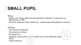
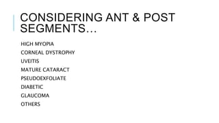
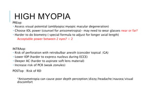
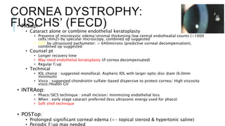
CATARACT SURGERY
IN SPECIAL
SITUATIONS
BY DR ZULFAHMI
CONSIDERING…
POTENTIAL VISUALIZATION PROBLEM
 CORNEAL OPACITIES
 SMALL PUPIL
ANTERIOR AND POSTERIOR SEGMENT
 HIGH MYOPIA
 CORNEAL DYSTROPHY
 UVEITIS
 MATURE CATARACT
 PSEUDOEXFOLIATE
 DIABETIC
 GLAUCOMA
 OTHERS
CORNEAL OPACITY
Difficult to see details : residual lens matter remained/
IOL placement under the iris.
Leukoma, pannus, corneal dystrophy, degeneration, and
band-shaped keratopathy, uveitis, glaucoma.
If there is a central corneal scar obscuring the pupil,
an optical sector iridectomy may be helpful.
SMALL PUPIL
PREop:
 Higher risk: Uveitis/DM/pseudoexfoliation S/Marfan’s S/glaucoma on
pilocarpine tx/ BPH
 Intensive dilatation 2Hour before op : tropicamide/phenylephrine/nevanac
INTRAop:
 Intracameral adrenaline
 Viscoelastics to dilate
 Iris hook & etc
 sphincter-ectomy (three small radial cuts on the sphincter pupillae, 120°
apart)
 radial iridotomy
CONSIDERING ANT & POST
SEGMENTS…
HIGH MYOPIA
CORNEAL DYSTROPHY
UVEITIS
MATURE CATARACT
PSEUDOEXFOLIATE
DIABETIC
GLAUCOMA
OTHERS
HIGH MYOPIA
PREop:
 Assess visual potential (amblyopia/myopic macular degeneration)
 Choose IOL power (counsel for anisometropia)- may need to wear glasses near or far?
 Harder to do biometry ( special formula to adjust for longer axial length)
Acceptable power between 2 eyes? < 2
INTRAop:
 Risk of perforation with retrobulbar anesth (consider topical /GA)
 Lower IOP (harder to express nucleus during ECCE)
 Deeper AC (harder to aspirate soft lens material)
 Increase risk of PCR (weak zonules)
POSTop : Risk of RD
*Anisometropia can cause poor depth perception/dizzy/headache/nausea/visual
discomfort
CORNEA DYSTROPHY:
FUSCHS’ (FECD)
• PREop:
• Cataract alone or combine endothelial keratoplasty
• Presence of microcystic edema/stromal thickening/low central endothealial counts (<1000
cells/mm2)–by specular microscopy, combined op suggested
• CCT by ultrasound pachymeter: > 640microns (predictive corneal decompensation),
combined op suggested
• Counsel pt
• Longer recovery time
• May need endothelial keratoplasty (if cornea decompensated)
• Regular f/up
• Technical
• IOL choice : suggested monofocal. Aspheric IOL with larger optic disc diam (6.0mm
minimum)
• Visco : suggested chondroitin sulfate-based dispersive to protect cornea/ High viscosity
visco/Healon GV
• INTRAop:
• Phaco/SICS technique : small incision/ minimizing endothelial loss
• When : early stage cataract preferred (less ultrasonic energy used for phaco)
• Soft shell technique
• POSTop:
• Prolonged significant corneal edema (+- topical steroid & hypertonic saline)
• Periodic f/up may needed
UVEITIS
PREop:
 Need to control inflammation
 Wait 2-3 months after quiet cell activity/ resolved
 Consider course of preop steroids & longer topical steroids postop (tab Pred 0.5mg/kg x3/7 prior & 6/52 topical
postop)
 Assess visual potential
 Dilate pupil in advance
 If synechiae severe superiorly, consider corneal section (gonio)
INTRAop:
 Small pupil problem
 Increase risk PCR (weak zonules)
 Increase inflammation (consider heparin coated IOL or aphakic)
 Increase risk of bleeding
POSTop:
 Higher risk for : corneal edema/ flare up inflam/glaucoma/hypotony/choroidal
effusion/CME
 Consider prophylaxis for infectious etiologies ( eg herpetic lesions)
MATURE CATARACT
Limitation to assess visual potential
 Pupil (optic nerve fx)
 Bscan (gross retinal anatomy)
 Light projection & color perception
 Potential acuity meter ( macular fx)
Poor view of capsulotomy/ capsulorrhexis edge
 Consider endocapsular technique/ air instead of visco/ capsular stain eg;trypan
High intra-capsular pressure
 CCC runs out/split easily
Floppy capsule d/t chronic bulky lens
 Viscoelastic tamponade
PSEUDOEXFOLIATION- RISK FOR
LOOSE ZONULES
PREop:
 Small pupil
 Shallow AC in normal AXL
 Phacodenesis
INTRAop:
 Higher risk for zonular dehiscence/lens sublux or dislocate/ PCR
POSTop
 IOP spikes
 Capsular phimosis
 Prolonged inflammation
 Corneal decompensation.
DIABETIC
Issues : difficult & progression of DR after op
PREop:
 Assess visual potential
 Laser PRP if necessary prior op
 Medical consult
 Treat macula oedema before op if fundal view permits
INTRAop:
 Protect corneal epithelium ( risk of abrasion & poor healing)
 Small pupil
 Consider stitching wound
POSTop:
 Control inflammation (esp in PDR eyes)
 Risk of PDR/CSME
 Risk of glaucoma
 Risk of PCO
GLAUCOMA
•Postoperative corneal edema may occur.
•May have pupils resistant to dilation. (Eg on long term pilocarpine)
•Complications like iris injury, capsular tear and zonular dialysis can all
aggravate pre-existing glaucoma.
•Previous trabeculectomy means that the functioning bleb must be preserved
during op, consider temporal approach,
•Synechiae and a shallow anterior chamber are often present.
OP: GLAUCOMA VS
CATARACT
If the glaucoma more severe, consider glaucoma surgery first or vice
versa.
If both are severe , consider combined cataract op & trabeculectomy
procedure.
Factors include:
 Severity & progression of glaucoma/cataract
 Patient factors: age,race,family hx of blindness from glaucoma, fellow eye blind
glaucoma, concomitant risk factor (dm/hpt/myopia etc), compliance to f/up &
OTHERS
DEEP SEATED EYE : Consider temporal approach
PTERYGIUM : Consider excised first if significant
SYNECHIAE: Consider change site of incision / synechiolysis
Back pain/ neck pain? Unable to lie flat
THANK Q
REFERENCES
Recognising ‘high-risk’ eyes before cataract surgery
https://www.ncbi.nlm.nih.gov/pmc/articles/PMC2377383/

Cataract surgery in special situations

  • 1.
  • 2.
    CONSIDERING… POTENTIAL VISUALIZATION PROBLEM CORNEAL OPACITIES  SMALL PUPIL ANTERIOR AND POSTERIOR SEGMENT  HIGH MYOPIA  CORNEAL DYSTROPHY  UVEITIS  MATURE CATARACT  PSEUDOEXFOLIATE  DIABETIC  GLAUCOMA  OTHERS
  • 3.
    CORNEAL OPACITY Difficult tosee details : residual lens matter remained/ IOL placement under the iris. Leukoma, pannus, corneal dystrophy, degeneration, and band-shaped keratopathy, uveitis, glaucoma. If there is a central corneal scar obscuring the pupil, an optical sector iridectomy may be helpful.
  • 4.
    SMALL PUPIL PREop:  Higherrisk: Uveitis/DM/pseudoexfoliation S/Marfan’s S/glaucoma on pilocarpine tx/ BPH  Intensive dilatation 2Hour before op : tropicamide/phenylephrine/nevanac INTRAop:  Intracameral adrenaline  Viscoelastics to dilate  Iris hook & etc  sphincter-ectomy (three small radial cuts on the sphincter pupillae, 120° apart)  radial iridotomy
  • 5.
    CONSIDERING ANT &POST SEGMENTS… HIGH MYOPIA CORNEAL DYSTROPHY UVEITIS MATURE CATARACT PSEUDOEXFOLIATE DIABETIC GLAUCOMA OTHERS
  • 6.
    HIGH MYOPIA PREop:  Assessvisual potential (amblyopia/myopic macular degeneration)  Choose IOL power (counsel for anisometropia)- may need to wear glasses near or far?  Harder to do biometry ( special formula to adjust for longer axial length) Acceptable power between 2 eyes? < 2 INTRAop:  Risk of perforation with retrobulbar anesth (consider topical /GA)  Lower IOP (harder to express nucleus during ECCE)  Deeper AC (harder to aspirate soft lens material)  Increase risk of PCR (weak zonules) POSTop : Risk of RD *Anisometropia can cause poor depth perception/dizzy/headache/nausea/visual discomfort
  • 7.
    CORNEA DYSTROPHY: FUSCHS’ (FECD) •PREop: • Cataract alone or combine endothelial keratoplasty • Presence of microcystic edema/stromal thickening/low central endothealial counts (<1000 cells/mm2)–by specular microscopy, combined op suggested • CCT by ultrasound pachymeter: > 640microns (predictive corneal decompensation), combined op suggested • Counsel pt • Longer recovery time • May need endothelial keratoplasty (if cornea decompensated) • Regular f/up • Technical • IOL choice : suggested monofocal. Aspheric IOL with larger optic disc diam (6.0mm minimum) • Visco : suggested chondroitin sulfate-based dispersive to protect cornea/ High viscosity visco/Healon GV • INTRAop: • Phaco/SICS technique : small incision/ minimizing endothelial loss • When : early stage cataract preferred (less ultrasonic energy used for phaco) • Soft shell technique • POSTop: • Prolonged significant corneal edema (+- topical steroid & hypertonic saline) • Periodic f/up may needed
  • 8.
    UVEITIS PREop:  Need tocontrol inflammation  Wait 2-3 months after quiet cell activity/ resolved  Consider course of preop steroids & longer topical steroids postop (tab Pred 0.5mg/kg x3/7 prior & 6/52 topical postop)  Assess visual potential  Dilate pupil in advance  If synechiae severe superiorly, consider corneal section (gonio) INTRAop:  Small pupil problem  Increase risk PCR (weak zonules)  Increase inflammation (consider heparin coated IOL or aphakic)  Increase risk of bleeding POSTop:  Higher risk for : corneal edema/ flare up inflam/glaucoma/hypotony/choroidal effusion/CME  Consider prophylaxis for infectious etiologies ( eg herpetic lesions)
  • 9.
    MATURE CATARACT Limitation toassess visual potential  Pupil (optic nerve fx)  Bscan (gross retinal anatomy)  Light projection & color perception  Potential acuity meter ( macular fx) Poor view of capsulotomy/ capsulorrhexis edge  Consider endocapsular technique/ air instead of visco/ capsular stain eg;trypan High intra-capsular pressure  CCC runs out/split easily Floppy capsule d/t chronic bulky lens  Viscoelastic tamponade
  • 10.
    PSEUDOEXFOLIATION- RISK FOR LOOSEZONULES PREop:  Small pupil  Shallow AC in normal AXL  Phacodenesis INTRAop:  Higher risk for zonular dehiscence/lens sublux or dislocate/ PCR POSTop  IOP spikes  Capsular phimosis  Prolonged inflammation  Corneal decompensation.
  • 11.
    DIABETIC Issues : difficult& progression of DR after op PREop:  Assess visual potential  Laser PRP if necessary prior op  Medical consult  Treat macula oedema before op if fundal view permits INTRAop:  Protect corneal epithelium ( risk of abrasion & poor healing)  Small pupil  Consider stitching wound POSTop:  Control inflammation (esp in PDR eyes)  Risk of PDR/CSME  Risk of glaucoma  Risk of PCO
  • 12.
    GLAUCOMA •Postoperative corneal edemamay occur. •May have pupils resistant to dilation. (Eg on long term pilocarpine) •Complications like iris injury, capsular tear and zonular dialysis can all aggravate pre-existing glaucoma. •Previous trabeculectomy means that the functioning bleb must be preserved during op, consider temporal approach, •Synechiae and a shallow anterior chamber are often present.
  • 13.
    OP: GLAUCOMA VS CATARACT Ifthe glaucoma more severe, consider glaucoma surgery first or vice versa. If both are severe , consider combined cataract op & trabeculectomy procedure. Factors include:  Severity & progression of glaucoma/cataract  Patient factors: age,race,family hx of blindness from glaucoma, fellow eye blind glaucoma, concomitant risk factor (dm/hpt/myopia etc), compliance to f/up &
  • 14.
    OTHERS DEEP SEATED EYE: Consider temporal approach PTERYGIUM : Consider excised first if significant SYNECHIAE: Consider change site of incision / synechiolysis Back pain/ neck pain? Unable to lie flat
  • 15.
  • 20.
    REFERENCES Recognising ‘high-risk’ eyesbefore cataract surgery https://www.ncbi.nlm.nih.gov/pmc/articles/PMC2377383/