Download as PDF, PPTX


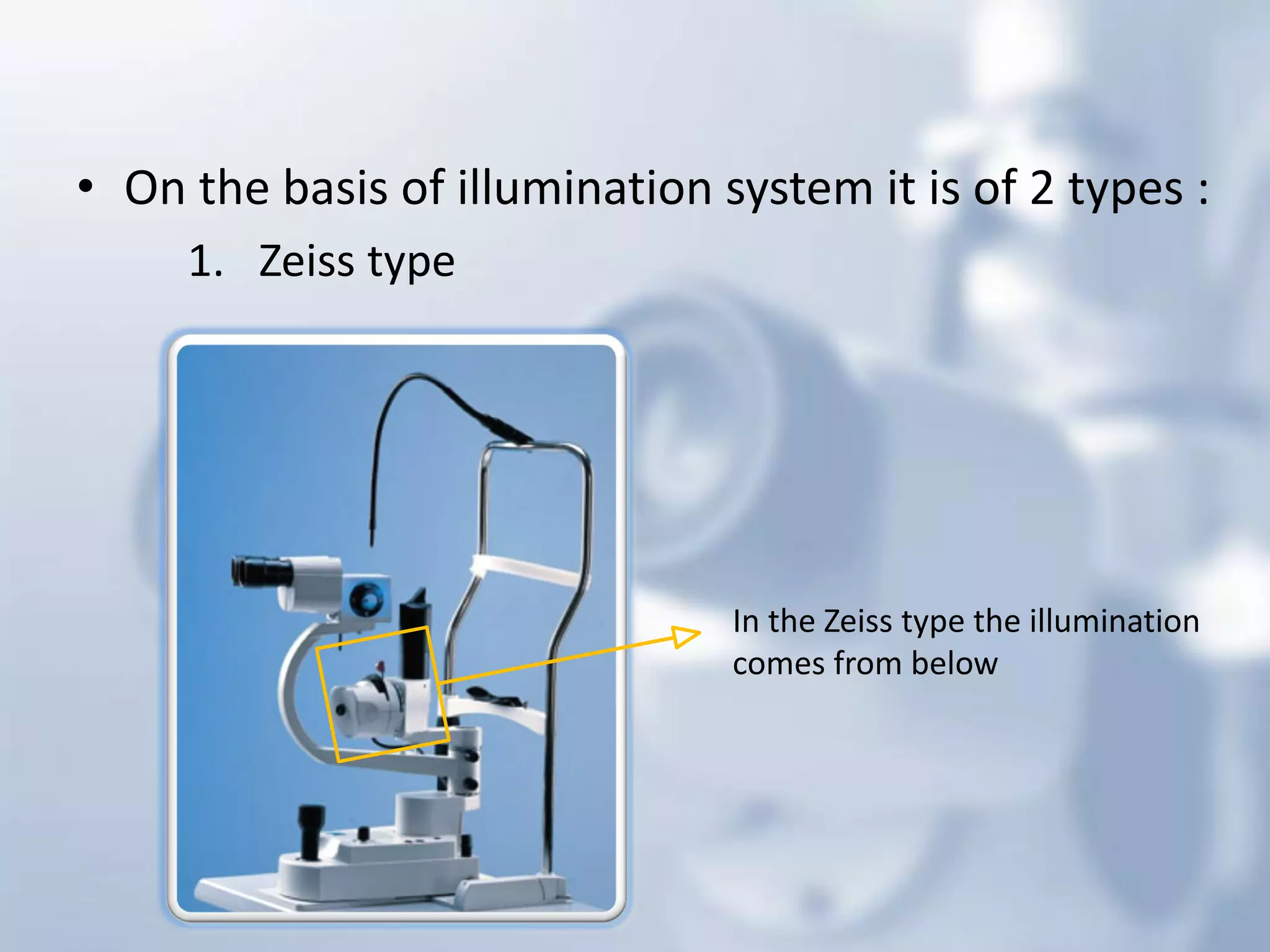

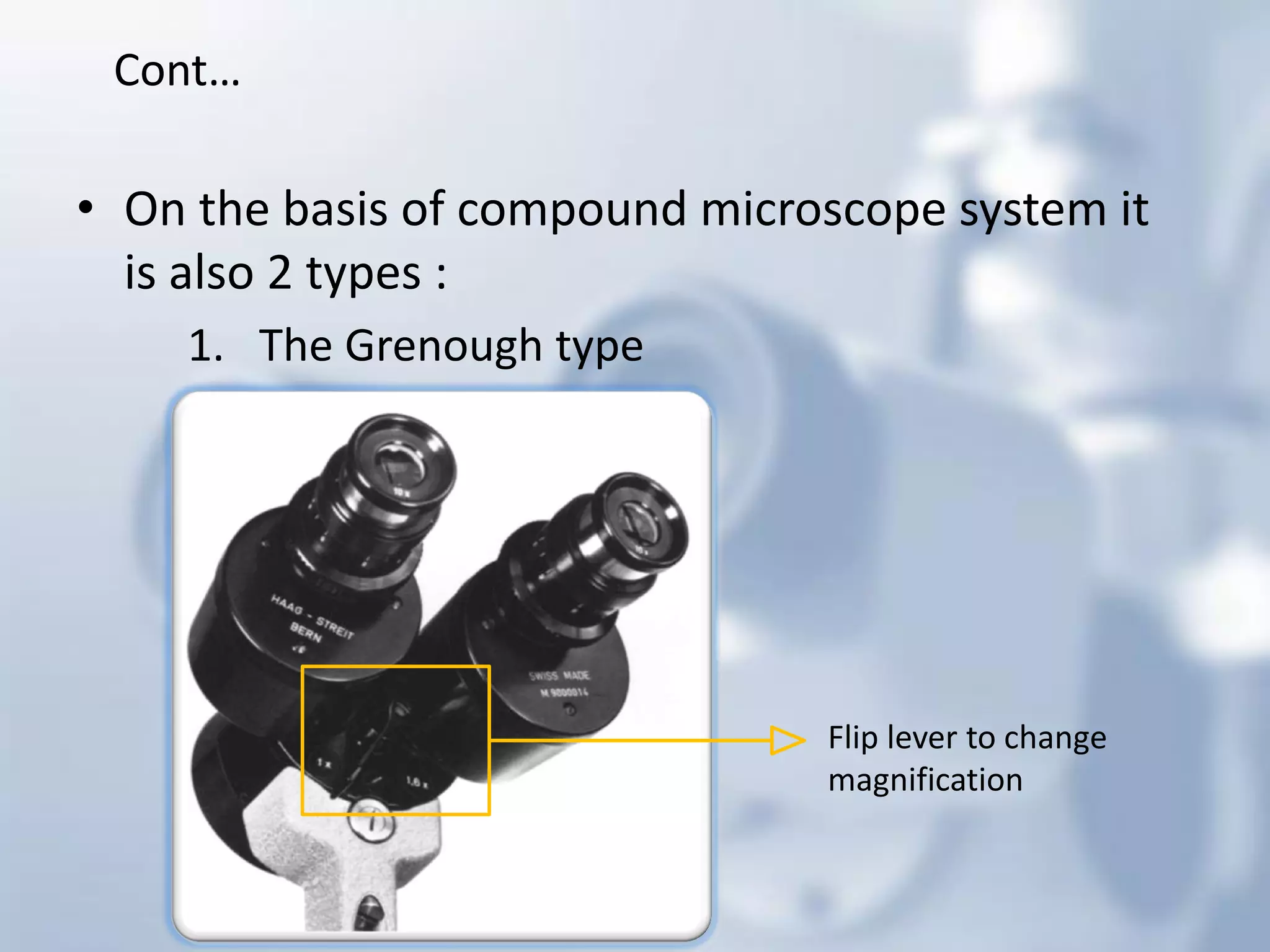
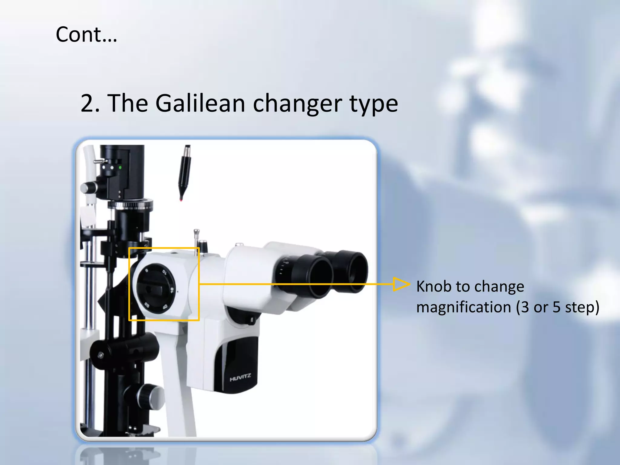
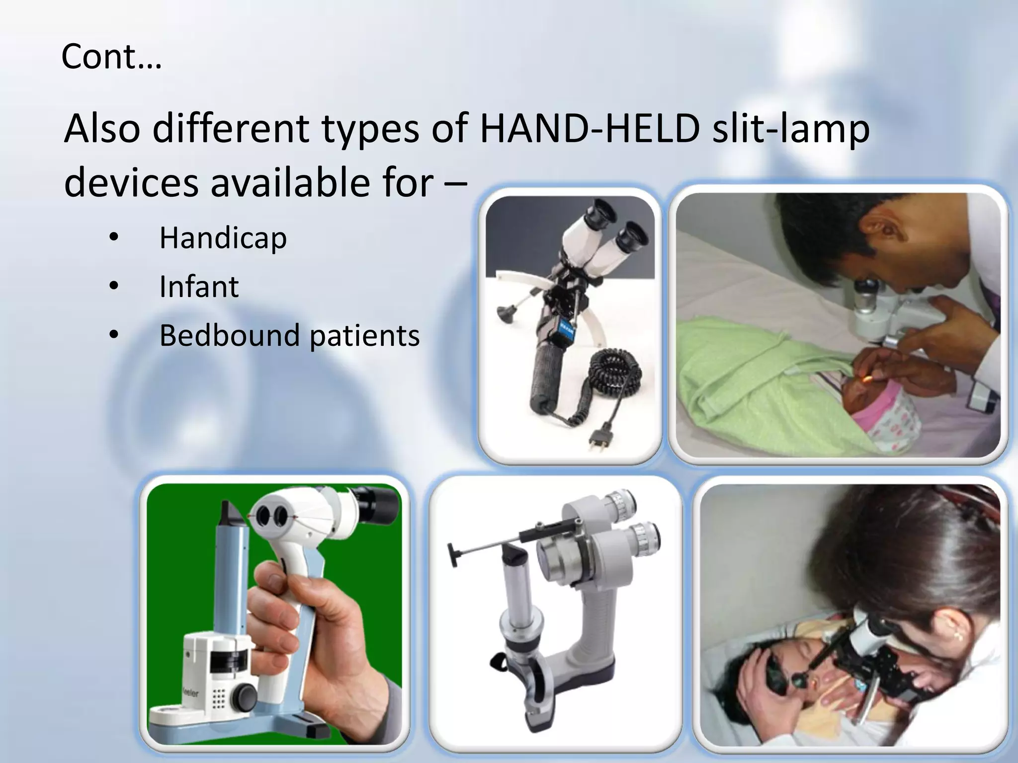


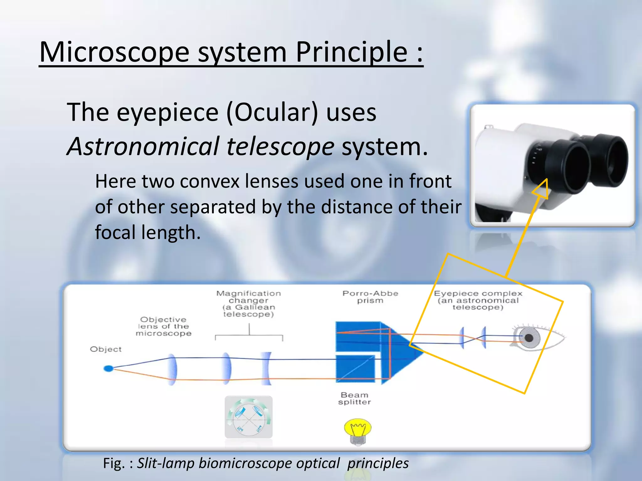
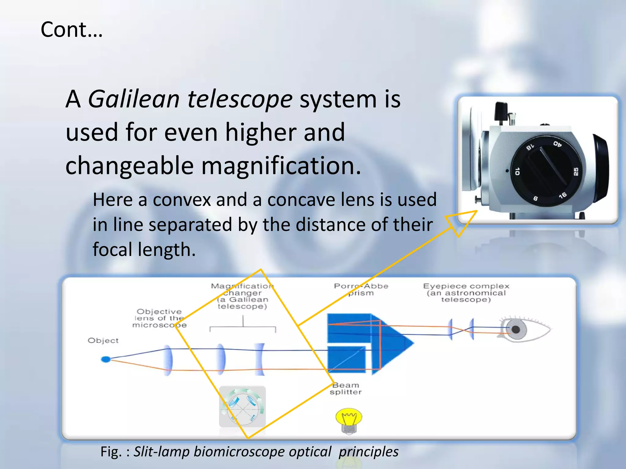
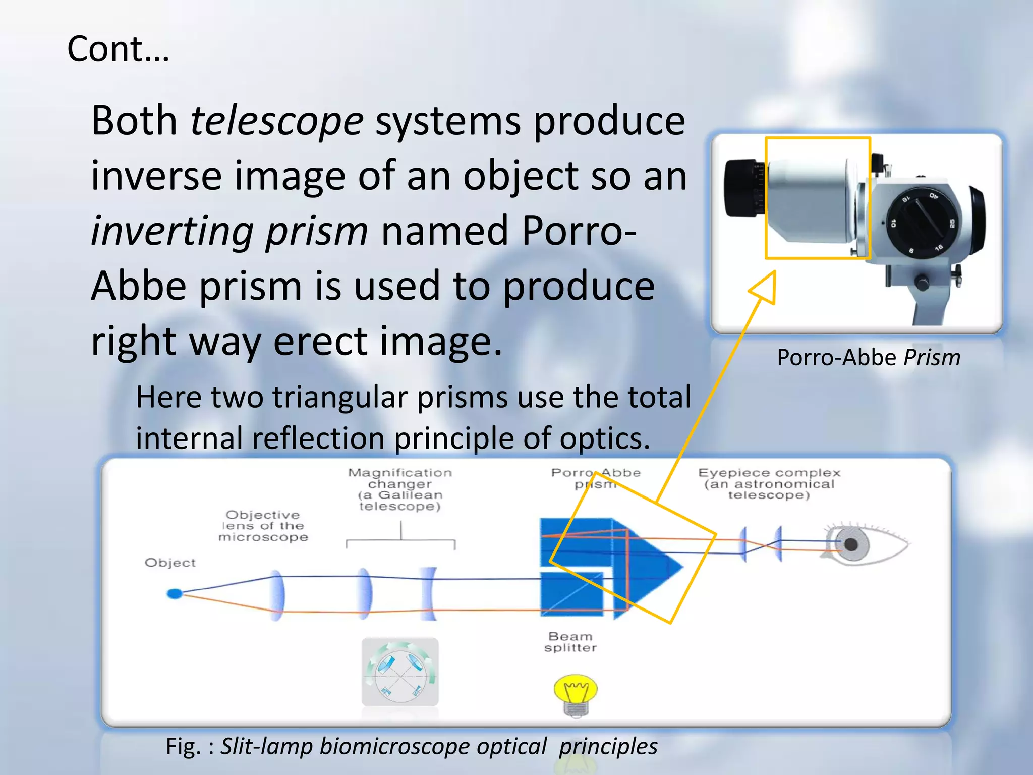
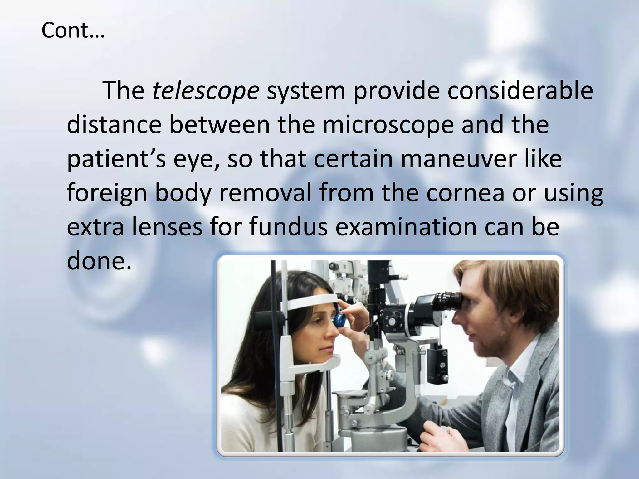


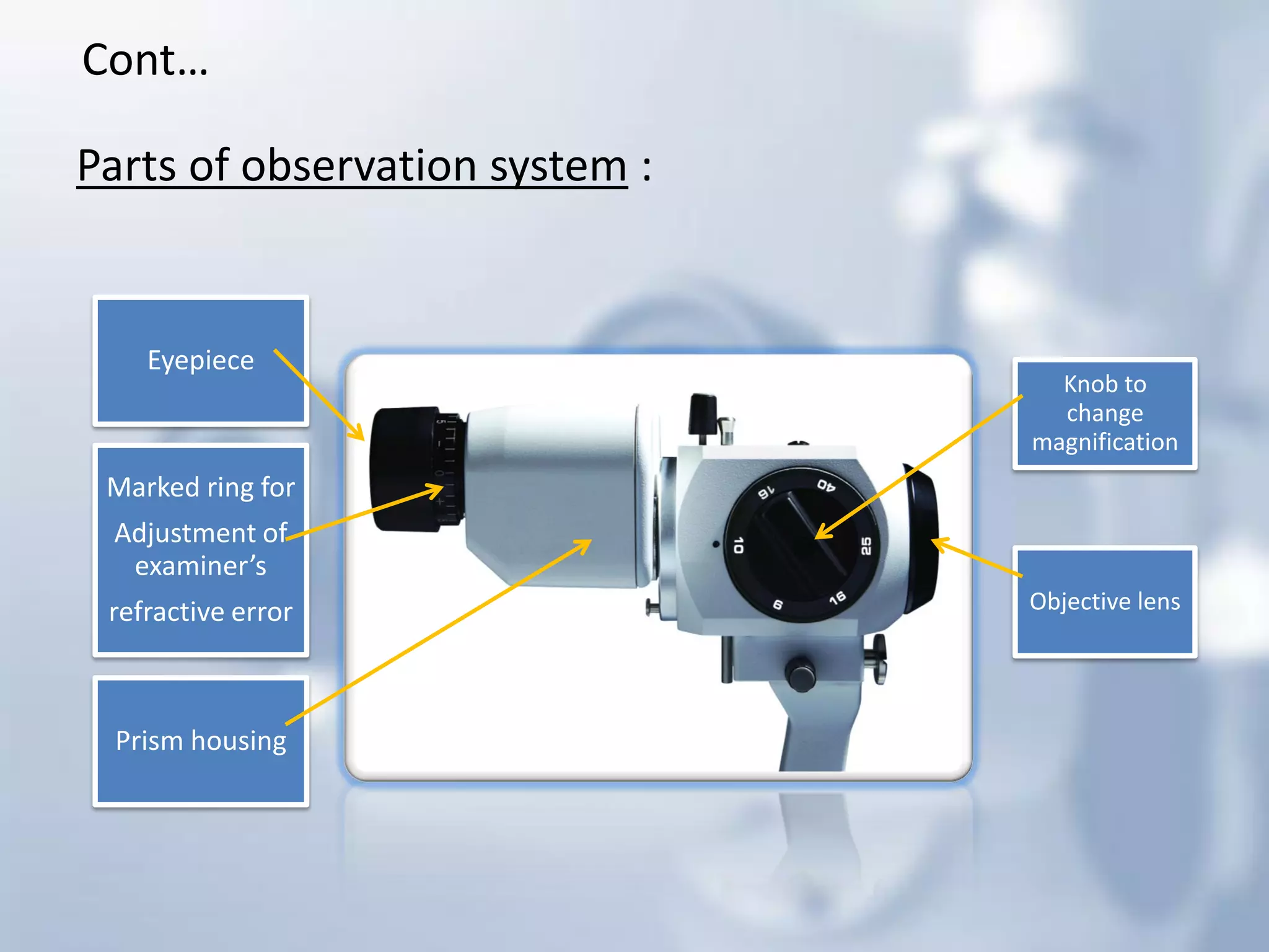


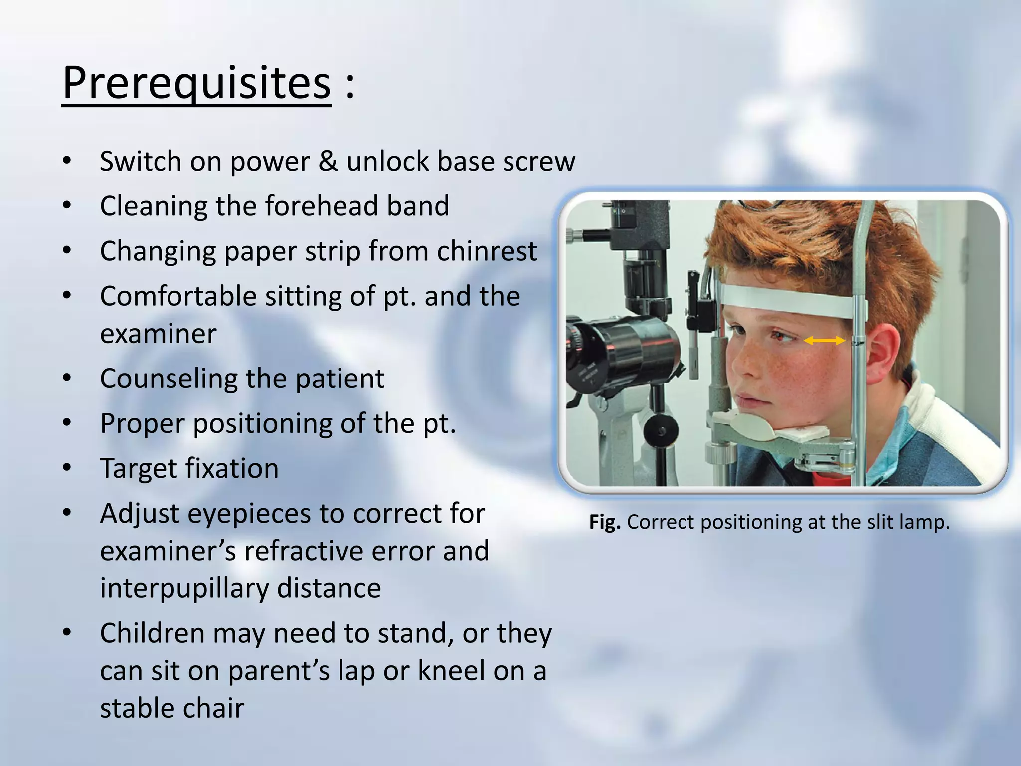

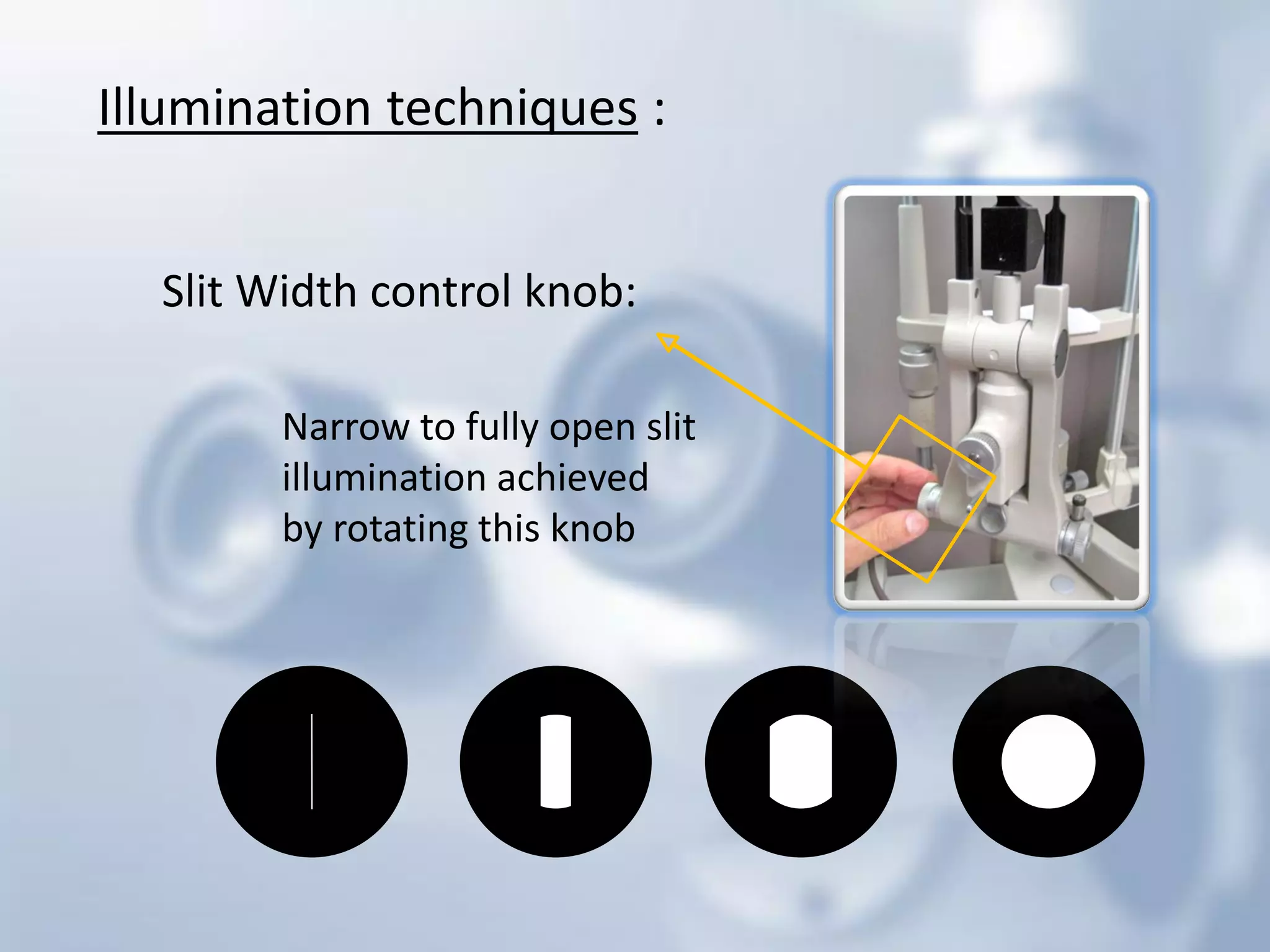

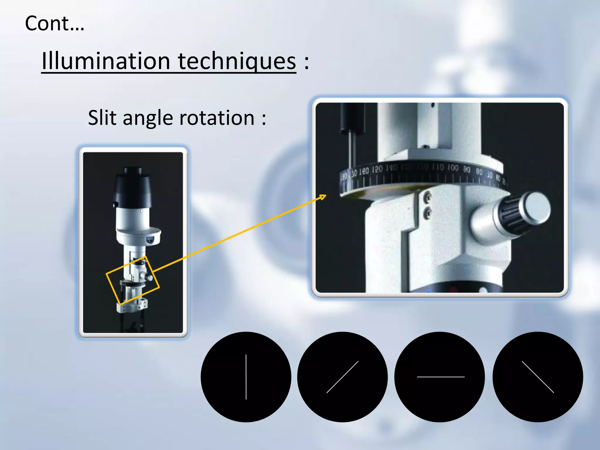
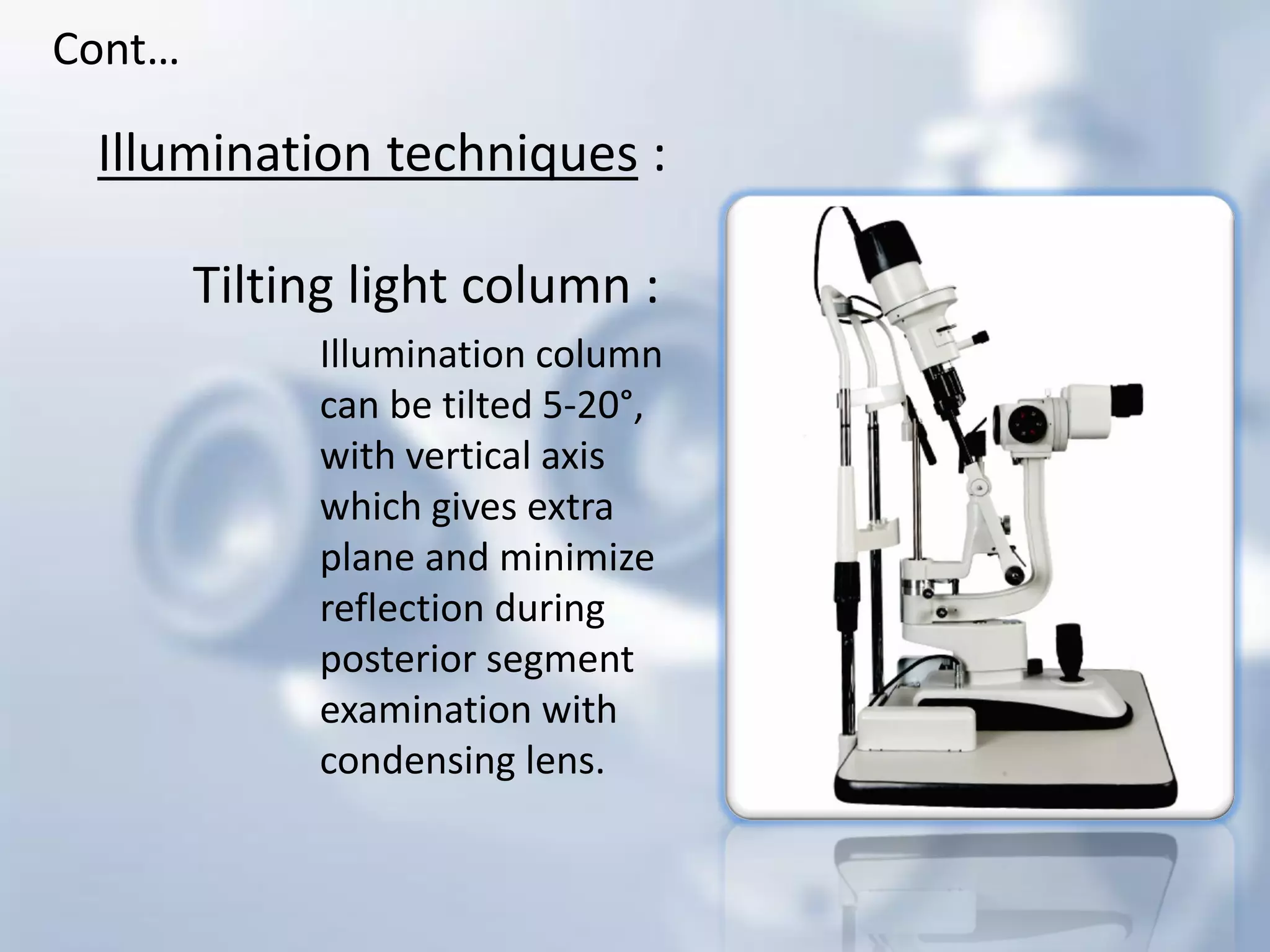
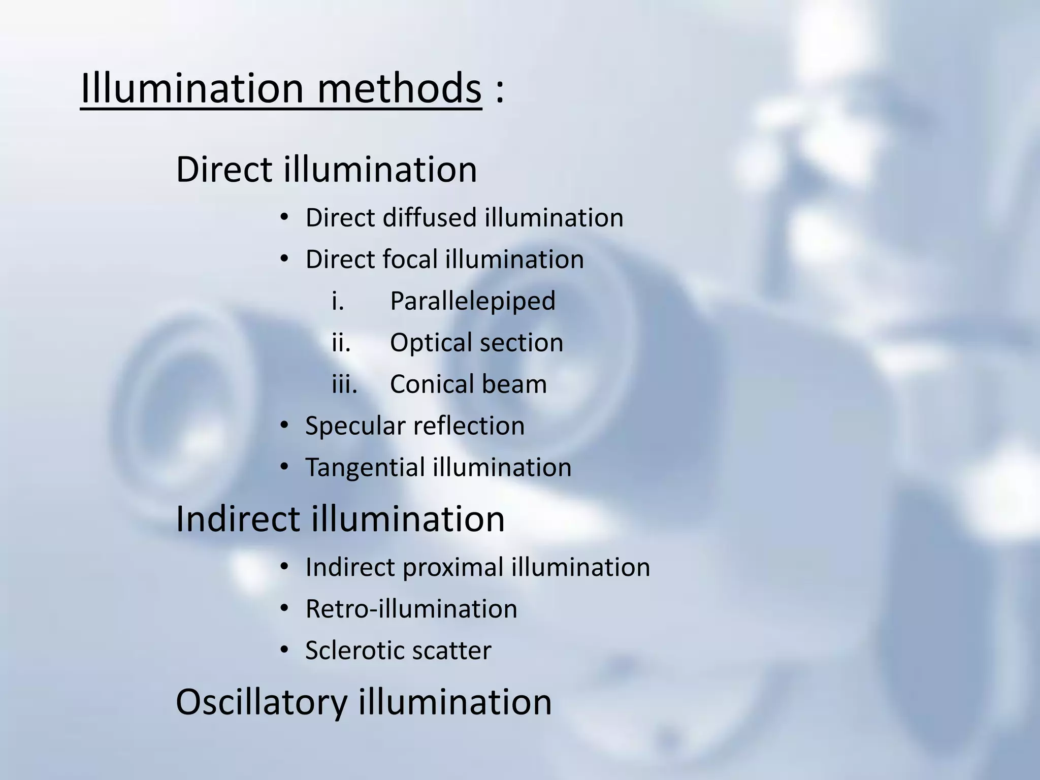


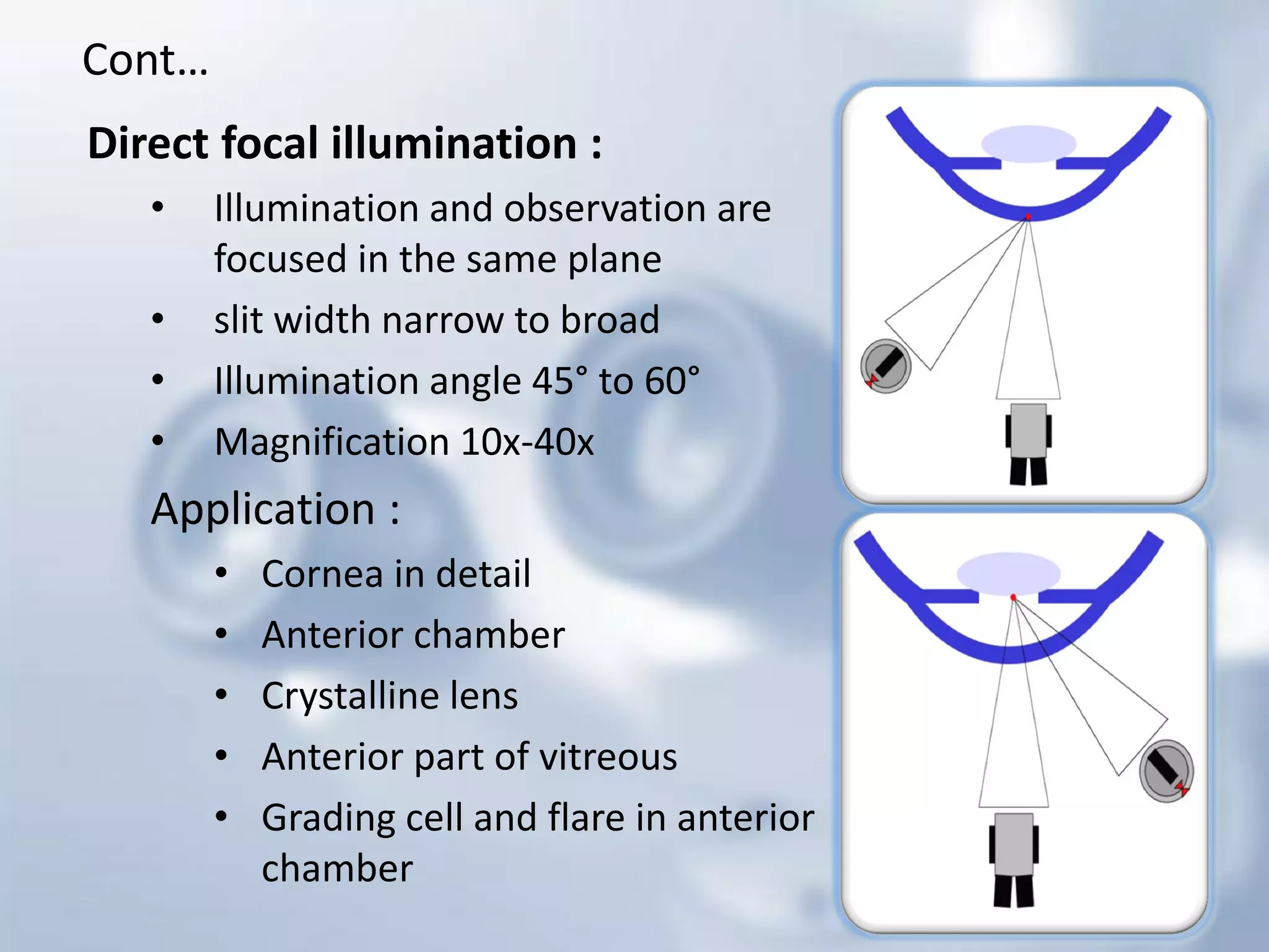

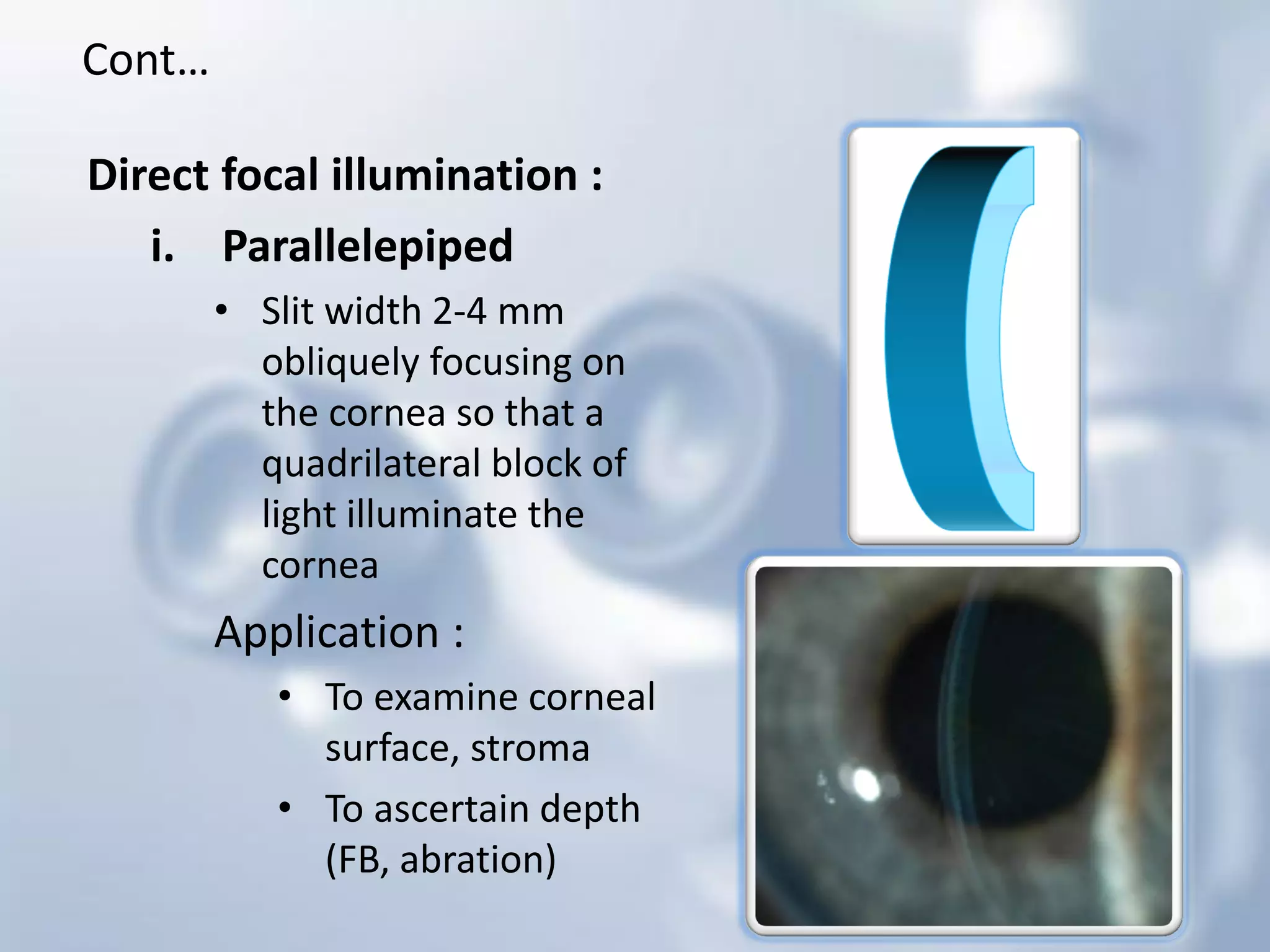

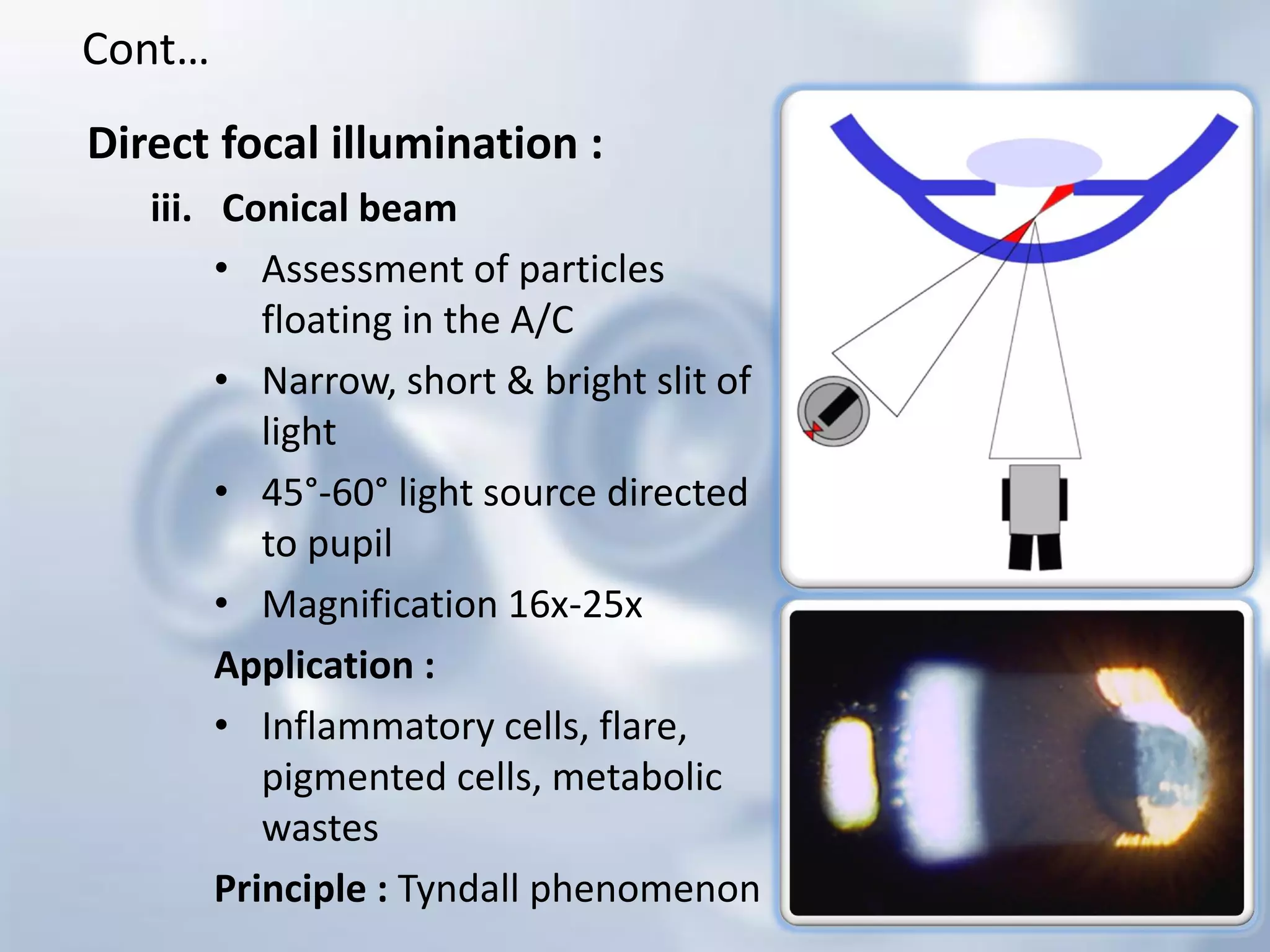
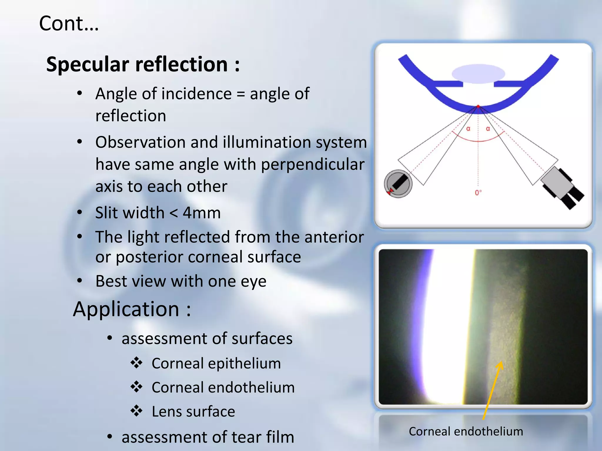

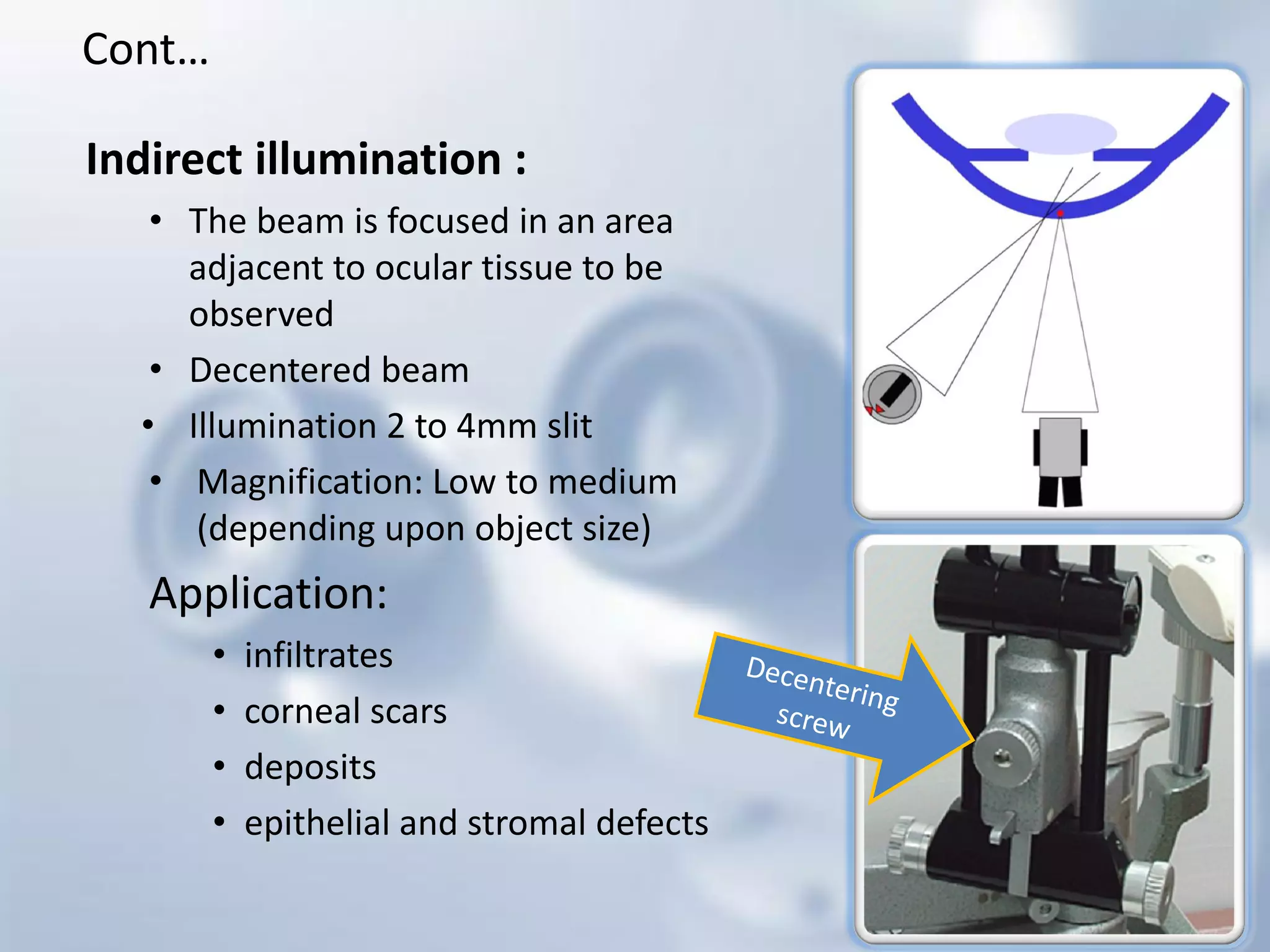

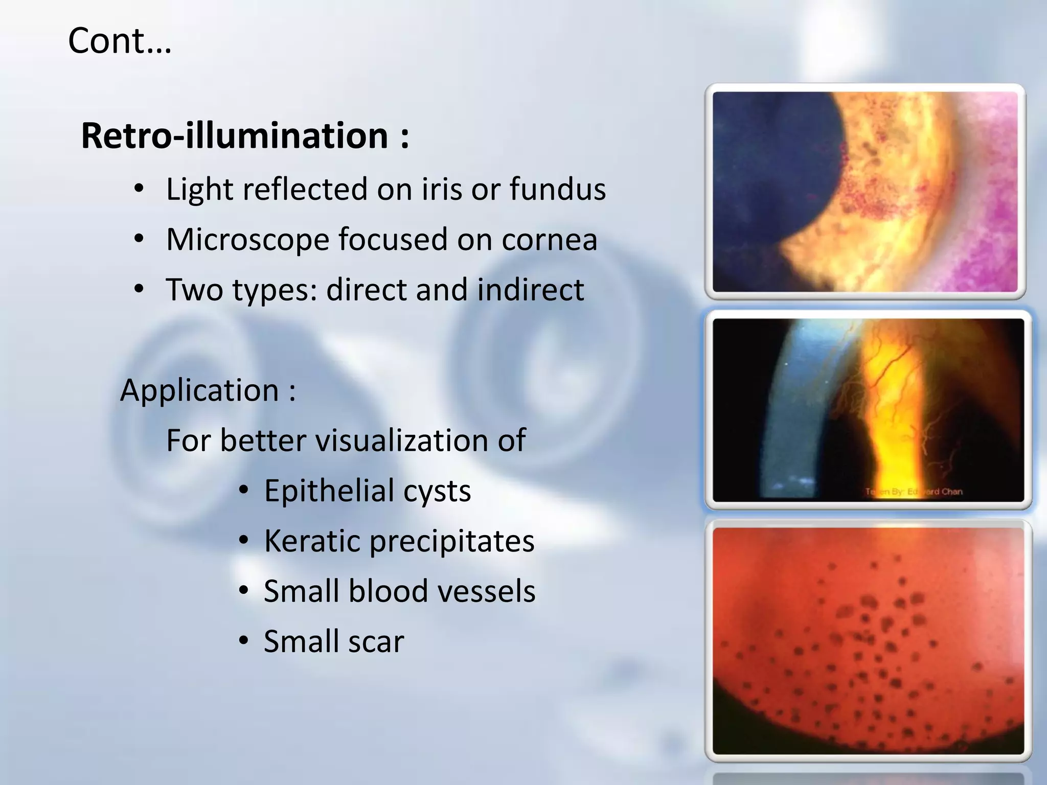

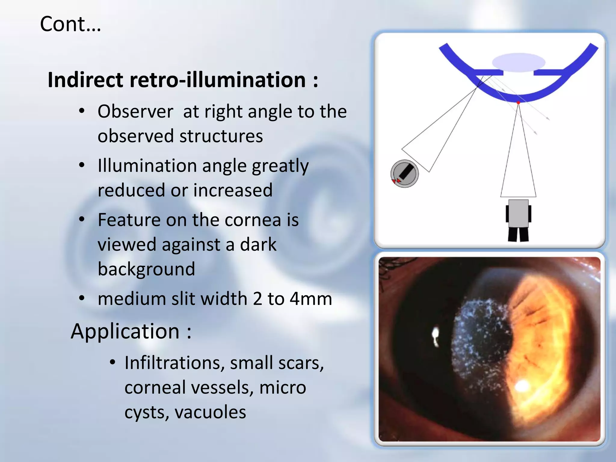

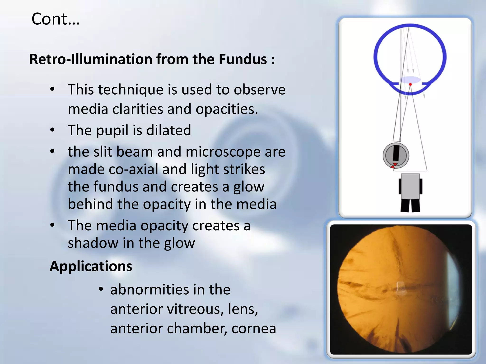


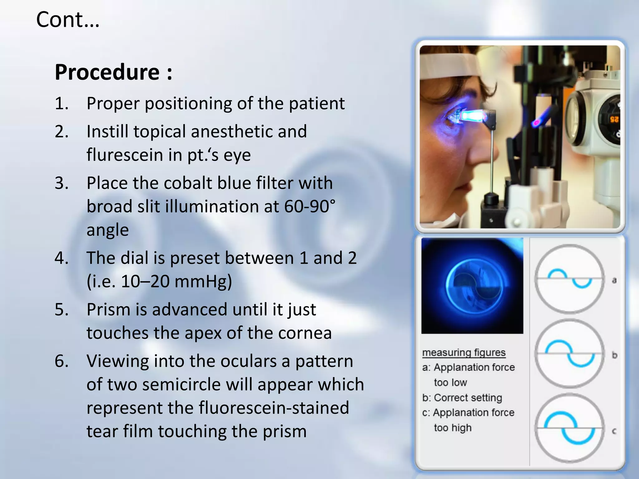


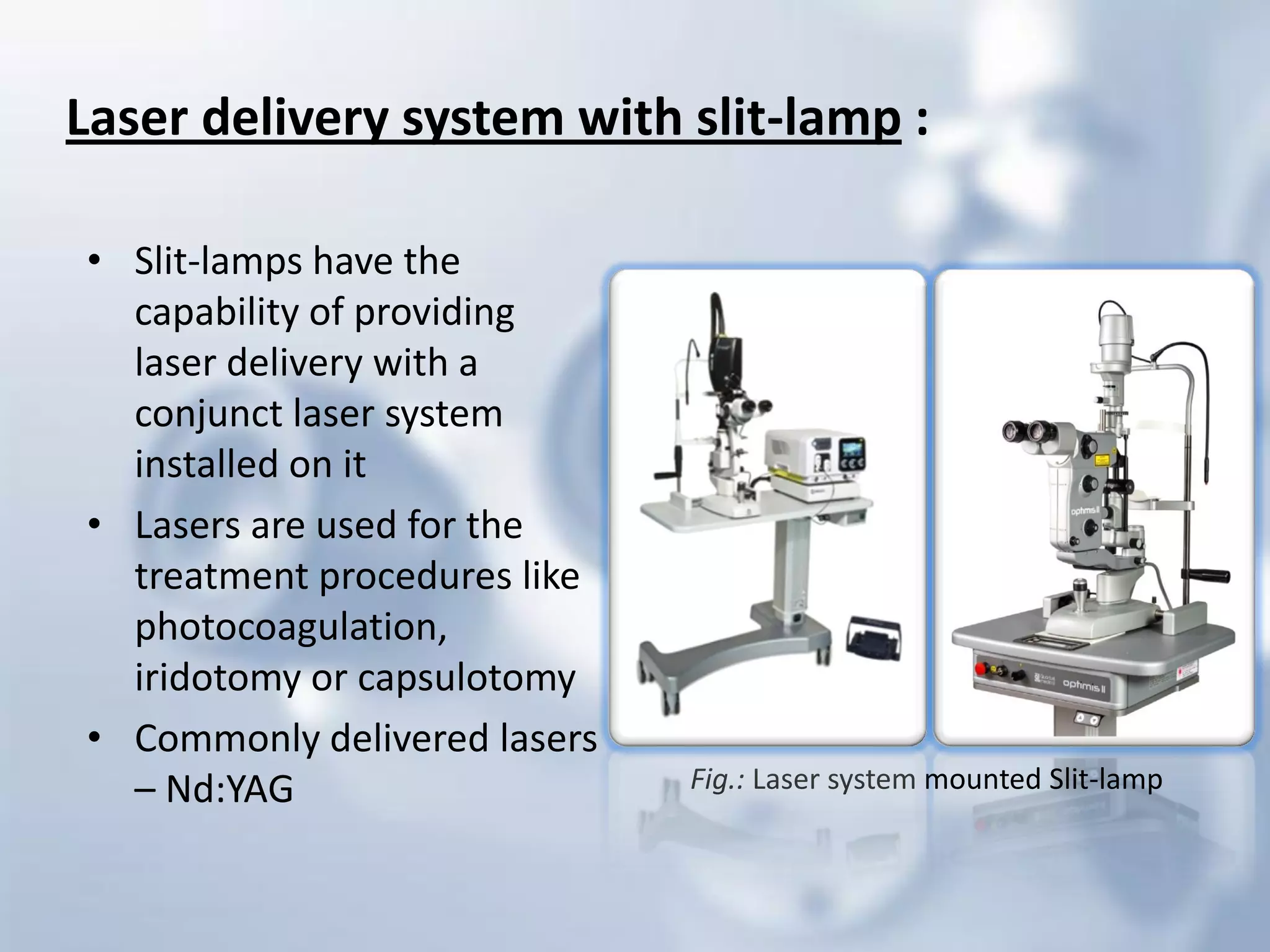


The document discusses the slit-lamp biomicroscope, which is used to examine the eye. It has three main components: the mechanical system to position the patient and microscope, the illumination system to provide a focused beam of light, and the observation system consisting of compound microscopes. Different illumination techniques such as direct, indirect, and focal illumination are used to examine different parts of the eye at various magnifications. The slit-lamp allows close examination of structures like the cornea, anterior chamber, and lens.


