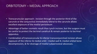
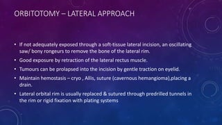
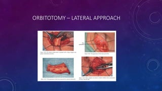
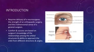
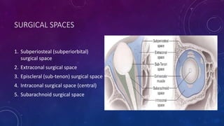
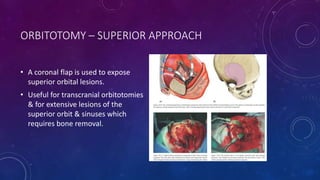
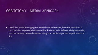
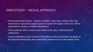
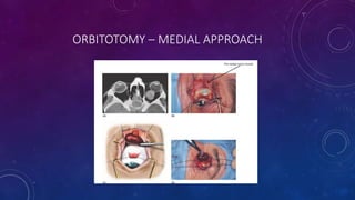
APPROACH TO ORBITAL
SURGERY
BIPIN BISTA
RESIDENT
OPHTHALMOLOGY
INTRODUCTION
• Requires delicacy of a neurosurgeon,
the strength of an orthopaedic surgery,
and the 3-dimensional sense of a
general surgeon.
• Comfort & success are based on
surgeon’s knowledge of the
relationships among the orbital
structures & ability to approach the
orbit from different directions & angles.
SURGICAL SPACES
1. Subperiosteal (subperiorbital)
surgical space
2. Extraconal surgical space
3. Episcleral (sub-tenon) surgical space
4. Intraconal surgical space (central)
5. Subarachnoid surgical space
INSTRUMENTS
Stryker saw with finger twitch, bony rongeurs
INSTRUMENTS
Sewall, wright
ORBITOTOMY – SUPERIOR APPROACH
1. Trancutaneous incisions
2. Transconjunctival incision
ORBITOTOMY – SUPERIOR APPROACH
• Transcutaneous incisions : an incision through the upper eyelid crease offers
good access to the superior orbital rim & periosteum, with a HIDDEN scar.
• Cosmetic result is better with an eyelid crease incision >> supraorbital rim.
• Eyelid crease incision leds to access to orbital rim by superior dissection in the
postorbicularis fascial plane anterior to orbital septum.
• After rim exposure, incision is made in arcus marginalis,then periosteum is
separated from the frontal bone of the orbital roof.
ORBITOTOMY – SUPERIOR APPROACH
• Upper eyelid crease may also be
used for entry into the medial
intraconal space, which requires
exposure of the medial edge of the
levator muscle & dissection through
the intermuscular septum
• Used for exposure & fenestration of
the retrobulbar ON, in case of IIH.
ORBITOTOMY – SUPERIOR APPROACH
• A coronal flap is used to expose
superior orbital lesions.
• Useful for transcranial orbitotomies
& for extensive lesions of the
superior orbit & sinuses which
requires bone removal.
ORBITOTOMY – SUPERIOR APPROACH
• Transconjunctival approach to reach the SN, episcleral, intraconal, or the
extraconal surgical spaces but dissection must be performed medial to levator
muscle to prevent ptosis.
• Vertical eyelid splitting of upper lid at the junction of the medial & central
thirds allows extended exposure to removal of SM intraconal tumors.
• Vertical incision of eyelid & levator aponeurosis to expose SM intraconal
space.
• Less chance postop ptosis & eyelid retraction syndrome.
ORBITOTOMY – INFERIOR APPROACH
• Suitable for masses that are visible
or palpable in the inferior
conjunctival fornix of the lower
eyelid, as well as for deeper
extraconal orbital masses.
• Access by dissecting between the
inferior & lateral recti.
• Also used for orbital floor # repair or
decompression.
ORBITOTOMY – INFERIOR APPROACH
• Trancutaneous approach :Minimal scarring by use of an infraciliary
blepharoplasty incision in the lower eyelid & dissection beneath the
orbicularis muscle to expose orbital septum & inferior orbital rim.
• Extended subciliary incision or an in incision in the lower lid crease allows
exposure to the rim.
• Orbital floor # are reached by the subperiosteal route.
ORBITOTOMY – INFERIOR APPROACH
• Transconjunctival incisions has largely replaced transcutaneous route.
• Incision made through the inferior conjunctiva & lower eyelid retractors.
• Exposure of the floor is optimised when incision is combined with lateral
canthotomy & cantholysis.
• Incision of the bulbar conjunctiva & tenon capsule allows to episcleral space.
• If Inferior Rectus is retracted, intraconal space can be accessed.
ORBITOTOMY – MEDIAL APPROACH
• Careful to avoid damaging the medial canthal tendon, lacrimal canaliculi &
sac, trochlea, superior oblique tendon & the muscle, inferior oblique muscle,
and the sensory nerves & vessels along the medial aspect of superior orbital
rim.
ORBITOTOMY – MEDIAL APPROACH
• Transcutaneous incision :Tumors
within or near the lacrimal sac, the
frontal or ethmoidal sinus & the
medial rectus can be
approached.(Lynch/
Frontoethmoidal incision) ; 9-10 mm
medial from medial canthal angle.
ORBITOTOMY – MEDIAL APPROACH
• Transconjunctival incision : incision in bulbar conjunctiva, allows entry into
extraconal or episcleral surgical space to expose the region of the ant. ON for
examination, biopsy, or sheath fenestration.
• If the posterior ON or muscle cone needs to be seen, a lateral/medial
orbitotomy.
• Lateral orbitotomy with removal of the lateral orbital wall allows the globe to
be displaced temporally, thus maximising medial access to the deeper orbit.
ORBITOTOMY – MEDIAL APPROACH
• Transcaruncular approach : incision through the posterior third of the
caruncle or the conjunctiva immediately lateral to the caruncle allows
excellent exposure of the medial periosteum
• Advantage of better cosmetic result than Lynch incision, but the surgeon must
be careful to protect the lacrimal canaliculi & remain posterior to lacrimal
apparatus.
• Combination of transcaruncular & inferior transconjunctival incision allows
exposure of the inferior & medial orbit : medial wall #, medial orbital bone
decompression, & for drainage of medial subperiosteal abscesses.
ORBITOTOMY – MEDIAL APPROACH
ORBITOTOMY – LATERAL APPROACH
• Used when a lesion is located within the lateral intraconal space, behind the
equator of the globe, or in the lacrimal gland fossa.
• Previously, traditional S-shaped Stallard-Wright skin incision, extending from
beneath the eyebrow laterally & curving down along the zygomatic arch,
allowed good exposure of the rim but a noticeable scar.
• Newer approach, upper eyelid crease incision or a lateral canthotomy : Both
allowed exposure of the lateral orbital rim & anterior portion of zygomatic
arch
• Dissecting through the periorbita & then intermuscular septum, above/below
lateral rectus posterior to globe provides access to the retrobulbar space.
STALLARD-WRIGHT SKIN INCISION
FRONTO –ZYGOMATIC APPROACH
ORBITOTOMY – LATERAL APPROACH
• If not adequately exposed through a soft-tissue lateral incision, an oscillating
saw/ bony rongeurs to remove the bone of the lateral rim.
• Good exposure by retraction of the lateral rectus muscle.
• Tumours can be prolapsed into the incision by gentle traction on eyelid.
• Maintain hemostasis – cryo , Allis, suture (cavernous hemangioma),placing a
drain.
• Lateral orbital rim is usually replaced & sutured through predrilled tunnels in
the rim or rigid fixation with plating systems
ORBITOTOMY – LATERAL APPROACH
ORBITAL DECOMPRESSION
• Surgical procedure to improve the volume- to- space discrepancy, occurs
primarily in TED.
• Goal is to allow the enlarged muscles & orbital fat to expand into periorbital
spaces
• Relieves pressure on the ON & its blood supply & reduces proptosis.
• Historically, removal of the medial orbital wall & much of orbital floor.
• Approach currently used is transconjunctival incision combined with a lateral
cantholysis
• Burring down the medial surface of the lateral wall further causes
decompression.
• Removal of retrobulbar fat further reduces proptosis.
ORBITAL DECOMPRESSION
POST OPERATIVE CARE FOR ORBITAL SURGERY
• Elevation of the head
• Ice compression
• Administration of steroids
• Placement of drain (24-36 hrs)
• Regular check-up of VA
• Avoid patching
SPECIAL SURGICAL TECHNIQUES
• Fine needle aspiration biopsy : lymphoid lesions, secondary tumours,
suspected metastatic tumors, blind eyes with ON tumors.
• Masses or traumatic injuries : frontal craniotomy or Frontotemporal OR
Orbitozygomatic approach.
COMPLICATIONS OF ORBITAL SURGERY
• Decreased or lost vision : excessive traction on the globe or ON, contusion of the ON, postop
infxn, hemorrhage which leads to increased intraorbital pressure & consequent ischaemic
injury to the ON.
• Severe pain should be evaluated for orbital hemorrhage.
• Decreased VA, proptosis, ecchymosis, increased IOP , afferent pupillary defect : Consideration
for reopening.
• Hypoaesthesia following orbital floor repair, along with downward displacement of globe &
postop exacerbation of upper eyelid retraction.
• Motility disorder
• 3rd CN injury : Superior orbital tumor resection, risk of ciliary ganglion injury.
• Other : ptosis, neuroparalytic keratopathy, pupillary changes, VH, detached retina, forehead
hypothesia, keratitis sicca, CSF fluid leak, & infection.
REFERENCES
1. AAO 2014-15 section 07 - Orbit, eyelids & lacrimal system.
2. Oculoplastic Surgery - Leatherbarrow_ Brian.
Approach to orbital surgery.

Approach to orbital surgery.

  • 1.
    APPROACH TO ORBITAL SURGERY BIPINBISTA RESIDENT OPHTHALMOLOGY
  • 2.
    INTRODUCTION • Requires delicacyof a neurosurgeon, the strength of an orthopaedic surgery, and the 3-dimensional sense of a general surgeon. • Comfort & success are based on surgeon’s knowledge of the relationships among the orbital structures & ability to approach the orbit from different directions & angles.
  • 3.
    SURGICAL SPACES 1. Subperiosteal(subperiorbital) surgical space 2. Extraconal surgical space 3. Episcleral (sub-tenon) surgical space 4. Intraconal surgical space (central) 5. Subarachnoid surgical space
  • 4.
    INSTRUMENTS Stryker saw withfinger twitch, bony rongeurs
  • 5.
  • 6.
    ORBITOTOMY – SUPERIORAPPROACH 1. Trancutaneous incisions 2. Transconjunctival incision
  • 7.
    ORBITOTOMY – SUPERIORAPPROACH • Transcutaneous incisions : an incision through the upper eyelid crease offers good access to the superior orbital rim & periosteum, with a HIDDEN scar. • Cosmetic result is better with an eyelid crease incision >> supraorbital rim. • Eyelid crease incision leds to access to orbital rim by superior dissection in the postorbicularis fascial plane anterior to orbital septum. • After rim exposure, incision is made in arcus marginalis,then periosteum is separated from the frontal bone of the orbital roof.
  • 8.
    ORBITOTOMY – SUPERIORAPPROACH • Upper eyelid crease may also be used for entry into the medial intraconal space, which requires exposure of the medial edge of the levator muscle & dissection through the intermuscular septum • Used for exposure & fenestration of the retrobulbar ON, in case of IIH.
  • 9.
    ORBITOTOMY – SUPERIORAPPROACH • A coronal flap is used to expose superior orbital lesions. • Useful for transcranial orbitotomies & for extensive lesions of the superior orbit & sinuses which requires bone removal.
  • 10.
    ORBITOTOMY – SUPERIORAPPROACH • Transconjunctival approach to reach the SN, episcleral, intraconal, or the extraconal surgical spaces but dissection must be performed medial to levator muscle to prevent ptosis. • Vertical eyelid splitting of upper lid at the junction of the medial & central thirds allows extended exposure to removal of SM intraconal tumors. • Vertical incision of eyelid & levator aponeurosis to expose SM intraconal space. • Less chance postop ptosis & eyelid retraction syndrome.
  • 11.
    ORBITOTOMY – INFERIORAPPROACH • Suitable for masses that are visible or palpable in the inferior conjunctival fornix of the lower eyelid, as well as for deeper extraconal orbital masses. • Access by dissecting between the inferior & lateral recti. • Also used for orbital floor # repair or decompression.
  • 12.
    ORBITOTOMY – INFERIORAPPROACH • Trancutaneous approach :Minimal scarring by use of an infraciliary blepharoplasty incision in the lower eyelid & dissection beneath the orbicularis muscle to expose orbital septum & inferior orbital rim. • Extended subciliary incision or an in incision in the lower lid crease allows exposure to the rim. • Orbital floor # are reached by the subperiosteal route.
  • 13.
    ORBITOTOMY – INFERIORAPPROACH • Transconjunctival incisions has largely replaced transcutaneous route. • Incision made through the inferior conjunctiva & lower eyelid retractors. • Exposure of the floor is optimised when incision is combined with lateral canthotomy & cantholysis. • Incision of the bulbar conjunctiva & tenon capsule allows to episcleral space. • If Inferior Rectus is retracted, intraconal space can be accessed.
  • 14.
    ORBITOTOMY – MEDIALAPPROACH • Careful to avoid damaging the medial canthal tendon, lacrimal canaliculi & sac, trochlea, superior oblique tendon & the muscle, inferior oblique muscle, and the sensory nerves & vessels along the medial aspect of superior orbital rim.
  • 15.
    ORBITOTOMY – MEDIALAPPROACH • Transcutaneous incision :Tumors within or near the lacrimal sac, the frontal or ethmoidal sinus & the medial rectus can be approached.(Lynch/ Frontoethmoidal incision) ; 9-10 mm medial from medial canthal angle.
  • 16.
  • 17.
    ORBITOTOMY – MEDIALAPPROACH • Transconjunctival incision : incision in bulbar conjunctiva, allows entry into extraconal or episcleral surgical space to expose the region of the ant. ON for examination, biopsy, or sheath fenestration. • If the posterior ON or muscle cone needs to be seen, a lateral/medial orbitotomy. • Lateral orbitotomy with removal of the lateral orbital wall allows the globe to be displaced temporally, thus maximising medial access to the deeper orbit.
  • 18.
  • 19.
    ORBITOTOMY – MEDIALAPPROACH • Transcaruncular approach : incision through the posterior third of the caruncle or the conjunctiva immediately lateral to the caruncle allows excellent exposure of the medial periosteum • Advantage of better cosmetic result than Lynch incision, but the surgeon must be careful to protect the lacrimal canaliculi & remain posterior to lacrimal apparatus. • Combination of transcaruncular & inferior transconjunctival incision allows exposure of the inferior & medial orbit : medial wall #, medial orbital bone decompression, & for drainage of medial subperiosteal abscesses.
  • 20.
  • 21.
    ORBITOTOMY – LATERALAPPROACH • Used when a lesion is located within the lateral intraconal space, behind the equator of the globe, or in the lacrimal gland fossa. • Previously, traditional S-shaped Stallard-Wright skin incision, extending from beneath the eyebrow laterally & curving down along the zygomatic arch, allowed good exposure of the rim but a noticeable scar. • Newer approach, upper eyelid crease incision or a lateral canthotomy : Both allowed exposure of the lateral orbital rim & anterior portion of zygomatic arch • Dissecting through the periorbita & then intermuscular septum, above/below lateral rectus posterior to globe provides access to the retrobulbar space.
  • 22.
  • 23.
  • 24.
    ORBITOTOMY – LATERALAPPROACH • If not adequately exposed through a soft-tissue lateral incision, an oscillating saw/ bony rongeurs to remove the bone of the lateral rim. • Good exposure by retraction of the lateral rectus muscle. • Tumours can be prolapsed into the incision by gentle traction on eyelid. • Maintain hemostasis – cryo , Allis, suture (cavernous hemangioma),placing a drain. • Lateral orbital rim is usually replaced & sutured through predrilled tunnels in the rim or rigid fixation with plating systems
  • 25.
  • 26.
    ORBITAL DECOMPRESSION • Surgicalprocedure to improve the volume- to- space discrepancy, occurs primarily in TED. • Goal is to allow the enlarged muscles & orbital fat to expand into periorbital spaces • Relieves pressure on the ON & its blood supply & reduces proptosis. • Historically, removal of the medial orbital wall & much of orbital floor. • Approach currently used is transconjunctival incision combined with a lateral cantholysis • Burring down the medial surface of the lateral wall further causes decompression. • Removal of retrobulbar fat further reduces proptosis.
  • 27.
  • 28.
  • 29.
    POST OPERATIVE CAREFOR ORBITAL SURGERY • Elevation of the head • Ice compression • Administration of steroids • Placement of drain (24-36 hrs) • Regular check-up of VA • Avoid patching
  • 30.
    SPECIAL SURGICAL TECHNIQUES •Fine needle aspiration biopsy : lymphoid lesions, secondary tumours, suspected metastatic tumors, blind eyes with ON tumors. • Masses or traumatic injuries : frontal craniotomy or Frontotemporal OR Orbitozygomatic approach.
  • 31.
    COMPLICATIONS OF ORBITALSURGERY • Decreased or lost vision : excessive traction on the globe or ON, contusion of the ON, postop infxn, hemorrhage which leads to increased intraorbital pressure & consequent ischaemic injury to the ON. • Severe pain should be evaluated for orbital hemorrhage. • Decreased VA, proptosis, ecchymosis, increased IOP , afferent pupillary defect : Consideration for reopening. • Hypoaesthesia following orbital floor repair, along with downward displacement of globe & postop exacerbation of upper eyelid retraction. • Motility disorder • 3rd CN injury : Superior orbital tumor resection, risk of ciliary ganglion injury. • Other : ptosis, neuroparalytic keratopathy, pupillary changes, VH, detached retina, forehead hypothesia, keratitis sicca, CSF fluid leak, & infection.
  • 32.
    REFERENCES 1. AAO 2014-15section 07 - Orbit, eyelids & lacrimal system. 2. Oculoplastic Surgery - Leatherbarrow_ Brian.

Editor's Notes

  • #5 Stryker saw with finger twitch, bony rongeurs
  • #6 Sewall, wright
  • #16 Coronal CT : large sup. Subperiosteal abscess, small medial abscess
  • #17 Left anterior ethmoidal ivory osteoma