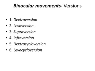
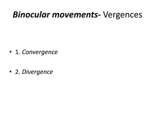
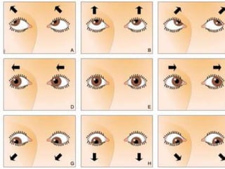
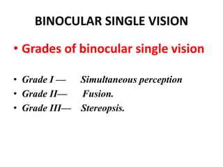
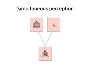
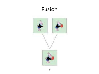
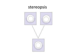
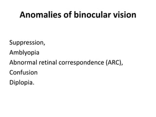
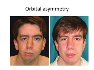
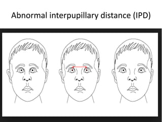
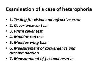
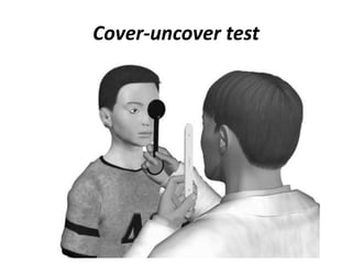
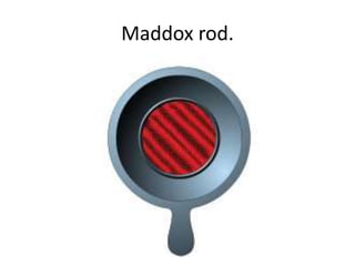
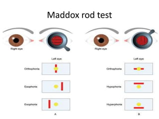
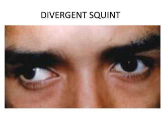
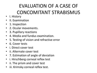
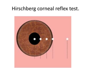
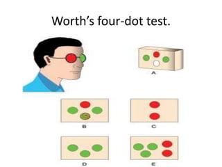
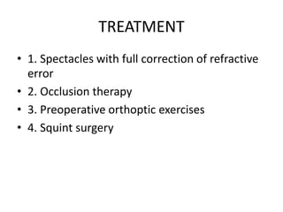
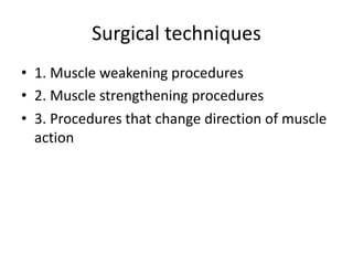
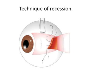
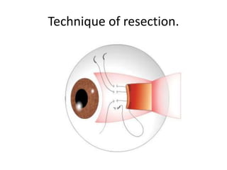
Strabismus and squint
DNYANESHWAR B. POTFODE
OPHTHALMIC OFFICER
Extraocular muscles
• Medial rectus : 5.5 mm
• Inferior rectus : 6.5 mm
• Lateral rectus : 6. 9 mm
• Superior rectus : 7.7 mm
• superior oblique muscle
• inferior oblique muscle
Extraocular muscles
Origin of the rectus muscles and the
superior oblique muscle
Nerve supply
• Oculomotor- superior, medial, inferior recti
and inferior oblique muscles.
• Trochlear- Superior oblique
• Abducent- Lateral rectus muscle.
Action of the extraocular muscles
OCULAR MOTILITY
• Uniocular movements- ‘ductions’
• Binocular movements-
versions
vergences.
Uniocular movements- ‘ductions’
• 1. Adduction
• 2. Abduction
• 3. Supraduction.
• 4. Infraduction
• 5. Incycloduction
• 6. Excycloduction
Binocular movements- Versions
• 1. Dextroversion
• 2. Levoversion.
• 3. Supraversion
• 4. Infraversion
• 5. Dextrocycloversion.
• 6. Levocycloversion
Binocular movements- Vergences
• 1. Convergence
• 2. Divergence
BINOCULAR SINGLE VISION
• Grades of binocular single vision
• Grade I — Simultaneous perception
• Grade II— Fusion.
• Grade III— Stereopsis.
Simultaneous perception
Fusion
stereopsis
Anomalies of binocular vision
Suppression,
Amblyopia
Abnormal retinal correspondence (ARC),
Confusion
Diplopia.
STRABISMUS
• A misalignment of the visual axes of
the two eyes is called squint or
strabismus.
Classification of strabismus
• I. Apparent squint or pseudostrabismus.
• II. Latent squint (Heterophoria)
• III. Manifest squint (Heterotropia)
1. Concomitant squint
2. Incomitant squint.
HETEROPHORIA
•Heterophoria also known as ‘latent
strabismus’, is a condition in which
the tendency of the eyes to deviate is
kept latent by fusion.
Types of heterophoria
• 1. Esophoria
• i Convergence
• ii Divergence
• 2. Exophoria
• 3. Hyperphoria
• 4. Cyclophoria
Etiology
1. Orbital asymmetry.
2. Abnormal interpupillary distance (IPD)
3. Faulty insertion of extraocular muscle.
4. A mild degree of extraocular muscleweakness.
5. Anomalous central distribution ofthe tonic
innervation of the two eyes
6. Anatomical variation in he position of the
macula
Orbital asymmetry
Abnormal interpupillary distance (IPD)
Symptoms
• 1. Symptoms of muscular fatigue.
• 2. Symptoms of failure to maintain binocular
single
• 3. Symptoms of defective postural sensations
cause
Examination of a case of heterophoria
• 1. Testing for vision and refractive error
• 2. Cover-uncover test.
• 3. Prism cover test
• 4. Maddox rod test
• 5. Maddox wing test.
• 6. Measurement of convergence and
accommodation
• 7. Measurement of fusional reserve
Cover-uncover test
Maddox rod.
Maddox rod test
Treatment
• 1. Correction of refractive error
• 2. Orthoptic treatment
• 3. Prescription of prism in glasses
• 4. Surgical treatment
Manifest squint
Etiology
• Refractive errors,
• Prolonged use of incorrect spectacles,
• Anisometropia,
• Corneal opacities,
• Lenticular opacities,
• Diseases of macula (e.g., central chorioretinitis),
• Optic atrophy, and
• Obstruction in the pupillary area due to
congenital ptosis.
CONVERGENT SQUINT
DIVERGENT SQUINT
EVALUATION OF A CASE OF
CONCOMITANT STRABISMUS
• I. History
• II. Examination
• 1. Inspection
• 2. Ocular movements.
• 3. Pupillary reactions
• 4. Media and fundus examination.
• 5. Testing of vision and refractive error
• 6. Cover tests
• i. Direct cover test
• ii. Alternate cover test
• 7. Estimation of angle of deviation
• i. Hirschberg corneal reflex test
• ii. The prism and cover test
• iii. Krimsky corneal reflex test.
Hirschberg corneal reflex test.
Worth’s four-dot test.
TREATMENT
• 1. Spectacles with full correction of refractive
error
• 2. Occlusion therapy
• 3. Preoperative orthoptic exercises
• 4. Squint surgery
Surgical techniques
• 1. Muscle weakening procedures
• 2. Muscle strengthening procedures
• 3. Procedures that change direction of muscle
action
Technique of recession.
Technique of resection.

Strabismus and squint