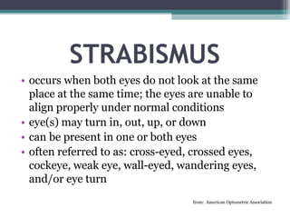
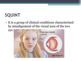
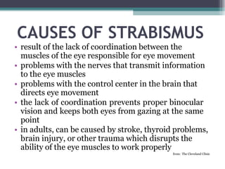
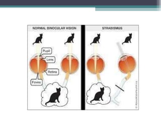
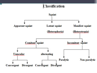
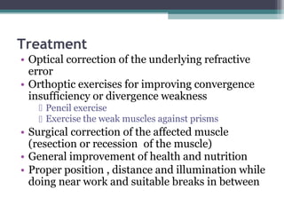
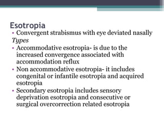
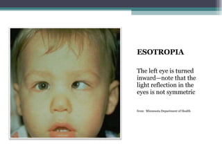
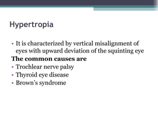
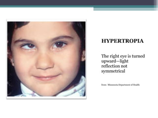
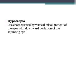
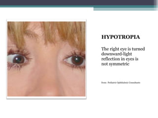
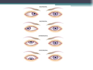
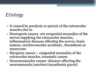
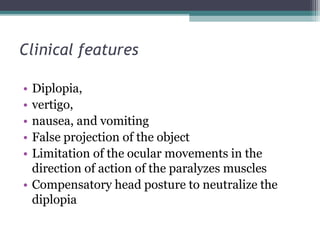
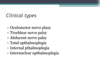
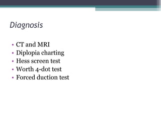
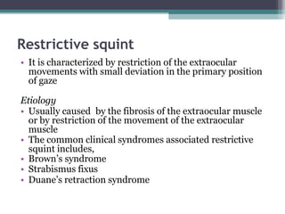
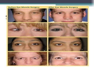
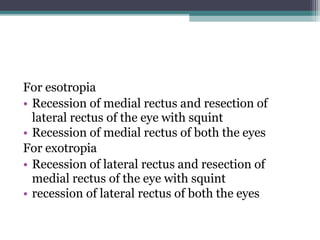
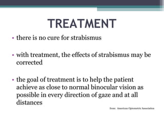
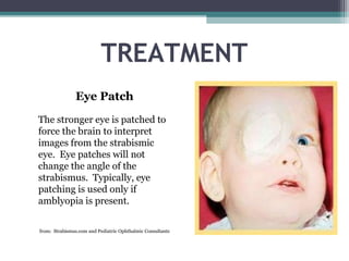
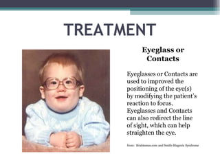
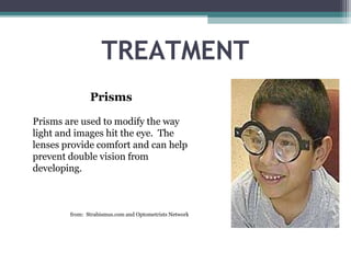
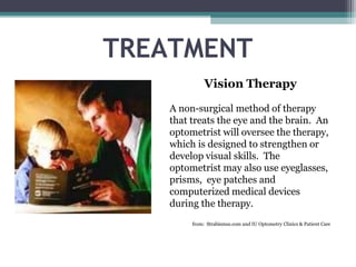
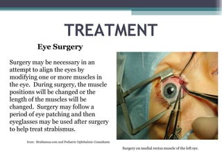
STRABISMUS
STRABISMUS
• occurs when both eyes do not look at the same
place at the same time; the eyes are unable to
align properly under normal conditions
• eye(s) may turn in, out, up, or down
• can be present in one or both eyes
• often referred to as: cross-eyed, crossed eyes,
cockeye, weak eye, wall-eyed, wandering eyes,
and/or eye turn
from: American Optometric Association
SQUINT
• It is a group of clinical conditions characterized
by misalignment of the visual axes of the two
eye.
CAUSES OF STRABISMUS
• result of the lack of coordination between the
muscles of the eye responsible for eye movement
• problems with the nerves that transmit information
to the eye muscles
• problems with the control center in the brain that
directs eye movement
• the lack of coordination prevents proper binocular
vision and keeps both eyes from gazing at the same
point
• in adults, can be caused by stroke, thyroid problems,
brain injury, or other trauma which disrupts the
ability of the eye muscles to work properly
from: The Cleveland Clinic
RISK FACTORS
• family history—if relatives have strabismus, a
person is more likely to develop it
• refractive errors—extreme farsightedness
(hyperopia) can develop strabismus due to the
amount of eye focusing necessary to keep vision
clear
• medical conditions—people with Down
syndrome or cerebral palsy and people who have
suffered a stroke or head injury are at higher risk
for developing strabismus
from: Strabismus.com
WHEN DOES IT OCCUR?
• congenital—developing during infancy; 50% of
children with strabismus are born with it
• acquired—developing in adulthood; can also
develop as a result of lack of treatment during
childhood
from: Strabismus.com
WHAT IT’S NOT
• Strabismus is NOT the same as Amblyopia!!
• Amblyopia is also called “lazy eye” and is a
condition where vision does not develop
normally during childhood
• Child may have one weak eye with poor vision
and one strong eye with normal vision
• Amblyopia DOES occur commonly with
Strabismus, but is a vision problem, where
strabismus is a muscle problem
from: Children’s Hospital Boston
DEVELOPMENT IN INFANTS
• a newborn’s eyes will typically move independently and
even drift outward
• it is not normal for an infant’s eyes to cross constantly
• by 3 to 4 months old, an infant should be able to focus on
objects and eyes should be straight, with no turning
• 30-50% of children with strabismus develop secondary
vision loss (amblyopia)
• the onset of strabismus is most common in children at 18
months to 6 years old
• if a parent notices their child’s eyes moving inward or
outward, the eyes seem to be crossed, or the child is not
focusing on objects, they should seek medical attention
from: Children’s Hospital Boston
DEVELOPMENT IN ADULTS
• most likely to develop as a result of injury or disease
• adult will most likely experience double vision, eye
fatigue, overlapped or blurred images, a pulling
sensation around the eyes, difficulty with reading,
and/or a loss of depth perception
• if loss of vision occurs, it is typically due to exotropia
(eye turns outward) rather than amblyopia because
vision is already developed in the adult brain and
the adult brain will not compensate for deviation the
way a child’s brain will
from: Eye Care America
DIAGNOSIS
• A comprehensive eye exam is necessary with focus on the
following:
• patient history—a comprehensive history is necessary
to assess symptoms, health problems, and medications
• visual acuity—measurements are taken to assess extent
to which vision is affected
• refraction—conducted to determine the lens power
necessary to compensate for any refractive error
• alignment and focusing—assessing how well your
eyes focus, move, and work together
• eye health exam—assessing the structures of the eye
(internal & external) to rule out possible disease
from: American Optometric Association
• APPARANT SQUINT
The presence of epicanthus and high errors of
refraction stimulate squint but in fact there is no
squint
• LATENT SQUINT
Is a clinical condition in which there is a
tendency of misalignment of the visual axes
which is corrected by the fusional capacity
Etiology
• Increased requirement for accommodation and
convergence results in esophoria
• Decreased requirement for accommodation and
convergence results in exophoria
• Occupations requiring too much close work
• General poor health, fatigue and advancing age
Symptoms
• Eye strain and head ache after prolonged work
• Blurring of prints and overlaping of words or
lines while reading
• Diplopia after close work for prolonged period
(intermittent squint ) or in the evening, on
reading or writing for an hour or two
• Crowding of words
• Symptoms of defective postural sensations
(difficulty to identify moving objects)
Diagnosis
• Patients case history regarding chief complaints,
onset and duration, previous treatment and
family history
• Visual acuity and monocular fixation pattern is
noted
• Cycloplegic refraction and fundus examination
• Ocular movements and head posture
• Measurement of convergence and
accommodation
• Assessment of binocular vision
• Cover- uncover test
• Maddox rod test
• Maddox wing test
• Prism vergence test
Treatment
• Optical correction of the underlying refractive
error
• Orthoptic exercises for improving convergence
insufficiency or divergence weakness
Pencil exercise
Exercise the weak muscles against prisms
• Surgical correction of the affected muscle
(resection or recession of the muscle)
• General improvement of health and nutrition
• Proper position , distance and illumination while
doing near work and suitable breaks in between
• MANIFEST SQUINT
• Dissociation of the eyes wherein the deviation
remains the same in all the directions of gaze
• Types
• Concomitant/ non paralytic squint
• Incomitant/ paralytic squint
Concomitant squint
• It is a type of squint characterized by normal
extraocular movements and constant degree of
squint in all the directions of gaze
• Types
• Esotropia (convergent squint)
• Exotropia (divergent squint)
• Hypertropia or hypotropia (Vertical squint)
Esotropia
• Convergent strabismus with eye deviated nasally
Types
• Accommodative esotropia- is due to the
increased convergence associated with
accommodation reflux
• Non accommodative esotropia- it includes
congenital or infantile esotropia and acquired
esotropia
• Secondary esotropia includes sensory
deprivation esotropia and consecutive or
surgical overcorrection related esotropia
ESOTROPIA
The left eye is turned
inward—note that the
light reflection in the
eyes is not symmetric
from: Minnesota Department of Health
Exotropia
• Divergent strabismus with eye deviated outward
temporally is called exotropia
• It can be infantile exotropia, primary exotropia,
and secondary exotropia
EXOTROPIA
The right eye is turned
outward—again, not
the light reflection in
the eyes is not
symmetrical
from: Minnesota Department of Health
Hypertropia
• It is characterized by vertical misalignment of
eyes with upward deviation of the squinting eye
The common causes are
• Trochlear nerve palsy
• Thyroid eye disease
• Brown’s syndrome
HYPERTROPIA
The right eye is turned
upward—light
reflection not
symmetrical
from: Minnesota Department of Health
• Hypotropia
• It is characterized by vertical misalignment of
the eyes with downward deviation of the
squinting eye
HYPOTROPIA
The right eye is turned
downward-light
reflection in eyes is
not symmetric
from: Pediatric Ophthalmic Consultants
Etiology
• Sensory problems- refractive error, cataract,
corneal opacity
• Motor problems- abnormalities of extra ocular
muscles, accommodation and convergence
• Central problems- defective fusion,
abnormalities in the cortical control of extra
ocular movements
• Incomitant squint
• It is characterized by limited or restricted
extraocular movements and varying degree of
squint in different directions of gaze
Types
• Paralytic and restrictive squint
Paralytic squint
• It is characterized by limited extraocular
movements in the direction of action of the
paralyzed muscle and varying degree of squint in
different directions of gaze
Etiology
• Is caused by paralysis or paresis of the extraocular
muscles due to
• Neurogenic causes- are congenital anomalies of the
nerves supplying the extraocular muscles,
inflammatory diseases affecting the nerves, brain
tumors, cerebrovascular accidents , thrombosis or
aneurysms
• Myogenic causes – congenital anomalies of the
extraocular muscles, traumatic causes
• Neuromauscular causes- diseases affecting the
neuromuscular junction (myasthenia gravis)
Clinical features
• Diplopia,
• vertigo,
• nausea, and vomiting
• False projection of the object
• Limitation of the ocular movements in the
direction of action of the paralyzes muscles
• Compensatory head posture to neutralize the
diplopia
Clinical types
• Oculomotor nerve plasy
• Trochlear nerve palsy
• Abducent nerve palsy
• Total opthalmoplegia
• Internal pthalmoplegia
• Internuclear opthalmoplegia
Diagnosis
• CT and MRI
• Diplopia charting
• Hess screen test
• Worth 4-dot test
• Forced duction test
Treatment
• Treating the underlying cause
• Treatment of diplopia by temporary occlusion of
the affected eye or the correction by opticals
• Surgical correction – recession and resection
muscle surgeries
• Injection of botulinum toxin to cause
chemodegeneration
Restrictive squint
• It is characterized by restriction of the extraocular
movements with small deviation in the primary position
of gaze
Etiology
• Usually caused by the fibrosis of the extraocular muscle
or by restriction of the movement of the extraocular
muscle
• The common clinical syndromes associated restrictive
squint includes,
• Brown’s syndrome
• Strabismus fixus
• Duane’s retraction syndrome
Diagnosis
• Assessment of visual acuity and refractive error
• Corneal reflux test
• Cover test
• Prism bar cover test
• Maddox rod test
• Maddox wing test
• Assessment of binocular vision
Treatment
• Optical correction of underlying refractive error
• Orthoptic exercises
• Occlusion therapy in case of amblyopia
• Surgical management
• Recession- done by making the muscle
slackening by shifting the insertion of the muscle
posteriorly towards its origin
• Resection- it is done by making the muscle taut
by shortening the muscle
Surgeries
The surgeries for the weakening of the muscle are,
• Recession
• Myotomy
• Tenotomy
Surgeries for strengthening the muscle are,
• Resection
• Tucking
• Transposition
For esotropia
• Recession of medial rectus and resection of
lateral rectus of the eye with squint
• Recession of medial rectus of both the eyes
For exotropia
• Recession of lateral rectus and resection of
medial rectus of the eye with squint
• recession of lateral rectus of both the eyes
TREATMENT
• there is no cure for strabismus
• with treatment, the effects of strabismus may be
corrected
• the goal of treatment is to help the patient
achieve as close to normal binocular vision as
possible in every direction of gaze and at all
distances
from: American Optometric Association
TREATMENT
Eye Patch
The stronger eye is patched to
force the brain to interpret
images from the strabismic
eye. Eye patches will not
change the angle of the
strabismus. Typically, eye
patching is used only if
amblyopia is present.
from: Strabismus.com and Pediatric Ophthalmic Consultants
TREATMENT
Eyeglass or
Contacts
Eyeglasses or Contacts are
used to improved the
positioning of the eye(s)
by modifying the patient’s
reaction to focus.
Eyeglasses and Contacts
can also redirect the line
of sight, which can help
straighten the eye.
from: Strabismus.com and Smith-Magenis Syndrome
TREATMENT
Prisms
Prisms are used to modify the way
light and images hit the eye. The
lenses provide comfort and can help
prevent double vision from
developing.
from: Strabismus.com and Optometrists Network
TREATMENT
Vision Therapy
A non-surgical method of therapy
that treats the eye and the brain. An
optometrist will oversee the therapy,
which is designed to strengthen or
develop visual skills. The
optometrist may also use eyeglasses,
prisms, eye patches and
computerized medical devices
during the therapy.
from: Strabismus.com and IU Optometry Clinics & Patient Care
TREATMENT
Eye Surgery
Surgery may be necessary in an
attempt to align the eyes by
modifying one or more muscles in
the eye. During surgery, the muscle
positions will be changed or the
length of the muscles will be
changed. Surgery may follow a
period of eye patching and then
eyeglasses may be used after surgery
to help treat strabismus.
from: Strabismus.com and Pediatric Ophthalmic Consultants
Surgery on medial rectus muscle of the left eye.
TREATMENT
The Bates Method
An alternative form of treatment that
uses natural remedies to correct vision
problems such as: relaxation, exercise,
games, and other activities. The Bates
Method claims to improve or restore
vision through the elimination of mental
strain. This is a very controversial
method in the medical field.
from: Strabismus.com and Seeing Without Glasses
TREATMENT
Botox Therapy
Used as an alternative to eye muscle
surgery. The idea is that the drug
will temporarily relax the eye
muscle, which will allow the
opposite eye to tighten and
straighten. The effects are short-
term—about 3 weeks. Eye muscle
exercises and eyeglasses may also be
used to help strengthen the eye.
from: Strabismus.com and IU Optometry Clinics & Patient Care

strabismus-190706104054.pptx

  • 1.
  • 2.
    STRABISMUS • occurs whenboth eyes do not look at the same place at the same time; the eyes are unable to align properly under normal conditions • eye(s) may turn in, out, up, or down • can be present in one or both eyes • often referred to as: cross-eyed, crossed eyes, cockeye, weak eye, wall-eyed, wandering eyes, and/or eye turn from: American Optometric Association
  • 3.
    SQUINT • It isa group of clinical conditions characterized by misalignment of the visual axes of the two eye.
  • 4.
    CAUSES OF STRABISMUS •result of the lack of coordination between the muscles of the eye responsible for eye movement • problems with the nerves that transmit information to the eye muscles • problems with the control center in the brain that directs eye movement • the lack of coordination prevents proper binocular vision and keeps both eyes from gazing at the same point • in adults, can be caused by stroke, thyroid problems, brain injury, or other trauma which disrupts the ability of the eye muscles to work properly from: The Cleveland Clinic
  • 5.
    RISK FACTORS • familyhistory—if relatives have strabismus, a person is more likely to develop it • refractive errors—extreme farsightedness (hyperopia) can develop strabismus due to the amount of eye focusing necessary to keep vision clear • medical conditions—people with Down syndrome or cerebral palsy and people who have suffered a stroke or head injury are at higher risk for developing strabismus from: Strabismus.com
  • 7.
    WHEN DOES ITOCCUR? • congenital—developing during infancy; 50% of children with strabismus are born with it • acquired—developing in adulthood; can also develop as a result of lack of treatment during childhood from: Strabismus.com
  • 8.
    WHAT IT’S NOT •Strabismus is NOT the same as Amblyopia!! • Amblyopia is also called “lazy eye” and is a condition where vision does not develop normally during childhood • Child may have one weak eye with poor vision and one strong eye with normal vision • Amblyopia DOES occur commonly with Strabismus, but is a vision problem, where strabismus is a muscle problem from: Children’s Hospital Boston
  • 9.
    DEVELOPMENT IN INFANTS •a newborn’s eyes will typically move independently and even drift outward • it is not normal for an infant’s eyes to cross constantly • by 3 to 4 months old, an infant should be able to focus on objects and eyes should be straight, with no turning • 30-50% of children with strabismus develop secondary vision loss (amblyopia) • the onset of strabismus is most common in children at 18 months to 6 years old • if a parent notices their child’s eyes moving inward or outward, the eyes seem to be crossed, or the child is not focusing on objects, they should seek medical attention from: Children’s Hospital Boston
  • 10.
    DEVELOPMENT IN ADULTS •most likely to develop as a result of injury or disease • adult will most likely experience double vision, eye fatigue, overlapped or blurred images, a pulling sensation around the eyes, difficulty with reading, and/or a loss of depth perception • if loss of vision occurs, it is typically due to exotropia (eye turns outward) rather than amblyopia because vision is already developed in the adult brain and the adult brain will not compensate for deviation the way a child’s brain will from: Eye Care America
  • 11.
    DIAGNOSIS • A comprehensiveeye exam is necessary with focus on the following: • patient history—a comprehensive history is necessary to assess symptoms, health problems, and medications • visual acuity—measurements are taken to assess extent to which vision is affected • refraction—conducted to determine the lens power necessary to compensate for any refractive error • alignment and focusing—assessing how well your eyes focus, move, and work together • eye health exam—assessing the structures of the eye (internal & external) to rule out possible disease from: American Optometric Association
  • 13.
    • APPARANT SQUINT Thepresence of epicanthus and high errors of refraction stimulate squint but in fact there is no squint • LATENT SQUINT Is a clinical condition in which there is a tendency of misalignment of the visual axes which is corrected by the fusional capacity
  • 14.
    Etiology • Increased requirementfor accommodation and convergence results in esophoria • Decreased requirement for accommodation and convergence results in exophoria • Occupations requiring too much close work • General poor health, fatigue and advancing age
  • 15.
    Symptoms • Eye strainand head ache after prolonged work • Blurring of prints and overlaping of words or lines while reading • Diplopia after close work for prolonged period (intermittent squint ) or in the evening, on reading or writing for an hour or two • Crowding of words • Symptoms of defective postural sensations (difficulty to identify moving objects)
  • 16.
    Diagnosis • Patients casehistory regarding chief complaints, onset and duration, previous treatment and family history • Visual acuity and monocular fixation pattern is noted • Cycloplegic refraction and fundus examination • Ocular movements and head posture • Measurement of convergence and accommodation
  • 17.
    • Assessment ofbinocular vision • Cover- uncover test • Maddox rod test • Maddox wing test • Prism vergence test
  • 18.
    Treatment • Optical correctionof the underlying refractive error • Orthoptic exercises for improving convergence insufficiency or divergence weakness Pencil exercise Exercise the weak muscles against prisms • Surgical correction of the affected muscle (resection or recession of the muscle) • General improvement of health and nutrition • Proper position , distance and illumination while doing near work and suitable breaks in between
  • 19.
    • MANIFEST SQUINT •Dissociation of the eyes wherein the deviation remains the same in all the directions of gaze • Types • Concomitant/ non paralytic squint • Incomitant/ paralytic squint
  • 20.
    Concomitant squint • Itis a type of squint characterized by normal extraocular movements and constant degree of squint in all the directions of gaze • Types • Esotropia (convergent squint) • Exotropia (divergent squint) • Hypertropia or hypotropia (Vertical squint)
  • 21.
    Esotropia • Convergent strabismuswith eye deviated nasally Types • Accommodative esotropia- is due to the increased convergence associated with accommodation reflux • Non accommodative esotropia- it includes congenital or infantile esotropia and acquired esotropia • Secondary esotropia includes sensory deprivation esotropia and consecutive or surgical overcorrection related esotropia
  • 22.
    ESOTROPIA The left eyeis turned inward—note that the light reflection in the eyes is not symmetric from: Minnesota Department of Health
  • 23.
    Exotropia • Divergent strabismuswith eye deviated outward temporally is called exotropia • It can be infantile exotropia, primary exotropia, and secondary exotropia
  • 24.
    EXOTROPIA The right eyeis turned outward—again, not the light reflection in the eyes is not symmetrical from: Minnesota Department of Health
  • 25.
    Hypertropia • It ischaracterized by vertical misalignment of eyes with upward deviation of the squinting eye The common causes are • Trochlear nerve palsy • Thyroid eye disease • Brown’s syndrome
  • 26.
    HYPERTROPIA The right eyeis turned upward—light reflection not symmetrical from: Minnesota Department of Health
  • 27.
    • Hypotropia • Itis characterized by vertical misalignment of the eyes with downward deviation of the squinting eye
  • 28.
    HYPOTROPIA The right eyeis turned downward-light reflection in eyes is not symmetric from: Pediatric Ophthalmic Consultants
  • 30.
    Etiology • Sensory problems-refractive error, cataract, corneal opacity • Motor problems- abnormalities of extra ocular muscles, accommodation and convergence • Central problems- defective fusion, abnormalities in the cortical control of extra ocular movements
  • 31.
    • Incomitant squint •It is characterized by limited or restricted extraocular movements and varying degree of squint in different directions of gaze Types • Paralytic and restrictive squint
  • 32.
    Paralytic squint • Itis characterized by limited extraocular movements in the direction of action of the paralyzed muscle and varying degree of squint in different directions of gaze
  • 33.
    Etiology • Is causedby paralysis or paresis of the extraocular muscles due to • Neurogenic causes- are congenital anomalies of the nerves supplying the extraocular muscles, inflammatory diseases affecting the nerves, brain tumors, cerebrovascular accidents , thrombosis or aneurysms • Myogenic causes – congenital anomalies of the extraocular muscles, traumatic causes • Neuromauscular causes- diseases affecting the neuromuscular junction (myasthenia gravis)
  • 34.
    Clinical features • Diplopia, •vertigo, • nausea, and vomiting • False projection of the object • Limitation of the ocular movements in the direction of action of the paralyzes muscles • Compensatory head posture to neutralize the diplopia
  • 35.
    Clinical types • Oculomotornerve plasy • Trochlear nerve palsy • Abducent nerve palsy • Total opthalmoplegia • Internal pthalmoplegia • Internuclear opthalmoplegia
  • 36.
    Diagnosis • CT andMRI • Diplopia charting • Hess screen test • Worth 4-dot test • Forced duction test
  • 37.
    Treatment • Treating theunderlying cause • Treatment of diplopia by temporary occlusion of the affected eye or the correction by opticals • Surgical correction – recession and resection muscle surgeries • Injection of botulinum toxin to cause chemodegeneration
  • 38.
    Restrictive squint • Itis characterized by restriction of the extraocular movements with small deviation in the primary position of gaze Etiology • Usually caused by the fibrosis of the extraocular muscle or by restriction of the movement of the extraocular muscle • The common clinical syndromes associated restrictive squint includes, • Brown’s syndrome • Strabismus fixus • Duane’s retraction syndrome
  • 39.
    Diagnosis • Assessment ofvisual acuity and refractive error • Corneal reflux test • Cover test • Prism bar cover test • Maddox rod test • Maddox wing test • Assessment of binocular vision
  • 40.
    Treatment • Optical correctionof underlying refractive error • Orthoptic exercises • Occlusion therapy in case of amblyopia • Surgical management • Recession- done by making the muscle slackening by shifting the insertion of the muscle posteriorly towards its origin • Resection- it is done by making the muscle taut by shortening the muscle
  • 41.
    Surgeries The surgeries forthe weakening of the muscle are, • Recession • Myotomy • Tenotomy Surgeries for strengthening the muscle are, • Resection • Tucking • Transposition
  • 42.
    For esotropia • Recessionof medial rectus and resection of lateral rectus of the eye with squint • Recession of medial rectus of both the eyes For exotropia • Recession of lateral rectus and resection of medial rectus of the eye with squint • recession of lateral rectus of both the eyes
  • 43.
    TREATMENT • there isno cure for strabismus • with treatment, the effects of strabismus may be corrected • the goal of treatment is to help the patient achieve as close to normal binocular vision as possible in every direction of gaze and at all distances from: American Optometric Association
  • 44.
    TREATMENT Eye Patch The strongereye is patched to force the brain to interpret images from the strabismic eye. Eye patches will not change the angle of the strabismus. Typically, eye patching is used only if amblyopia is present. from: Strabismus.com and Pediatric Ophthalmic Consultants
  • 45.
    TREATMENT Eyeglass or Contacts Eyeglasses orContacts are used to improved the positioning of the eye(s) by modifying the patient’s reaction to focus. Eyeglasses and Contacts can also redirect the line of sight, which can help straighten the eye. from: Strabismus.com and Smith-Magenis Syndrome
  • 46.
    TREATMENT Prisms Prisms are usedto modify the way light and images hit the eye. The lenses provide comfort and can help prevent double vision from developing. from: Strabismus.com and Optometrists Network
  • 47.
    TREATMENT Vision Therapy A non-surgicalmethod of therapy that treats the eye and the brain. An optometrist will oversee the therapy, which is designed to strengthen or develop visual skills. The optometrist may also use eyeglasses, prisms, eye patches and computerized medical devices during the therapy. from: Strabismus.com and IU Optometry Clinics & Patient Care
  • 48.
    TREATMENT Eye Surgery Surgery maybe necessary in an attempt to align the eyes by modifying one or more muscles in the eye. During surgery, the muscle positions will be changed or the length of the muscles will be changed. Surgery may follow a period of eye patching and then eyeglasses may be used after surgery to help treat strabismus. from: Strabismus.com and Pediatric Ophthalmic Consultants Surgery on medial rectus muscle of the left eye.
  • 49.
    TREATMENT The Bates Method Analternative form of treatment that uses natural remedies to correct vision problems such as: relaxation, exercise, games, and other activities. The Bates Method claims to improve or restore vision through the elimination of mental strain. This is a very controversial method in the medical field. from: Strabismus.com and Seeing Without Glasses
  • 50.
    TREATMENT Botox Therapy Used asan alternative to eye muscle surgery. The idea is that the drug will temporarily relax the eye muscle, which will allow the opposite eye to tighten and straighten. The effects are short- term—about 3 weeks. Eye muscle exercises and eyeglasses may also be used to help strengthen the eye. from: Strabismus.com and IU Optometry Clinics & Patient Care