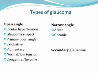
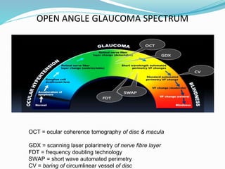
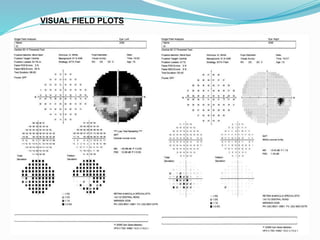
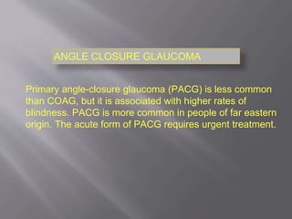
Glaucoma
Open angle & Closed Angle
Dnyaneshwar bhagwan potfode
Types of glaucoma
Open angle
Ocular hypertension
Glaucoma suspect
Primary open angle
Exfoliative
Pigmentary
Normal/low tension
Congenital/Juvenile
Narrow angle
Acute
Chronic
Secondary glaucoma
OPEN ANGLE GLAUCOMA SPECTRUM
OCT = ocular coherence tomography of disc & macula
GDX = scanning laser polarimetry of nerve fibre layer
FDT = frequency doubling technology
SWAP = short wave automated perimetry
CV = baring of circumlinear vessel of disc
OPEN ANGLE GLAUCOMA
Approximately 10% of UK blindness registrations are related
to glaucoma.
Around half a million people are currently affected by COAG
in England and there are over a million glaucoma-related
outpatient visits in the hospital eye service (HES) annually.
On average a person diagnosed with glaucoma will have one
initial visit and 40 follow up visits.
Fifty percent of glaucoma in the community in UK remains
undiagnosed; previously undetected cases are largely
identified at routine sight tests by community optometrists.
The number of individuals affected with COAG is expected
to rise due to changes in population demographics.
Standard Examination
Visual Acuity
Visual field testing (Humphrey)
Central corneal thickness
Intraocular pressure (Goldmann)
Gonioscopy
Dilated optic disc and fundus examination
Digital optic disc photos and imaging
(OCT disc & macula)
HUMPHREY FRIEDMANN
CONFRONTATION
BJERRUM SCREEN
FIELD APPARATUS
VISUAL FIELD PLOTS
VISUAL FIELD PLOTS
Intraocular pressure (tonometry)
 Normal is between 10mmHg and 21mmHg
(above atmospheric)
 Is some people, even this statistically normal
pressure too high for that individual person =
Normal Pressure Glaucoma (NPG)
 Measure the pressure indirectly:
Applanation
Tonopen
Icare
Pulsair
Schiotz
Other apparatus to check IOP
PULSAIR
TONOPEN
SCHIOTZ
iCARE
Check optic discs for pathological cupping
Direct ophthalmoscope
Slit lamp and 90 D lens
Imaging:
Disc photos
Depth of nerve fibre layer
OCT OF OPTIC DISCS FOR NERVE
FIBRE LAYER DEPTH
OCT of MACULA
Secondary glaucoma
Traumatic
Post surgery
Uveitis/Inflammatory
Steroid responsive
Rubeotic
Extraocular pathology
(carotid cavernous fistula etc.)
TREATMENT FOR PEOPLE WITH OHT OR SUSPECTED COAG
CENTRAL
CORNEAL
THICKNESS
>590 micrometres 555-590 micrometres <555 micrometres
ANY
UNTREATED
IOP (mmHg)
>21-25 25-32 >21-25 25-32 >21-25 25-32 >32
AGE (yrs) ANY ANY ANY
TREAT UNTIL
60
TREAT UNTIL
65
TREAT UNTIL
80
ANY
TREATMENT
NO
TREATMENT
NO
TREATMENT
NO
TREATMENT
PGA PGA PGA PGA
Antiglaucoma medications
Prostaglandin analogues
Latanoprost/ Travaprost
Prostamides( Bimatoprost)
Carbonic anhydrase inhibitors (Brinzolamide, Dorzolamide)
Alpha 2 agonists (Brimonidine)
Beta Blockers (Timolol)
Pilocarpine
Combinations of the above
Oral acetazolamide
After start or change in treatment, check IOP at 1-4 months
Monitoring intervals for patients on treatment for OHT or suspected COAG
Clinical assessment Monitoring interval (mnths
IOP at target
Risk of
conversion to
COAG
Outcome IOP alone
IOP, optic
nerve head
visual field
Yes Low
No change in
treat plan
Not
applicable
12-24
Yes High
No change in
treat plan
Not
applicable
6-12
No Low
Review target
IOP or
change treat
plan
1-4 6-12
No High
Review target
IOP or
change treat
plan
1-4 4-6
Monitoring intervals for patients on treatment for COAG
Clinical assessment Monitoring interval (months)
IOP at target Progression Outcome IOP alone
IOP, optic nerve
head, visual field
Yes No
No change in
treatment plan
Not
applicable
6-12
Yes Yes
Review target
IOP and change
treat plan
1-4 2-6
Yes Uncertain
No change in
treatment plan
Not
applicable
2-6
No No
Review target
IOP or change
treatment plan
1-4 6-12
No Yes/Uncertain
Change treatment
plan 1-2 2-6
Glaucoma treatment
Laser
Laser iridotomy in narrow
angle (prevention and
therapy)
Laser trabeculoplasty in
open angle glaucoma
Selective (SLT)
Argon Laser (ALT)
Cyclophotocoagulation
Surgery
Trabeculectomy
with mitomycin-C (MMC)
with 5-fluorouracil (5FU)
Non penetrating glaucoma
surgery
Drainage devices
Minimally invasive glaucoma
surgery (MIGS)
Trabectome
Primary angle-closure glaucoma (PACG) is less common
than COAG, but it is associated with higher rates of
blindness. PACG is more common in people of far eastern
origin. The acute form of PACG requires urgent treatment.
ANGLE CLOSURE GLAUCOMA
GONIOSCOPY

GLAUCOMA

  • 1.
    Glaucoma Open angle &Closed Angle Dnyaneshwar bhagwan potfode
  • 2.
    Types of glaucoma Openangle Ocular hypertension Glaucoma suspect Primary open angle Exfoliative Pigmentary Normal/low tension Congenital/Juvenile Narrow angle Acute Chronic Secondary glaucoma
  • 3.
    OPEN ANGLE GLAUCOMASPECTRUM OCT = ocular coherence tomography of disc & macula GDX = scanning laser polarimetry of nerve fibre layer FDT = frequency doubling technology SWAP = short wave automated perimetry CV = baring of circumlinear vessel of disc
  • 4.
    OPEN ANGLE GLAUCOMA Approximately10% of UK blindness registrations are related to glaucoma. Around half a million people are currently affected by COAG in England and there are over a million glaucoma-related outpatient visits in the hospital eye service (HES) annually. On average a person diagnosed with glaucoma will have one initial visit and 40 follow up visits. Fifty percent of glaucoma in the community in UK remains undiagnosed; previously undetected cases are largely identified at routine sight tests by community optometrists. The number of individuals affected with COAG is expected to rise due to changes in population demographics.
  • 5.
    Standard Examination Visual Acuity Visualfield testing (Humphrey) Central corneal thickness Intraocular pressure (Goldmann) Gonioscopy Dilated optic disc and fundus examination Digital optic disc photos and imaging (OCT disc & macula)
  • 6.
  • 7.
  • 8.
  • 9.
    Intraocular pressure (tonometry) Normal is between 10mmHg and 21mmHg (above atmospheric)  Is some people, even this statistically normal pressure too high for that individual person = Normal Pressure Glaucoma (NPG)  Measure the pressure indirectly: Applanation Tonopen Icare Pulsair Schiotz
  • 10.
    Other apparatus tocheck IOP PULSAIR TONOPEN SCHIOTZ iCARE
  • 11.
    Check optic discsfor pathological cupping Direct ophthalmoscope Slit lamp and 90 D lens Imaging: Disc photos Depth of nerve fibre layer
  • 13.
    OCT OF OPTICDISCS FOR NERVE FIBRE LAYER DEPTH
  • 14.
  • 15.
    Secondary glaucoma Traumatic Post surgery Uveitis/Inflammatory Steroidresponsive Rubeotic Extraocular pathology (carotid cavernous fistula etc.)
  • 16.
    TREATMENT FOR PEOPLEWITH OHT OR SUSPECTED COAG CENTRAL CORNEAL THICKNESS >590 micrometres 555-590 micrometres <555 micrometres ANY UNTREATED IOP (mmHg) >21-25 25-32 >21-25 25-32 >21-25 25-32 >32 AGE (yrs) ANY ANY ANY TREAT UNTIL 60 TREAT UNTIL 65 TREAT UNTIL 80 ANY TREATMENT NO TREATMENT NO TREATMENT NO TREATMENT PGA PGA PGA PGA
  • 17.
    Antiglaucoma medications Prostaglandin analogues Latanoprost/Travaprost Prostamides( Bimatoprost) Carbonic anhydrase inhibitors (Brinzolamide, Dorzolamide) Alpha 2 agonists (Brimonidine) Beta Blockers (Timolol) Pilocarpine Combinations of the above Oral acetazolamide After start or change in treatment, check IOP at 1-4 months
  • 18.
    Monitoring intervals forpatients on treatment for OHT or suspected COAG Clinical assessment Monitoring interval (mnths IOP at target Risk of conversion to COAG Outcome IOP alone IOP, optic nerve head visual field Yes Low No change in treat plan Not applicable 12-24 Yes High No change in treat plan Not applicable 6-12 No Low Review target IOP or change treat plan 1-4 6-12 No High Review target IOP or change treat plan 1-4 4-6
  • 19.
    Monitoring intervals forpatients on treatment for COAG Clinical assessment Monitoring interval (months) IOP at target Progression Outcome IOP alone IOP, optic nerve head, visual field Yes No No change in treatment plan Not applicable 6-12 Yes Yes Review target IOP and change treat plan 1-4 2-6 Yes Uncertain No change in treatment plan Not applicable 2-6 No No Review target IOP or change treatment plan 1-4 6-12 No Yes/Uncertain Change treatment plan 1-2 2-6
  • 20.
    Glaucoma treatment Laser Laser iridotomyin narrow angle (prevention and therapy) Laser trabeculoplasty in open angle glaucoma Selective (SLT) Argon Laser (ALT) Cyclophotocoagulation Surgery Trabeculectomy with mitomycin-C (MMC) with 5-fluorouracil (5FU) Non penetrating glaucoma surgery Drainage devices Minimally invasive glaucoma surgery (MIGS) Trabectome
  • 21.
    Primary angle-closure glaucoma(PACG) is less common than COAG, but it is associated with higher rates of blindness. PACG is more common in people of far eastern origin. The acute form of PACG requires urgent treatment. ANGLE CLOSURE GLAUCOMA
  • 22.