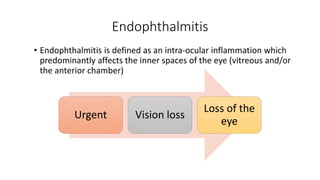
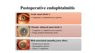
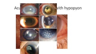
Endophthalmitis
3rd year resident, MNUMS
B.Bulganchimeg MD
Endophthalmitis
• Endophthalmitis is defined as an intra-ocular inflammation which
predominantly affects the inner spaces of the eye (vitreous and/or
the anterior chamber)
Urgent Vision loss
Loss of the
eye
Classification
Infectious
Exogenous
Post
operative
Acute
Chronic
Traumatic
Bleb
associated
After IV
injection
Endogenous
Noninfectious
Types of endophthalmitis
Exogenous (>85%)
• Acute Postoperative
• Chronic Postoperative
• Traumatic
• Filtering Bleb-Associated
• After Intravitreal
Injections
Endogenous (<15%)
• Originates from sources
within the body
• 2-8% of endophthalmitis
cases
• Recent intravenous
infusion, DM, HIV, IV drug
abuse, renal failure on
dialysis, cardiac disease,
malignancy,
immunosuppressive
therapy, or indwelling
catheters
• Liver abscess
Postoperative endophtalmitis
Acute onset (6wk>)
• Coagulase (-) staphylococcus species
Chronic -delayed onset (6wk<)
• Coagulase (-) staphylococcus species
• Fungi, propioni bacterium acnes
Bleb-associated (months,years after)
• Streptococcus species
• Haemophilus species
• Gram (+) organisms
Bacterial Fungal
Viral Parasitic
Common organism causing endophthalmitis
A. Exogenous
Acute onset Postsurgical Gram positive : S. epidermidis, S. aureus , Streptococcus spp.
Gram negative : Pseudomonas, Proteus, H. influenzae, Klebsiella, E. coli,
Enterobacter
Delayed onset Postsurgical Fungi : aspergillus, fusarium, candid, penicillum
Bacteria : P. acnes S. epidermidis
Post-traumatic Bacillus spp. S. epidermidis Streptococcus spp.
fungi (fusarium)
B. Endogenous Bacteria : B. cereus ( IV abusers), Streptococcus spp.,
S. aureus, Meningococci , H. influenzae
Fungi : mucor, candida
Bacterial entry into eye
Cascade of inflammatory products
Inflammatory cell recruitment
Tissue destruction
Release of digestive enzymes by the cells&
toxins by bacteria
PATHOGENESIS
Post operative endophthalmitis
• Cataract surgery 0.08-0.7%
Intravitreal injections
Penetrating keratoplasty 0.11-0.18%
 Trabeculectomy and glaucoma drainage device implantation 0.2%–9.6%
 Pars Plana Vitrectomy 0.03-0.05%
Postoperative endophthalmitis
Acute • (<6 weeks)
Chronic • (>6 weeks)
Acute Postoperative Endophthalmitis
• Shortly after ocular surgery /within 1-2wk, usually 3-5 days/
Epidemiology:
• APE most common form of endophthalmitis
• Following cataract surgery 0.08% - 0.68%
• Rates increasing since clear corneal incisions
• Highest risk after 2ndary IOL (0.2-0.367%),
• Lowest risk after PPV (0.03-0.046%).
10
0.12
0.3-0.5
0
2
4
6
8
10
12
ICCE ECCE Phaco (CCI)
The incidence of endophthalmitis after cataract surgery
Acute endophthalmitis with hypopyon
Source of infection
Risk factors
Preoperative
Blepharitis
Active conjunctivitis
LD infection, obstruction
Operative
Wound abnormalities
Vitreous loss
Prolonged surgery
Endophthalmitis Vitrectomy Study (EVS)
94.3% 82.1% 74%
34.5% Photophobia Purulent discharge
Symptoms
85% hypopyon 79% hazy media 26 % LP
Clinical signs
• Decreased visual acuity
• Lids- edema
• Conjunctiva- congestion and
chemosis
• Cornea edema, ring
abscess(Pseudomonas and
Bacillus)
• Anterior chambercells and flare
• Hypopyon (in 86%patients in
EVS)
• Iris muddy and boggy
• Pupil-fibrinous exudate
• IOL-may be covered by fibrin
• Fundus-
 vitreous exudates,
scattered retinal haemorrhages
Periphlebitis
 loss of red reflex
Presenting Visual Acuity
• HM vs LP - Important
• Pars plana vitrectomy recommended in patients with visual acuity of
light perception (LP).
• Hand motion (HM) or better, tap and inject intravitreal antibiotics
• When measuring VA, the technique of differentiating LP vs. HM vision
is most important
Differential Diagnosis of Acute
Endophthalmitis
• Occult retention of lens cortex or nucleus
• Hypopyon uveitis (Behcet’s or rifabutin)
• Inflammatory reaction to intravitreal drug
• Blebitis
• Keratitis
• Toxic anterior segment syndrome (TASS)
Toxic anterior segment syndrome (TASS)
Sterile postoperative
endophthalmitis
• Rapid onset ( 12-24 hours,
limbus to limbus corneal
edema)
• Noninfectious substance the
enters AS
• Increased intraocular pressure
• Absent vitreous inflammation
• Iris damage
• Response to steroid
Diagnosis
• Clicinal signs, symptoms
,evaluation
• Ultrasound
• Microbiological testing
• Aqueous and Vitreous Specimen
• PCR
Ultrasound
• Should be performed if significant media opacification prevents
adequate view of the fundus
• Findings c/w endophthalmitis:
• Dispersed vitreous opacities with vitritis
• Chorioretinal thickening
• Rule out: RD or choroidals, dislocated lens material, retained foreign
bodies
• Retinal or choroidal detachment are poor prognostic factors
Aqueous tap (40% Gr +)
• An anterior
chamber
paracentesis is
performed using a
25 or 27 gauge
needle and 0.1 ml
of aqeous material
is aspirated.
Vitreous tap (60% Gr +)
• A trans pars plana aspiration
with a 23 gauge needle
• 3mm posterior to limbus in
pseudophakic and aphakic
eyes.
• 4mm posterior to limbus in
phakic eyes
• 0.2 ml of vitreous aspirated.
Microbiological Characteristics (EVS)
CNS (Staph.epi)
70%
Staph.aureus
10%
Streptococcus
9%
Enterococcus
2%
Other gr +
3%
Gr -
6%
CNS (Staph.epi)
Staph.aureus
Streptococcus
Enterococcus
Other gr +
Gr -
Treatment goals
1. Retention of useful vision
2. Minimize the infection with antimicrobial agents.
3. Limit the inflammation.
4. Symptomatic relief.
Treatment
• PPV
• Intravitreal AB
• Systemic
antibiotics
• Cotricosteroids
• Supportive
treatment
Endophthalmitis
Is there good reflex ?
Yes
No
Initiate medical therapy and observe
patient closely for the 1st 24 hours
Vitrectomy
Is there improvement?
Yes No
Continue the medical
therapy and close
observation
Treatment algorithm of CEVE for
acute postoperative endohthalmitis
TREATMENT
For VA that is "hand motions" or better:
 Tap and inject. First tap the eye for samples:
o Anterior chamber aqueous tap with 30g needle on a tuberculin syringe
sent for cultures
o Vitreous sample obtained through the pars plana with a 25 gauge, 1
inch needle directed toward the mid-vitreous cavity and then sent for
cultures
 Inject the vitreous cavity with antibiotics (ie. Vancomycin and amikacin
were used in the EVS. Ceftazidine is often used in clinical practice in the
place of amikacin, primarily due to the risk of aminoglycoside toxicity with
amikacin)
For VA that is worse than "hand motions":
 Pars plana vitrectomy. Send aqueous and vitrectomy cultures taken during
surgery.
 Inject intravitreal antibiotics at the time of surgery
Follow cultures and adjust antibiotics accordingly, if necessary
The EVS
Conducted between 1990 and January
1994, the EVS was a prospective, randomized,
multicenter trial on 420 eyes to determine
whether it is necessary in acute postoperative
endophthalmitis to use systemic antibiotics
or perform routine immediate vitrectomy
Materials and method
• 24 centers across the US
• 1990.02-1994.01
Patient selection
• Patients had clinical signs and symptoms of bacterial endophthalmitis
within 6 weeks after cataract surgery or secondary intraocular lens
implantation.
Inclusion criteria
• ETDRS- VA=LP↑ or
20/50↓
• Cornea, AC-clear
• Cornea clear to perform
VIT
• Hypopyon or sufficient
clouding of the AC or
vitreous to obscure a
view of second-order
retinal arterioles.
Exclusion criteria
• VA<0.2 before cataract
• Intraocular surgery
• Penetrating trauma
• Injection of IV AB
• Prior PPV
• RD, CD
• strong suspicion of
fungal endophthalmitis
• 18 age ↓
Screened 1283
patients
855 patients /6wk-
endoph diagnosed/
510 met eligibilty criteria
420 agreed to
participate
Study groups
Initial
VIT+IV
antibiotics
Initial VIT
without IV
antibiotics
Initial TAP
with IV
antibiotics
Initial TAP
without IV
antibiotics
Initial procedure (Eyelid culture, 0.1ml AC sample)
VIT group
• 3 port pars plana VIT
• When necessary
vizualization, AC was
cleared
TAP group
• Vitreous spicemen collected
either PP vitreous needle
aspiration or vitreous biopsy
• Vitreous sample 0.1-0.3mL
Study medication
• After initial VIT or TAP procedure all patients: intravitreal inj of
amikacin 0.4mg/0.1ml, vancomycin 1.0mg/0.1ml
• Vanco 25mg/0.5ml+ceftazidime 100mg/0.5ml+dexa
6mg/0.25ml→subconj
• Topical AB 1d/4h
• Topical cycloplegics, corticosteroids
• Systemic CS (prednisoloni 30mg*2→5-10days)
Results
• Systemic antibiotics do not improve the
outcome
• Vitrectomy is indicated only in eyes with
light perception vision.
1. There was no difference in final visual acuity
or media clarity with or without the use of
systemic antibiotics.
2. HM ↑ - immediate VIT not influenced
3. LP – VIT increase VA.
a. Three times more likely to achieve ≥20/40
(33% vs 11%)
b. Two times more likely to achieve ≥20/100
(56% vs 30%)
c. Less likely to incur <5/200 (20% vs 47%)
Prevention
1. Pre-operative
oPovidone iodine drops
oTopical AB
oMeticulous draping
2. Intra-operative
oIrrigation of A/C with vancomycin
3. Post-operative
• Intra cameral cefuroxime
• Post op subconj AB injection
• Post op AB drops
Chronic Postoperative Endophthalmitis
(> 6 weeks)
• Usually manifest several weeks
or months after surgery
• Less common than acute variety
• Organisms isolated are less
virulent bacteria and fungal
66%
17%
17%
Pathogens
Pro.acnes
Staph.epidermidis
Candida parapsilosis
Chronic saccular endophthalmitis (Note
whitish plaque)
SYMPTOMS-
• Photophobia
• Blurred vision
• Mild pain
SIGNS-
• KPs in AC
• Vitreous flare and cells
• Capsular plaque(in
Propionibacterium
acnesendophthalmitis after
Nd:Yagcapsulotomy)
• Granuloma formation in the
pupillaryarea or near the section(in
fungalendophthalmitis)
DDx of postoperative endophthalmitis
• Noninfectious uveitis
• Retained lens material
• Triamcinolone acetonide particles
• Longstanding (dehemoglobinized) vitreous hemorrhage
Diagnosis
• Clinical diagnosis
• B scan (RD, SCH, retained lens fragment)
• Cultures (VS>AS)
Commonly used cuture media
5% blood agar
Chocolate agar
Sabouraud agar
Anaerobic blood agar
Treatment for Chronic Cases
• Obtain A/C and vitreous samples
• Identification of infectious organism is key in management of these
infections Intravitreal antibiotics
• However, Vanco is often inadequate for P acnes
• Often need PPV and removal of capsule with IOL
Bleb related endophthalmitis
• Occurs as a result of pathogenic organisms gaining entry to intraocular
tissue through the conjunctival filtering bleb
• Mean time b/w surgery and endophthalmitis is 19.1 months (range 3-9
years)
• 0.2 – 9.6% of glaucoma filtering procedures
• Increased incidence with use of antifibrotic agents
• Early PPV
• Thin, cystic, avascular conjunctiva
• Clinical signs infected
 White bleb
 Vitritis
 Hypopyon
Endophthalmitis after Intravitreal Injection
• Increasing use of these agents, therefore concern for risk of
endophthalmitis
• Like acute postop endophthalmitis, CNS is the most common cause
• No infectious agent is identified in many cases
• Triamcinolone acetonide crystals can migrate into A/C and mimic
hypopyon
• 1.4%/injection for IVK
• 0.2%/injection for ranibizumab
• Intravitreal triamcinolone may play a role in endophthalmitis
potentiation
Non-infectious Endophthalmitis after
Triamcinolone Injection or intravitreal anti-VEGF
(aflibercept)
• Crystals in A/C or vitreous cavity can be difficult to distinguish from
inflammatory reaction
• Use gravity-induced shifting of layered material and absence of A/C
flare or fibrin as a sign of a non-inflammatory cause
• Triamcinolone crystals may shift from the inferior anterior chamber
angle when the pt is placed on his or her side, a phenomenon that
does not occur with sticky, fibrin-laden material in a true
inflammatory hypopyon
• Management options
• Close observation, topical steroids alone, oral levofloxacin alone
Post traumatic endophthalmitis
• Approximately 25% of
endophthalmitis cases
• After open globe injury, chance
of developing endophthalmitis
is approx 7%
• Injuries including IOFB have
higher rates
Risk factors
• Dirty wound
• Lens capsule rupture
• older age(>50)
• Initial presentation with a delay of more than 24 hours
• The presence of intraocular foreign bodies.
The incidence of endophthalmitis in cases of
• penetrating ocular trauma: 3.3% to 30% and
• after intraocular foreign body: 1.3% to 61%.
Managemant
• Primary repair and removal of IOFB as soon as possible
• Exclude the possibility of occult, retained IOFB
• CT scan with thin 1mm cuts
• Obtain cultures
• Intravitreal Vanco and Ceftaz
• Some advocate Gent + Clinda for synergistic effect against Bacillus and
Staph
Post-traumatic endophthalmitis has worst visual prognosis than other
categories
Endogenous endophthalmitis (2-8%)
• Hematogenous spread of organism from distant source
• It occurs when microorganisms in the bloodstream get into the eye, cross
the blood–retinal barrier, and infect the ocular tissue.
• Retina and choroid primarily involved due to high vascularity.
• Predisposing factors
 Diabetes - immunosuppresion(AIDS,malignancies medications)
 recent major abdominal surgery –
 prolong indwelling catheter ( intravenous , TPN)
 intravenous drug abuser
 distant infection ( endocarditis, meningitis, septicemia etc)
no structural defect in globe
Management of Endogenous Endophthalmitis
• Cultures are essential and may be lifesaving
• Vitreous sample should be obtained in all cases
• ID of causatic organism is made by systemic culture of blood, urine or
CSF in 75-80% of cases
• In contrast to postoperative endophthalmitis, systemic antibiotics are
central to the treatment of endogenous endophthalmitis
• Focal chorioretinitis and associated mild vitritis can respond to
systemic therapy alone
Symptoms
• Decreased or blurred vision
• ( sudden / severe – acute)
• ( slowly / mild—chronic)
• Pain
• Photophobia
• Redness of eyes
• Swollen eyelids Discharge
• White lesion in black part of the eye
• Floaters Fever
Fungal endophthalmitis
• Chronic infection (Rarely acute)
• Late onset (Redness + floaters)
• Fixed cheesy hypopyon
• Fibrinous mesh like exudation
• Snowball and fluffy opacities in the vitreous
Candidal endophthalmitis
• The two most characteristic
1. creamy white, well
circumscribed chorioretinal
lesions , most common in the
posterior pole
2. yellow or white fluffy vitreous
opacities.
“string of pearls”
Aspergillus endophthalmitis
• Most patients with endogenous Aspergillus
• Acute or subacute ocular symptoms.
• AS inflammatory signs: AC cells , KPs, hypopyon
• In most cases the organism initially affects the posterior segment,
forming a yellowish macular infiltrate .
• It frequently involves the macula, and this probably accounts for the
rapid onset of visual loss in many cases.
• A frequent clinical feature is the
pseudohypopyon in either the
preretinal (subhyaloid) or
subretinalspace .
• Vitritis
• Other features include occlusive
vasculitis and retinal hemorrhages.
• After treatment the area of
chorioretinitis progresses to
formation of a subretinal scar.
• The visual outcome is generally poor
Endophthalmitis

Endophthalmitis

  • 1.
    Endophthalmitis 3rd year resident,MNUMS B.Bulganchimeg MD
  • 2.
    Endophthalmitis • Endophthalmitis isdefined as an intra-ocular inflammation which predominantly affects the inner spaces of the eye (vitreous and/or the anterior chamber) Urgent Vision loss Loss of the eye
  • 3.
  • 4.
    Types of endophthalmitis Exogenous(>85%) • Acute Postoperative • Chronic Postoperative • Traumatic • Filtering Bleb-Associated • After Intravitreal Injections Endogenous (<15%) • Originates from sources within the body • 2-8% of endophthalmitis cases • Recent intravenous infusion, DM, HIV, IV drug abuse, renal failure on dialysis, cardiac disease, malignancy, immunosuppressive therapy, or indwelling catheters • Liver abscess
  • 5.
    Postoperative endophtalmitis Acute onset(6wk>) • Coagulase (-) staphylococcus species Chronic -delayed onset (6wk<) • Coagulase (-) staphylococcus species • Fungi, propioni bacterium acnes Bleb-associated (months,years after) • Streptococcus species • Haemophilus species • Gram (+) organisms
  • 6.
  • 7.
    Common organism causingendophthalmitis A. Exogenous Acute onset Postsurgical Gram positive : S. epidermidis, S. aureus , Streptococcus spp. Gram negative : Pseudomonas, Proteus, H. influenzae, Klebsiella, E. coli, Enterobacter Delayed onset Postsurgical Fungi : aspergillus, fusarium, candid, penicillum Bacteria : P. acnes S. epidermidis Post-traumatic Bacillus spp. S. epidermidis Streptococcus spp. fungi (fusarium) B. Endogenous Bacteria : B. cereus ( IV abusers), Streptococcus spp., S. aureus, Meningococci , H. influenzae Fungi : mucor, candida
  • 8.
    Bacterial entry intoeye Cascade of inflammatory products Inflammatory cell recruitment Tissue destruction Release of digestive enzymes by the cells& toxins by bacteria PATHOGENESIS
  • 9.
    Post operative endophthalmitis •Cataract surgery 0.08-0.7% Intravitreal injections Penetrating keratoplasty 0.11-0.18%  Trabeculectomy and glaucoma drainage device implantation 0.2%–9.6%  Pars Plana Vitrectomy 0.03-0.05%
  • 10.
    Postoperative endophthalmitis Acute •(<6 weeks) Chronic • (>6 weeks)
  • 11.
    Acute Postoperative Endophthalmitis •Shortly after ocular surgery /within 1-2wk, usually 3-5 days/ Epidemiology: • APE most common form of endophthalmitis • Following cataract surgery 0.08% - 0.68% • Rates increasing since clear corneal incisions • Highest risk after 2ndary IOL (0.2-0.367%), • Lowest risk after PPV (0.03-0.046%).
  • 12.
    10 0.12 0.3-0.5 0 2 4 6 8 10 12 ICCE ECCE Phaco(CCI) The incidence of endophthalmitis after cataract surgery
  • 13.
  • 14.
  • 15.
    Risk factors Preoperative Blepharitis Active conjunctivitis LDinfection, obstruction Operative Wound abnormalities Vitreous loss Prolonged surgery
  • 16.
    Endophthalmitis Vitrectomy Study(EVS) 94.3% 82.1% 74% 34.5% Photophobia Purulent discharge
  • 17.
    Symptoms 85% hypopyon 79%hazy media 26 % LP
  • 18.
    Clinical signs • Decreasedvisual acuity • Lids- edema • Conjunctiva- congestion and chemosis • Cornea edema, ring abscess(Pseudomonas and Bacillus) • Anterior chambercells and flare • Hypopyon (in 86%patients in EVS)
  • 19.
    • Iris muddyand boggy • Pupil-fibrinous exudate • IOL-may be covered by fibrin • Fundus-  vitreous exudates, scattered retinal haemorrhages Periphlebitis  loss of red reflex
  • 20.
    Presenting Visual Acuity •HM vs LP - Important • Pars plana vitrectomy recommended in patients with visual acuity of light perception (LP). • Hand motion (HM) or better, tap and inject intravitreal antibiotics • When measuring VA, the technique of differentiating LP vs. HM vision is most important
  • 21.
    Differential Diagnosis ofAcute Endophthalmitis • Occult retention of lens cortex or nucleus • Hypopyon uveitis (Behcet’s or rifabutin) • Inflammatory reaction to intravitreal drug • Blebitis • Keratitis • Toxic anterior segment syndrome (TASS)
  • 22.
    Toxic anterior segmentsyndrome (TASS) Sterile postoperative endophthalmitis • Rapid onset ( 12-24 hours, limbus to limbus corneal edema) • Noninfectious substance the enters AS • Increased intraocular pressure • Absent vitreous inflammation • Iris damage • Response to steroid
  • 24.
    Diagnosis • Clicinal signs,symptoms ,evaluation • Ultrasound • Microbiological testing • Aqueous and Vitreous Specimen • PCR
  • 25.
    Ultrasound • Should beperformed if significant media opacification prevents adequate view of the fundus • Findings c/w endophthalmitis: • Dispersed vitreous opacities with vitritis • Chorioretinal thickening • Rule out: RD or choroidals, dislocated lens material, retained foreign bodies • Retinal or choroidal detachment are poor prognostic factors
  • 26.
    Aqueous tap (40%Gr +) • An anterior chamber paracentesis is performed using a 25 or 27 gauge needle and 0.1 ml of aqeous material is aspirated.
  • 27.
    Vitreous tap (60%Gr +) • A trans pars plana aspiration with a 23 gauge needle • 3mm posterior to limbus in pseudophakic and aphakic eyes. • 4mm posterior to limbus in phakic eyes • 0.2 ml of vitreous aspirated.
  • 28.
    Microbiological Characteristics (EVS) CNS(Staph.epi) 70% Staph.aureus 10% Streptococcus 9% Enterococcus 2% Other gr + 3% Gr - 6% CNS (Staph.epi) Staph.aureus Streptococcus Enterococcus Other gr + Gr -
  • 29.
    Treatment goals 1. Retentionof useful vision 2. Minimize the infection with antimicrobial agents. 3. Limit the inflammation. 4. Symptomatic relief.
  • 30.
    Treatment • PPV • IntravitrealAB • Systemic antibiotics • Cotricosteroids • Supportive treatment
  • 31.
    Endophthalmitis Is there goodreflex ? Yes No Initiate medical therapy and observe patient closely for the 1st 24 hours Vitrectomy Is there improvement? Yes No Continue the medical therapy and close observation Treatment algorithm of CEVE for acute postoperative endohthalmitis
  • 32.
    TREATMENT For VA thatis "hand motions" or better:  Tap and inject. First tap the eye for samples: o Anterior chamber aqueous tap with 30g needle on a tuberculin syringe sent for cultures o Vitreous sample obtained through the pars plana with a 25 gauge, 1 inch needle directed toward the mid-vitreous cavity and then sent for cultures  Inject the vitreous cavity with antibiotics (ie. Vancomycin and amikacin were used in the EVS. Ceftazidine is often used in clinical practice in the place of amikacin, primarily due to the risk of aminoglycoside toxicity with amikacin) For VA that is worse than "hand motions":  Pars plana vitrectomy. Send aqueous and vitrectomy cultures taken during surgery.  Inject intravitreal antibiotics at the time of surgery Follow cultures and adjust antibiotics accordingly, if necessary
  • 33.
    The EVS Conducted between1990 and January 1994, the EVS was a prospective, randomized, multicenter trial on 420 eyes to determine whether it is necessary in acute postoperative endophthalmitis to use systemic antibiotics or perform routine immediate vitrectomy
  • 34.
    Materials and method •24 centers across the US • 1990.02-1994.01 Patient selection • Patients had clinical signs and symptoms of bacterial endophthalmitis within 6 weeks after cataract surgery or secondary intraocular lens implantation.
  • 35.
    Inclusion criteria • ETDRS-VA=LP↑ or 20/50↓ • Cornea, AC-clear • Cornea clear to perform VIT • Hypopyon or sufficient clouding of the AC or vitreous to obscure a view of second-order retinal arterioles. Exclusion criteria • VA<0.2 before cataract • Intraocular surgery • Penetrating trauma • Injection of IV AB • Prior PPV • RD, CD • strong suspicion of fungal endophthalmitis • 18 age ↓
  • 36.
    Screened 1283 patients 855 patients/6wk- endoph diagnosed/ 510 met eligibilty criteria 420 agreed to participate
  • 37.
    Study groups Initial VIT+IV antibiotics Initial VIT withoutIV antibiotics Initial TAP with IV antibiotics Initial TAP without IV antibiotics
  • 38.
    Initial procedure (Eyelidculture, 0.1ml AC sample) VIT group • 3 port pars plana VIT • When necessary vizualization, AC was cleared TAP group • Vitreous spicemen collected either PP vitreous needle aspiration or vitreous biopsy • Vitreous sample 0.1-0.3mL
  • 39.
    Study medication • Afterinitial VIT or TAP procedure all patients: intravitreal inj of amikacin 0.4mg/0.1ml, vancomycin 1.0mg/0.1ml • Vanco 25mg/0.5ml+ceftazidime 100mg/0.5ml+dexa 6mg/0.25ml→subconj • Topical AB 1d/4h • Topical cycloplegics, corticosteroids • Systemic CS (prednisoloni 30mg*2→5-10days)
  • 40.
    Results • Systemic antibioticsdo not improve the outcome • Vitrectomy is indicated only in eyes with light perception vision. 1. There was no difference in final visual acuity or media clarity with or without the use of systemic antibiotics. 2. HM ↑ - immediate VIT not influenced 3. LP – VIT increase VA. a. Three times more likely to achieve ≥20/40 (33% vs 11%) b. Two times more likely to achieve ≥20/100 (56% vs 30%) c. Less likely to incur <5/200 (20% vs 47%)
  • 41.
    Prevention 1. Pre-operative oPovidone iodinedrops oTopical AB oMeticulous draping 2. Intra-operative oIrrigation of A/C with vancomycin 3. Post-operative • Intra cameral cefuroxime • Post op subconj AB injection • Post op AB drops
  • 42.
    Chronic Postoperative Endophthalmitis (>6 weeks) • Usually manifest several weeks or months after surgery • Less common than acute variety • Organisms isolated are less virulent bacteria and fungal 66% 17% 17% Pathogens Pro.acnes Staph.epidermidis Candida parapsilosis
  • 43.
    Chronic saccular endophthalmitis(Note whitish plaque)
  • 44.
    SYMPTOMS- • Photophobia • Blurredvision • Mild pain SIGNS- • KPs in AC • Vitreous flare and cells • Capsular plaque(in Propionibacterium acnesendophthalmitis after Nd:Yagcapsulotomy) • Granuloma formation in the pupillaryarea or near the section(in fungalendophthalmitis)
  • 45.
    DDx of postoperativeendophthalmitis • Noninfectious uveitis • Retained lens material • Triamcinolone acetonide particles • Longstanding (dehemoglobinized) vitreous hemorrhage
  • 46.
    Diagnosis • Clinical diagnosis •B scan (RD, SCH, retained lens fragment) • Cultures (VS>AS) Commonly used cuture media 5% blood agar Chocolate agar Sabouraud agar Anaerobic blood agar
  • 47.
    Treatment for ChronicCases • Obtain A/C and vitreous samples • Identification of infectious organism is key in management of these infections Intravitreal antibiotics • However, Vanco is often inadequate for P acnes • Often need PPV and removal of capsule with IOL
  • 48.
    Bleb related endophthalmitis •Occurs as a result of pathogenic organisms gaining entry to intraocular tissue through the conjunctival filtering bleb • Mean time b/w surgery and endophthalmitis is 19.1 months (range 3-9 years) • 0.2 – 9.6% of glaucoma filtering procedures • Increased incidence with use of antifibrotic agents • Early PPV • Thin, cystic, avascular conjunctiva • Clinical signs infected  White bleb  Vitritis  Hypopyon
  • 49.
    Endophthalmitis after IntravitrealInjection • Increasing use of these agents, therefore concern for risk of endophthalmitis • Like acute postop endophthalmitis, CNS is the most common cause • No infectious agent is identified in many cases • Triamcinolone acetonide crystals can migrate into A/C and mimic hypopyon • 1.4%/injection for IVK • 0.2%/injection for ranibizumab • Intravitreal triamcinolone may play a role in endophthalmitis potentiation
  • 50.
    Non-infectious Endophthalmitis after TriamcinoloneInjection or intravitreal anti-VEGF (aflibercept) • Crystals in A/C or vitreous cavity can be difficult to distinguish from inflammatory reaction • Use gravity-induced shifting of layered material and absence of A/C flare or fibrin as a sign of a non-inflammatory cause • Triamcinolone crystals may shift from the inferior anterior chamber angle when the pt is placed on his or her side, a phenomenon that does not occur with sticky, fibrin-laden material in a true inflammatory hypopyon • Management options • Close observation, topical steroids alone, oral levofloxacin alone
  • 51.
    Post traumatic endophthalmitis •Approximately 25% of endophthalmitis cases • After open globe injury, chance of developing endophthalmitis is approx 7% • Injuries including IOFB have higher rates
  • 52.
    Risk factors • Dirtywound • Lens capsule rupture • older age(>50) • Initial presentation with a delay of more than 24 hours • The presence of intraocular foreign bodies. The incidence of endophthalmitis in cases of • penetrating ocular trauma: 3.3% to 30% and • after intraocular foreign body: 1.3% to 61%.
  • 53.
    Managemant • Primary repairand removal of IOFB as soon as possible • Exclude the possibility of occult, retained IOFB • CT scan with thin 1mm cuts • Obtain cultures • Intravitreal Vanco and Ceftaz • Some advocate Gent + Clinda for synergistic effect against Bacillus and Staph Post-traumatic endophthalmitis has worst visual prognosis than other categories
  • 54.
    Endogenous endophthalmitis (2-8%) •Hematogenous spread of organism from distant source • It occurs when microorganisms in the bloodstream get into the eye, cross the blood–retinal barrier, and infect the ocular tissue. • Retina and choroid primarily involved due to high vascularity. • Predisposing factors  Diabetes - immunosuppresion(AIDS,malignancies medications)  recent major abdominal surgery –  prolong indwelling catheter ( intravenous , TPN)  intravenous drug abuser  distant infection ( endocarditis, meningitis, septicemia etc) no structural defect in globe
  • 55.
    Management of EndogenousEndophthalmitis • Cultures are essential and may be lifesaving • Vitreous sample should be obtained in all cases • ID of causatic organism is made by systemic culture of blood, urine or CSF in 75-80% of cases • In contrast to postoperative endophthalmitis, systemic antibiotics are central to the treatment of endogenous endophthalmitis • Focal chorioretinitis and associated mild vitritis can respond to systemic therapy alone
  • 56.
    Symptoms • Decreased orblurred vision • ( sudden / severe – acute) • ( slowly / mild—chronic) • Pain • Photophobia • Redness of eyes • Swollen eyelids Discharge • White lesion in black part of the eye • Floaters Fever
  • 57.
    Fungal endophthalmitis • Chronicinfection (Rarely acute) • Late onset (Redness + floaters) • Fixed cheesy hypopyon • Fibrinous mesh like exudation • Snowball and fluffy opacities in the vitreous
  • 58.
    Candidal endophthalmitis • Thetwo most characteristic 1. creamy white, well circumscribed chorioretinal lesions , most common in the posterior pole 2. yellow or white fluffy vitreous opacities. “string of pearls”
  • 59.
    Aspergillus endophthalmitis • Mostpatients with endogenous Aspergillus • Acute or subacute ocular symptoms. • AS inflammatory signs: AC cells , KPs, hypopyon • In most cases the organism initially affects the posterior segment, forming a yellowish macular infiltrate . • It frequently involves the macula, and this probably accounts for the rapid onset of visual loss in many cases.
  • 60.
    • A frequentclinical feature is the pseudohypopyon in either the preretinal (subhyaloid) or subretinalspace . • Vitritis • Other features include occlusive vasculitis and retinal hemorrhages. • After treatment the area of chorioretinitis progresses to formation of a subretinal scar. • The visual outcome is generally poor

Editor's Notes

  • #21 EVS gave the standard criteria to evaluate visual acuity in endophthalmitis : 'If no letters could be read on the (ETDRS) chart at 4 m, then at 1 m, vision was tested for the ability to count fingers. If the patient was unable to count fingers, vision was tested for the ability to recognize hand motions. For this, the patient's opposite eye was occluded, and a light source, such as a lamp used for near vision, was directed from behind the patient to the examiner's hand that either was stationary or was moved at one motion per second in a horizontal or vertical direction at a distance of 60 cm from the eye. The patient was asked to identify whether the examiner's hand was still, moving sideways, or moving up and down. The presentation was repeated five times, and hand-motion visual acuity was considered present if the patient was able to identify the examiner's action on at least four of the presentations. If the examiner was not convinced that hand motions could be detected, LP was tested at 0.9 m with an indirect ophthalmoscope set at maximum intensity.'