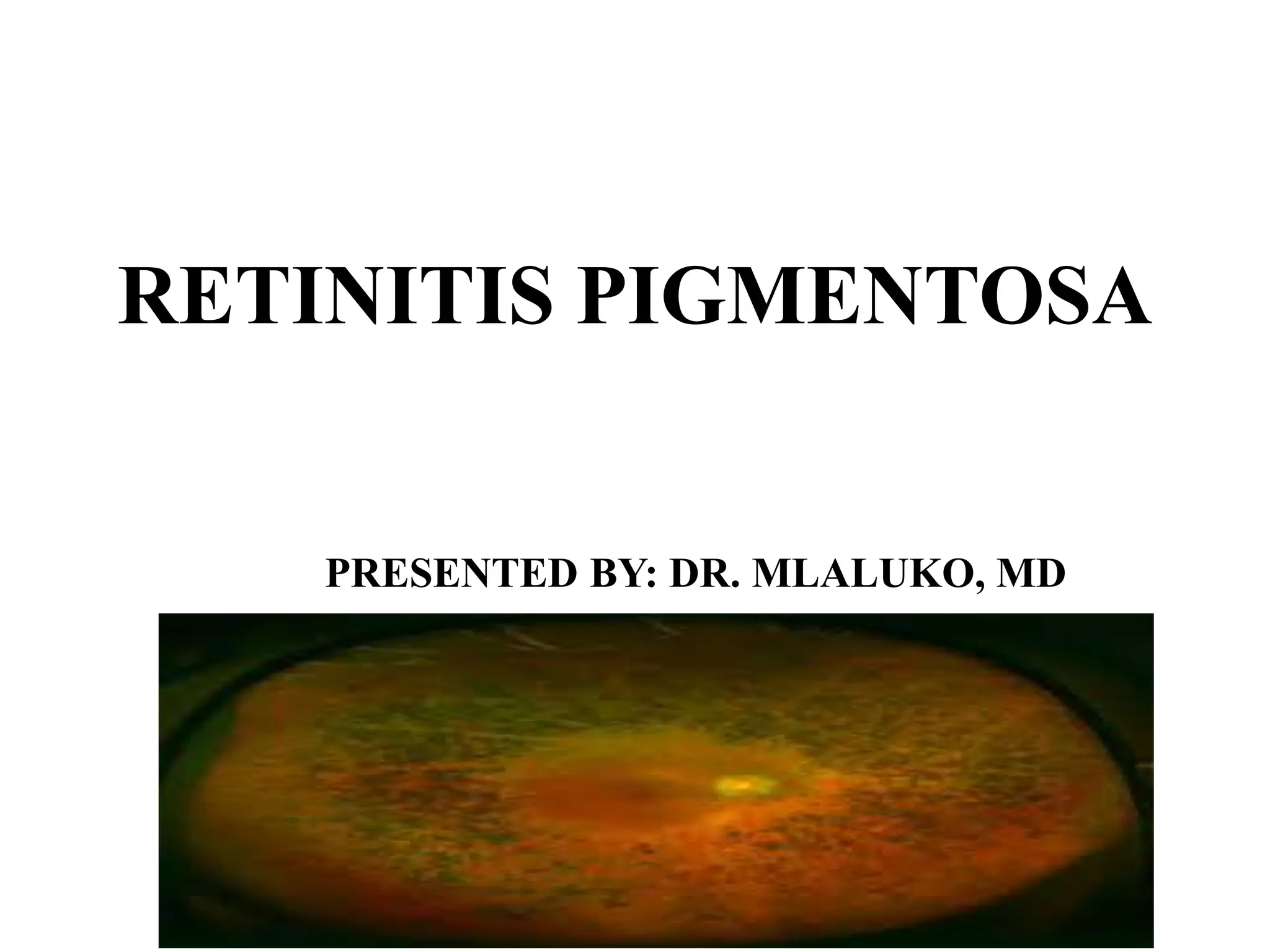
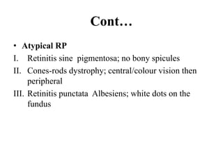
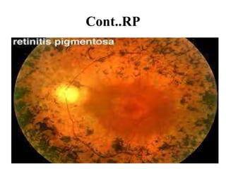
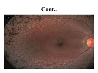
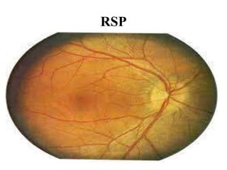
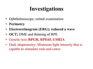
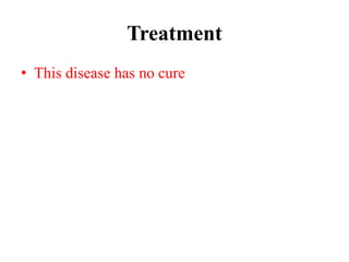
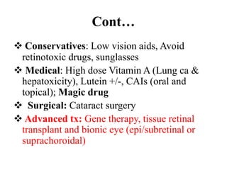
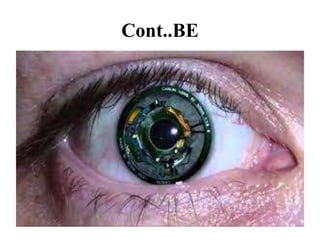
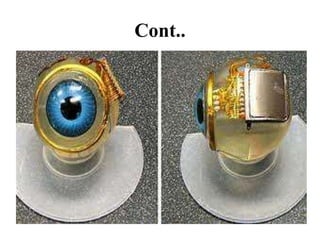
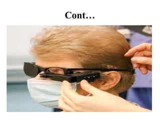
RETINITIS PIGMENTOSA
PRESENTED BY: DR. MLALUKO, MD
Retinitis pigmentosa
• Is rare inherited degenerative eye disease that
cause severe vision impairment. In this disease,
the retina is damaged and lead to loss of
photoreceptors (cones and rods) and progressive
vision loss.
• Primary it start to rods as primary degeneration
then follows cones as secondary degeneration.
• Later it affect the central vision , which is needed
for reading, driving, recognizing faces then in
adulthood the patient become blind.
Cont…
• 1:3,000-5,000
• a.k.a Rod-cone dystrophy, Pigmentary
retinopathy/Tapetoretinal generation
Pathophysiology
• Photoreceptors apoptosis
• Retinal degeneration due to failure RPE to
phagocytose the shed rod outer segment discs results
in accumulation of rod outer segment debris
• Defect in the accumulation of cGMP-
phosphosiesterase which leads to accumulation of
toxic level of cGMP
Mode of inheritance
• Sporadic: It is called simplex/isolated RP; most
common (50%); only one person in the family
presents with features
• Inherited:
i. AD (25%); best prognosis
ii. AR (25%)
iii. XLR (10%); worst prognosis
Clinical symptoms and signs
• Poor vision in dim light (Nyctalopia)
• Progress loss of vision at night (night
blindness)
• Delayed adaptation to bright light
• Flashes of light
• Mild to severe loss of peripheral visual field
• Poor visual acuity (reduced central vision)
• Glares
Cont…
Diagnosis
• History taking; night blindness, familial
history
• Fundal findings; bone spicules, arterioriolar
attenuation and pallor disc+/- macula edema
• Ocular association of RP; OCHTN/POAG,
posterior subcapsular/cortical cataract, myopia,
microphthalmia, keratoconus, PVD, optic disc
atrophy, epiretinal membrane
Cont…
• Systemic (syndromic) association
i. Lawrence-Moon-Bandet-Biedl syndrome;
Obesity, MR, polydactyl, polyuria, polydypsia,
hirschsprung disease, renal anomalies
ii. Cockayne syndrome; Cataract, optic atrophy,
dry eyes, squint, nystagmus, corneal opacity
iii. Refsum’s syndrome; hearing loss, loss of smell
and ataxia
iv. Usher’s syndrome; hearing loss
Cont…
• Atypical RP
I. Retinitis sine pigmentosa; no bony spicules
II. Cones-rods dystrophy; central/colour vision then
peripheral
III. Retinitis punctata Albesiens; white dots on the
fundus
Cont..RP
Cont..
Cont..
RSP
Investigations
• Ophthalmoscope; retinal examination
• Perimetry
• Electroretinogram (ERG); reduced a wave
• OCT; DME and thinning of RPE
• Genetic test; RPGR, RPE65, USH2A
• Dark adaptometry: Minimum light intensity that is
capable to stimulate rods and cones
Treatment
• This disease has no cure
Cont…
 Conservatives: Low vision aids, Avoid
retinotoxic drugs, sunglasses
 Medical: High dose Vitamin A (Lung ca &
hepatoxicity), Lutein +/-, CAIs (oral and
topical); Magic drug
 Surgical: Cataract surgery
 Advanced tx: Gene therapy, tissue retinal
transplant and bionic eye (epi/subretinal or
suprachoroidal)
Cont..BE
Cont..
Cont…
Differential diagnoses
• Hypovitaminosis A
AKSANTEEEEEENI

RETINITIS PIGMENTOSA.pptx

  • 1.
  • 2.
    Retinitis pigmentosa • Israre inherited degenerative eye disease that cause severe vision impairment. In this disease, the retina is damaged and lead to loss of photoreceptors (cones and rods) and progressive vision loss. • Primary it start to rods as primary degeneration then follows cones as secondary degeneration. • Later it affect the central vision , which is needed for reading, driving, recognizing faces then in adulthood the patient become blind.
  • 3.
    Cont… • 1:3,000-5,000 • a.k.aRod-cone dystrophy, Pigmentary retinopathy/Tapetoretinal generation
  • 4.
    Pathophysiology • Photoreceptors apoptosis •Retinal degeneration due to failure RPE to phagocytose the shed rod outer segment discs results in accumulation of rod outer segment debris • Defect in the accumulation of cGMP- phosphosiesterase which leads to accumulation of toxic level of cGMP
  • 5.
    Mode of inheritance •Sporadic: It is called simplex/isolated RP; most common (50%); only one person in the family presents with features • Inherited: i. AD (25%); best prognosis ii. AR (25%) iii. XLR (10%); worst prognosis
  • 6.
    Clinical symptoms andsigns • Poor vision in dim light (Nyctalopia) • Progress loss of vision at night (night blindness) • Delayed adaptation to bright light • Flashes of light • Mild to severe loss of peripheral visual field • Poor visual acuity (reduced central vision) • Glares
  • 7.
  • 8.
    Diagnosis • History taking;night blindness, familial history • Fundal findings; bone spicules, arterioriolar attenuation and pallor disc+/- macula edema • Ocular association of RP; OCHTN/POAG, posterior subcapsular/cortical cataract, myopia, microphthalmia, keratoconus, PVD, optic disc atrophy, epiretinal membrane
  • 9.
    Cont… • Systemic (syndromic)association i. Lawrence-Moon-Bandet-Biedl syndrome; Obesity, MR, polydactyl, polyuria, polydypsia, hirschsprung disease, renal anomalies ii. Cockayne syndrome; Cataract, optic atrophy, dry eyes, squint, nystagmus, corneal opacity iii. Refsum’s syndrome; hearing loss, loss of smell and ataxia iv. Usher’s syndrome; hearing loss
  • 10.
    Cont… • Atypical RP I.Retinitis sine pigmentosa; no bony spicules II. Cones-rods dystrophy; central/colour vision then peripheral III. Retinitis punctata Albesiens; white dots on the fundus
  • 11.
  • 12.
  • 13.
  • 14.
  • 15.
    Investigations • Ophthalmoscope; retinalexamination • Perimetry • Electroretinogram (ERG); reduced a wave • OCT; DME and thinning of RPE • Genetic test; RPGR, RPE65, USH2A • Dark adaptometry: Minimum light intensity that is capable to stimulate rods and cones
  • 16.
  • 17.
    Cont…  Conservatives: Lowvision aids, Avoid retinotoxic drugs, sunglasses  Medical: High dose Vitamin A (Lung ca & hepatoxicity), Lutein +/-, CAIs (oral and topical); Magic drug  Surgical: Cataract surgery  Advanced tx: Gene therapy, tissue retinal transplant and bionic eye (epi/subretinal or suprachoroidal)
  • 18.
  • 19.
  • 20.
  • 21.
  • 22.