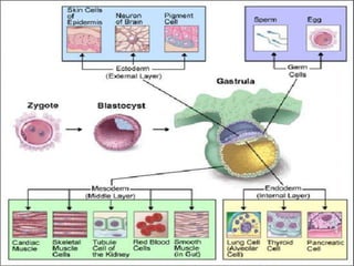
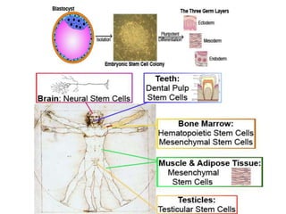
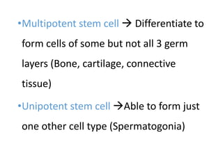
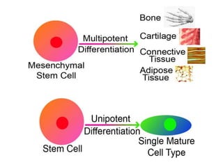
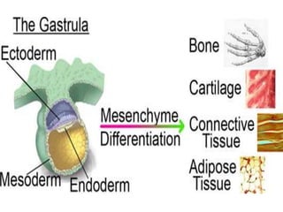
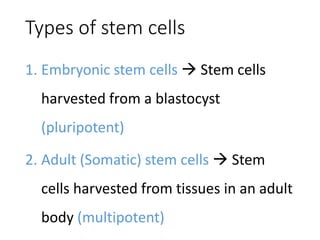
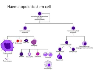
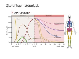
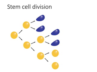
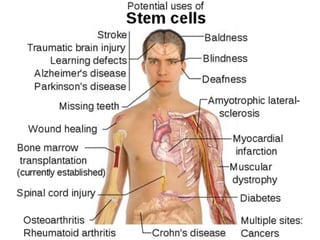
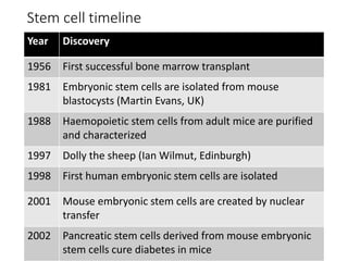
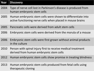
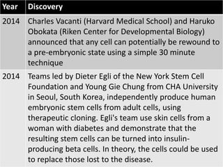
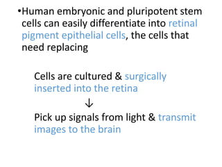
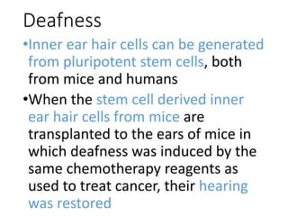
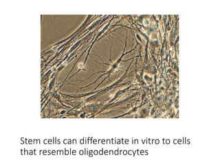
This document discusses stem cell therapy and the properties and types of stem cells. It outlines the history of key stem cell discoveries from the 1950s to present. Stem cells can be embryonic, adult, hematopoietic, or other types. Clinical trials are exploring using stem cells to treat conditions like macular degeneration, multiple sclerosis, spinal cord injuries, diabetes, and more. Challenges include developing cell types that can properly integrate and replacing lost or damaged tissues.
What are stem cells? This presentation provides an overview of multiple different stem cells including embryonic stem cells, mesenchymal stem cells, cancer stem cells, induced pluripotent stem cells, hematopoietic stem cells and neural stem cells.
For better view, press F5.
As we go through our lives each of us will have very different needs for our own healthcare.
Scientist's are constantly researching to make medical care treatment more personalized.
One way they are doing this is by-
Stem Cells therapy
Stem-cell therapy is the use of stem cells to treat or prevent a disease or condition.
It is also known as regenerative medicine, promotes the reparative response of diseased, dysfunctional or injured tissue using stem cells or their derivatives.
It is the next chapter of organ transplantation and uses cells instead of donor organs, which are limited in supply.
What are Stem cells?
Stem cells are called “master cells”
Stem cells are cells that are undifferentiated.
What are Stem cells?
Steam cells have the potential to become all other kinds of cells in our body.
What are Stem cells?
Types of Stem cells
How stem cell therapy works?
Disease cured by stem cell therapy.
Spinal Cord Injuries
Stem cell treatment of Diabetes mellitus type 1 & 2
Stem cell treatment of Stroke
Cancer treatment
Heart damage
Baldness
Tooth implanting
Deafness and blindness
Have stem cells already been used to treat diseases?
Ethical Consideration of Stem Cell Therapy
As the research method mainly focused on Embryonic Stem Cells, which involves taking tissue from an aborted embryo to get proper material to study. This is typically done just days after conception or between the 5th and 9th week.
Since then, researchers have moved on to more ethical study methods, such as Induced Pluripotent Stem Cells (iPS). iPS is artificially derived from a non-pluripotent cell, such as adult somatic cells.
Nowadays stem cell treatment has been spreaded throughout the world. It has also been grown commercially in developed countries.
It is thought that one day it may be the major key to treat various diseases.
Using stem cells to conduct medical research and treat disease is acceptable?
Don’t know
No
Yes
Do you approve of the extraction of stem cells from human embryos for medical research?
Don’t know
No
Yes
iPSCs are pluripotent; unlike ESC, iPSCs are not derived from the embryo, but instead created from differentiated cells in the lab through a process – cellular reprogramming.
WHAT IS EMBRYONIC STEM CELL?
TYPES OF EMBRYONIC STEM CELL
RECENT RESEARCH OF EMBRYONIC STEM CELL
ADVANTAGES OF EMBRYONIC STEM CELL
LIMITATION OF EMBRYONIC STEM CELL.
In our latest SlideShare, we discuss the exciting new field of stem cells in regenerative medicine. These slides cover briefly the sources and types of stem cells, as well as the recent figures and growing interest in this sector. Recent R&D highlights would focus on the vast potential of stem cells; for blood disorders, multiple sclerosis, diabetes as well as brain injury.
Imagine that you have been told you have an illness that cannot be cured or what if your body has been irreversibly paralysed. There is no hope. But there is a science that could change that. It’s Called Stem Cell Research and it’s an important step in the medical revolution. But it comes with controversies as it uses Human Embryos’ as Raw Material.
But something astounding happened in the year 2006 that removed the usage of surplus embryos from the equation altogether. It’s about a brand new technology that can turn back the clock on your body cells. This is cutting edge of science where new developments are happing all the time. The iPSCs could be the potential medicine of 21st century. So what are stem cells? Why do they Matter? What are iPSCs and how it changed the biological rules?
If the cell is able to form all cell types of the embryo & adult (Fertilized egg cell) Totipotent stem cell
Stem cell able to differentiate into all 3 germ layers Pluripotent stem cell (Embryonic stem cell)
Multipotent stem cell Differentiate to form cells of some but not all 3 germ layers (Bone, cartilage, connective tissue)
Unipotent stem cell Able to form just one other cell type (Spermatogonia)
Embryos created in vitro fertilization
Aborted embryos
Limited tissues (bone marrow, muscle, brain)
Discrete populations of adult stem cells generate replacements for cells that are lost through normal wear and tear, injury or disease
Placental cord
Baby teeth
Diabetes patients lose the function of their insulin-producing beta cells of the pancreas
Human embryonic stem cells may be grown in cell cultures and stimulate to form insulin-producing cells , that can be transplanted into the patients
Pancreas is digested with collagenase that frees islets from surrounding cells
Centrifugation of isolates containing mainly alpha and beta cells, purified islets beta cells
Transplanted through a catheter into the liver where they become permanently established Caused when key brain cells that produce message carrying chemical/neurotransmitter (dopamine) die off.
Symptoms start with the patients trembling and can end up paralyzed
Harvesting of stem cells from patients bone marrow, foetus or any other source
Culturing of harvested stem cells in lab conditions - to get high concentrations of stem cells
Then purified and high concentration of stem cells are surgically injected in the brain of patient.
A stem cell is a "blank" cell that can give rise to multiple tissue types such as a skin, muscle, or nerve cell.
Under certain physiologic or experimental conditions, they can be induced to become tissue- or organ-specific cells with special functions.
This presentation describes in detail the various types and sources of stem cells. it also describes the stem cell therapies used to treat various diseases.
Introduction.
Properties of Stem Cells.
Key Research events.
Embryonic Stem Cell.
Stem cell Cultivation.
Stem cells are central to three processes in an organism.
Research & Clinical Application of stem cell.
Research patents.
Conclusion.
Reference.
youtube link : https://www.youtube.com/watch?v=da69DB6dU58&lc=z13osnvyfnnryny2z22qh3y4rs2bd3h2d
Stem cells can be defined simply as cells which are not specialized in any specific tissue or organs.
In other words, stem cells have not differentiated into other cell types to form tissues and organs.
They are the base or foundational cells to develop into cells which specialized in certain functions.
Another distinguishing characteristics of stem cells is their ability to undergo division, giving rise to more stem cells.
The significance of stem cells in their application to the human body and human health boils down to the two important characteristics of differentiation and self-regeneration.
Imagine how powerful they can be if stems cells can be developed into heart cells, especially when someone’s heart is doomed to fail Or, for someone with damaged brain cells or nerve cells, wouldn’t it be extremely great news if stem cells can develop new brain cells or nerve cells for the person.
Indeed, the potential and possibilities of exploiting stem cells for medical science and health science are enormous.
Many untreatable diseases and ailments may in the near future become curable.
Stem cells are classified into various types based on their ability to undergo differentiation into different cell types.
In other words, their classification, and hence their name, is derived from their potential to develop into one, two or several other cell types.
In my presentation I’ll discuss the principals of formation the stem cell and its applications .
stem cell research is yet to be advanced , once fully developed can alleviate human suffering, this ppt reviews the contemporary evidence, pitfalls and challenges
Enhancing Innovation in STEM by Exploring Aesthetics Derek Ham
This presentation was presented at the 2nd Annual Bridging the Gap STEM Conference in Raleigh, NC. Discover how K-16 STEM curricula should readily embrace aesthetics as a core component of their pedagogy. By doing so, it opens a new world of creativity and innovation for STEM inquiry. We present a compelling argument for pulling aesthetics out of art education curricula to be placed right at the center of STEM education. This session was hands-on, allowing attendees to participate in learning concepts through an interactive educational game called SHAPE.
PRP Platelet Rich Plasma is a non-surgical, non-invasive treatment for hair loss. “Cell therapy” is a relatively new frontier in medical technology which involves introducing cells into a tissue in order to treat a disease or condition. Areas of research such as stem cell processing and activation, cell multiplication, cytokines, growth factors, extracellular matrix products, bioscaffolds, platelet rich plasma (PRP), gene therapy, tissue repair and more are some of the exciting new ways in which scientists are investigating in the use of cell therapy to physicians improve outcomes and treat a variety of conditions.
Adjunctive therapies which impact wound healing like the use of hyperbaric oxygen, copper peptide solutions, phototherapy, extracellular matrix (ECM), protein soaks, etc. have been a part of Dr. Alan Bauman’s post-operative treatment regimens after hair transplantation for years. Helping the body safely “help itself” is the cornerstone of these types of treatments which often utilize natural components found in the human body. Along those lines, PRP has already been found to be a helpful adjunct successfully in use in many areas of medicine, including orthopedics, dentistry and plastic surgery for tissue repair, tissue regeneration and wound healing.
What is Platelet Rich Plasma (PRP)?
Produced in just minutes from a small quantity of your own blood, PRP contains a wide variety of the body’s own natural growth factors which may be helpful in accelerating tissue regeneration, improving healing time and stimulating hair growth. Platelets are cell fragments that are normally found in the blood and play a key role in blood clotting and are a natural source of growth factors. Special medical equipment is used by your physician to separate a platelets from a small sample of your own blood and prepare them for use. Once applied and activated, platelets release their stores of powerful growth factors and other molecules.
What Growth Factors are found in Platelet Rich Plasma (PRP)? The platelets collected in PRP are activated by thrombin and calcium chloride to induce the release of these growth factor proteins and other cytokines:
• platelet-derived growth factor
• transforming growth factor beta
• fibroblast growth factor
• insulin-like growth factor 1
• insulin-like growth factor 2
• vascular endothelial growth factor
• epidermal growth factor
• interleukin-8
• keratinocyte growth factor
• connective tissue growth factor
HOW IS PRP MADE?
PRP is made from a small sample of your own blood. The FDA-approved process concentrates the platelets found in the blood sample and separates them from other blood components. These concentrated platelets contain huge reservoirs of growth factors and wound healing factors. Growth factors are natural components of your body whose potential benefits can now be harnessed using modern medical technology. For hands-on PRP training classes v
Cell and gene therapy for Parkinson’s disease - part 1Parkinson's UK
Presentation by Prof Deniz Kirik, MD, PhD at the Parkinson's UK Research Conference, November 2010 in York.
With introduction by Dr Oliver Bandmann.
Part 2: http://www.slideshare.net/ParkinsonsResearchUK/cell-and-gene-therapy-for-parkinsons-disease-part-2.
Part 3: http://www.slideshare.net/ParkinsonsResearchUK/cell-and-gene-therapy-for-parkinsons-disease-part-3
Medical Management of Hair Loss for the Integrative Medicine Practice - Dr Al...Bauman Medical Group, P.A.
Medical Management of Hair Loss
For the Integrative Medicine Practice 5/15/14
Workshop Director:
Alan J. Bauman, M.D. – Diplomate, American Board of Hair Restoration Surgery
Course Description:
Hair loss affects roughly 35 million men and 21 million women in the United States. Forty percent of men will have noticeable hair loss by age thirty‐five and sixty‐five percent of women will have noticeable hair loss by the time they are sixty years of age. Hereditary hair loss, or Androgenetic Alopecia, has cosmetic implications often associated with the aging process, can also be an indicator of other imbalances or disease within the human body. Because chronic and progressive hair loss often deeply impacts the quality of life of its sufferers, the negative psychosocial implications for the hair loss sufferer as well as the positive benefits of treatment should be strongly taken into consideration.
Learning Objectives:
In this one-day module, the participants will learn about the etiology, diagnosis, psychosocial impact and non-surgical management of androgenetic alopecia from hair loss expert, Alan J. Bauman, M.D. The anatomy and physiology of hair follicle structure and function and phases of the hair cycle will be explained in detail. Participants will learn about hair loss risk factors such as genetics, the effects of hormones, hair loss associated with the use of specific medications and supplements, in addition to medical conditions that influence hair follicle function and how to identify them. Participants will learn how to consult with the hair loss sufferer, evaluate and scientifically measure hair loss via cross-sectional trichometry, high-powered dermoscopy and global photography. They will learn to diagnose using the standard Norwood-Hamilton and Ludwig-Savin scales as well as identify and rule out non-hereditary hair loss with the purpose of developing a realistic short and long-term treatment plan. Exploration of the various modalities to combat hair loss will be discussed including the importance of hormone balancing, basic knowledge of herbal/natural remedies, mechanism and protocols of laser and light-based devices, pros and cons of traditional and pharmacy-compounded medical therapy, as well as the latest information on the application of cell therapy, growth factors and stem cells will be discussed. Instruction will be given on how to create a customized results-oriented treatment, tracking and follow-up/compliance protocol for patients suffering from common female and male pattern hair loss. Attendees will also leave this session with suggested office space plans, patient flow and staffing recommendations for incorporating the medical management of hair loss into their practice as either a stand-alone service or as an integrated part of their existing treatment offerings.
Join us for the preconference workshop on May 15, 2014 at A4M Conference held at the Orlando Gaylord Palms. Register at hai.rs/a4mhair514
Dr. Kenneth Dickie from Royal Centre of Plastic Surgery in Barrie, Ontario explained the use of stem cells technology in plastic surgery.
If you have any questions, please contact Dr. Kenneth Dickie at http://royalcentreofplasticsurgery.com/
Stem cell technology | Presented by pranjali V. Bhadanepranjali bhadane
This presentation is related to biological topic stem cell technology. stem cell technology plays an important role in life science. stem cell technology is most important for future researchers on personal genomics, personal drug and Complex Disease Research...
The complete, compiled presentation on stem cell research. The contents include background history along with the introduction, different stem cell types, cultivation process, stem cell cloning and potential uses, the negative aspects and ethical concerns regarding stem cell therapy. Different examples of the useful work in stem cell therapy field has also been mentioned.
Hello friends. In this PPT I am talking about drugs used in the treatment of type 2 diabetes mellitus. If you like it, please do let me know in the comments section. A single word of appreciation from you will encourage me to make more of such videos. Thanks. Enjoy and welcome to the beautiful world of pharmacology where pharmacology comes to life. This video is intended for MBBS, BDS, paramedical and any person who wishes to have a basic understanding of the subject in the simplest way.
AMA General considerations 12.6.23.pptxKarun Kumar
Hello friends. In this PPT I am talking about Antimicrobial drugs-General Considerations. If you like it, please do let me know in the comments section. A single word of appreciation from you will encourage me to make more of such videos. Thanks. Enjoy and welcome to the beautiful world of pharmacology where pharmacology comes to life. This video is intended for MBBS, BDS, paramedical and any person who wishes to have a basic understanding of the subject in the simplest way.
Hello friends. In this PPT I am talking about general anaesthetics and skeletal muscle relaxants. If you like it, please do let me know in the comments section. A single word of appreciation from you will encourage me to make more of such videos. Thanks. Enjoy and welcome to the beautiful world of pharmacology where pharmacology comes to life. This video is intended for MBBS, BDS, paramedical and any person who wishes to have a basic understanding of the subject in the simplest way.
Hello friends. In this PPT I am talking about drugs affecting blood. If you like it, please do let me know in the comments section. A single word of appreciation from you will encourage me to make more of such videos. Thanks. Enjoy and welcome to the beautiful world of pharmacology where pharmacology comes to life. This video is intended for MBBS, BDS, paramedical and any person who wishes to have a basic understanding of the subject in the simplest way.
Drugs & Aids with specific applications in dentalKarun Kumar
Hello friends. In this PPT I am talking about drugs & aids with specific applications in dental disorders. If you like it, please do let me know in the comments section. A single word of appreciation from you will encourage me to make more of such videos. Thanks. Enjoy and welcome to the beautiful world of pharmacology where pharmacology comes to life. This video is intended for MBBS, BDS, paramedical and any person who wishes to have a basic understanding of the subject in the simplest way.
Hello friends. In this PPT I am talking about diuretics. If you like it, please do let me know in the comments section. A single word of appreciation from you will encourage me to make more of such videos. Thanks. Enjoy and welcome to the beautiful world of pharmacology where pharmacology comes to life. This video is intended for MBBS, BDS, paramedical and any person who wishes to have a basic understanding of the subject in the simplest way.
Hello friends. In this PPT I am talking about Cardiovascular system drugs. If you like it, please do let me know in the comments section. A single word of appreciation from you will encourage me to make more of such videos. Thanks. Enjoy and welcome to the beautiful world of pharmacology where pharmacology comes to life. This video is intended for MBBS, BDS, paramedical and any person who wishes to have a basic understanding of the subject in the simplest way.
Hello friends. In this PPT I am talking about CNS drugs. If you like it, please do let me know in the comments section. A single word of appreciation from you will encourage me to make more of such videos. Thanks. Enjoy and welcome to the beautiful world of pharmacology where pharmacology comes to life. This video is intended for MBBS, BDS, paramedical and any person who wishes to have a basic understanding of the subject in the simplest way.
Hello friends. In this PPT I am talking about Autacoids. If you like it, please do let me know in the comments section. A single word of appreciation from you will encourage me to make more of such videos. Thanks. Enjoy and welcome to the beautiful world of pharmacology where pharmacology comes to life. This video is intended for MBBS, BDS, paramedical and any person who wishes to have a basic understanding of the subject in the simplest way.
Hello friends. In this PPT I am talking about Anti-viral drugs drugs. If you like it, please do let me know in the comments section. A single word of appreciation from you will encourage me to make more of such videos. Thanks. Enjoy and welcome to the beautiful world of pharmacology where pharmacology comes to life. This video is intended for MBBS, BDS, paramedical and any person who wishes to have a basic understanding of the subject in the simplest way.
Hello friends. In this PPT I am talking about Anti-TB & anti leprosy drugs. If you like it, please do let me know in the comments section. A single word of appreciation from you will encourage me to make more of such videos. Thanks. Enjoy and welcome to the beautiful world of pharmacology where pharmacology comes to life. This video is intended for MBBS, BDS, paramedical and any person who wishes to have a basic understanding of the subject in the simplest way.
Hello friends. In this PPT I am talking about antiseptics, disinfectants. If you like it, please do let me know in the comments section. A single word of appreciation from you will encourage me to make more of such videos. Thanks. Enjoy and welcome to the beautiful world of pharmacology where pharmacology comes to life. This video is intended for MBBS, BDS, paramedical and any person who wishes to have a basic understanding of the subject in the simplest way.
Hello friends. In this PPT I am talking about antiprotozoal drugs. If you like it, please do let me know in the comments section. A single word of appreciation from you will encourage me to make more of such videos. Thanks. Enjoy and welcome to the beautiful world of pharmacology where pharmacology comes to life. This video is intended for MBBS, BDS, paramedical and any person who wishes to have a basic understanding of the subject in the simplest way.
Hello friends. In this PPT I am talking about anti-fungal drugs. If you like it, please do let me know in the comments section. A single word of appreciation from you will encourage me to make more of such videos. Thanks. Enjoy and welcome to the beautiful world of pharmacology where pharmacology comes to life. This video is intended for MBBS, BDS, paramedical and any person who wishes to have a basic understanding of the subject in the simplest way.
Hello friends. In this PPT I am talking about anti-cancer drugs. If you like it, please do let me know in the comments section. A single word of appreciation from you will encourage me to make more of such videos. Thanks. Enjoy and welcome to the beautiful world of pharmacology where pharmacology comes to life. This video is intended for MBBS, BDS, paramedical and any person who wishes to have a basic understanding of the subject in the simplest way.
Hello friends. In this PPT I am talking about autonomic nervous system. If you like it, please do let me know in the comments section. A single word of appreciation from you will encourage me to make more of such videos. Thanks. Enjoy and welcome to the beautiful world of pharmacology where pharmacology comes to life. This video is intended for MBBS, BDS, paramedical and any person who wishes to have a basic understanding of the subject in the simplest way.
AETCOM (Attitude, Ethics and Communication module)Karun Kumar
Hello friends. In this PPT I am talking about AETCOM (Attitude, Ethics and Communication module) of Pharmacology. If you like it, please do let me know in the comments section. A single word of appreciation from you will encourage me to make more of such videos. Thanks. Enjoy and welcome to the beautiful world of pharmacology where pharmacology comes to life. This video is intended for MBBS, BDS, paramedical and any person who wishes to have a basic understanding of the subject in the simplest way
Seminar of U.V. Spectroscopy by SAMIR PANDASAMIR PANDA
Spectroscopy is a branch of science dealing the study of interaction of electromagnetic radiation with matter.
Ultraviolet-visible spectroscopy refers to absorption spectroscopy or reflect spectroscopy in the UV-VIS spectral region.
Ultraviolet-visible spectroscopy is an analytical method that can measure the amount of light received by the analyte.
Cancer cell metabolism: special Reference to Lactate PathwayAADYARAJPANDEY1
Normal Cell Metabolism:
Cellular respiration describes the series of steps that cells use to break down sugar and other chemicals to get the energy we need to function.
Energy is stored in the bonds of glucose and when glucose is broken down, much of that energy is released.
Cell utilize energy in the form of ATP.
The first step of respiration is called glycolysis. In a series of steps, glycolysis breaks glucose into two smaller molecules - a chemical called pyruvate. A small amount of ATP is formed during this process.
Most healthy cells continue the breakdown in a second process, called the Kreb's cycle. The Kreb's cycle allows cells to “burn” the pyruvates made in glycolysis to get more ATP.
The last step in the breakdown of glucose is called oxidative phosphorylation (Ox-Phos).
It takes place in specialized cell structures called mitochondria. This process produces a large amount of ATP. Importantly, cells need oxygen to complete oxidative phosphorylation.
If a cell completes only glycolysis, only 2 molecules of ATP are made per glucose. However, if the cell completes the entire respiration process (glycolysis - Kreb's - oxidative phosphorylation), about 36 molecules of ATP are created, giving it much more energy to use.
IN CANCER CELL:
Unlike healthy cells that "burn" the entire molecule of sugar to capture a large amount of energy as ATP, cancer cells are wasteful.
Cancer cells only partially break down sugar molecules. They overuse the first step of respiration, glycolysis. They frequently do not complete the second step, oxidative phosphorylation.
This results in only 2 molecules of ATP per each glucose molecule instead of the 36 or so ATPs healthy cells gain. As a result, cancer cells need to use a lot more sugar molecules to get enough energy to survive.
introduction to WARBERG PHENOMENA:
WARBURG EFFECT Usually, cancer cells are highly glycolytic (glucose addiction) and take up more glucose than do normal cells from outside.
Otto Heinrich Warburg (; 8 October 1883 – 1 August 1970) In 1931 was awarded the Nobel Prize in Physiology for his "discovery of the nature and mode of action of the respiratory enzyme.
WARNBURG EFFECT : cancer cells under aerobic (well-oxygenated) conditions to metabolize glucose to lactate (aerobic glycolysis) is known as the Warburg effect. Warburg made the observation that tumor slices consume glucose and secrete lactate at a higher rate than normal tissues.
(May 29th, 2024) Advancements in Intravital Microscopy- Insights for Preclini...Scintica Instrumentation
Intravital microscopy (IVM) is a powerful tool utilized to study cellular behavior over time and space in vivo. Much of our understanding of cell biology has been accomplished using various in vitro and ex vivo methods; however, these studies do not necessarily reflect the natural dynamics of biological processes. Unlike traditional cell culture or fixed tissue imaging, IVM allows for the ultra-fast high-resolution imaging of cellular processes over time and space and were studied in its natural environment. Real-time visualization of biological processes in the context of an intact organism helps maintain physiological relevance and provide insights into the progression of disease, response to treatments or developmental processes.
In this webinar we give an overview of advanced applications of the IVM system in preclinical research. IVIM technology is a provider of all-in-one intravital microscopy systems and solutions optimized for in vivo imaging of live animal models at sub-micron resolution. The system’s unique features and user-friendly software enables researchers to probe fast dynamic biological processes such as immune cell tracking, cell-cell interaction as well as vascularization and tumor metastasis with exceptional detail. This webinar will also give an overview of IVM being utilized in drug development, offering a view into the intricate interaction between drugs/nanoparticles and tissues in vivo and allows for the evaluation of therapeutic intervention in a variety of tissues and organs. This interdisciplinary collaboration continues to drive the advancements of novel therapeutic strategies.
The increased availability of biomedical data, particularly in the public domain, offers the opportunity to better understand human health and to develop effective therapeutics for a wide range of unmet medical needs. However, data scientists remain stymied by the fact that data remain hard to find and to productively reuse because data and their metadata i) are wholly inaccessible, ii) are in non-standard or incompatible representations, iii) do not conform to community standards, and iv) have unclear or highly restricted terms and conditions that preclude legitimate reuse. These limitations require a rethink on data can be made machine and AI-ready - the key motivation behind the FAIR Guiding Principles. Concurrently, while recent efforts have explored the use of deep learning to fuse disparate data into predictive models for a wide range of biomedical applications, these models often fail even when the correct answer is already known, and fail to explain individual predictions in terms that data scientists can appreciate. These limitations suggest that new methods to produce practical artificial intelligence are still needed.
In this talk, I will discuss our work in (1) building an integrative knowledge infrastructure to prepare FAIR and "AI-ready" data and services along with (2) neurosymbolic AI methods to improve the quality of predictions and to generate plausible explanations. Attention is given to standards, platforms, and methods to wrangle knowledge into simple, but effective semantic and latent representations, and to make these available into standards-compliant and discoverable interfaces that can be used in model building, validation, and explanation. Our work, and those of others in the field, creates a baseline for building trustworthy and easy to deploy AI models in biomedicine.
Bio
Dr. Michel Dumontier is the Distinguished Professor of Data Science at Maastricht University, founder and executive director of the Institute of Data Science, and co-founder of the FAIR (Findable, Accessible, Interoperable and Reusable) data principles. His research explores socio-technological approaches for responsible discovery science, which includes collaborative multi-modal knowledge graphs, privacy-preserving distributed data mining, and AI methods for drug discovery and personalized medicine. His work is supported through the Dutch National Research Agenda, the Netherlands Organisation for Scientific Research, Horizon Europe, the European Open Science Cloud, the US National Institutes of Health, and a Marie-Curie Innovative Training Network. He is the editor-in-chief for the journal Data Science and is internationally recognized for his contributions in bioinformatics, biomedical informatics, and semantic technologies including ontologies and linked data.
THE IMPORTANCE OF MARTIAN ATMOSPHERE SAMPLE RETURN.Sérgio Sacani
The return of a sample of near-surface atmosphere from Mars would facilitate answers to several first-order science questions surrounding the formation and evolution of the planet. One of the important aspects of terrestrial planet formation in general is the role that primary atmospheres played in influencing the chemistry and structure of the planets and their antecedents. Studies of the martian atmosphere can be used to investigate the role of a primary atmosphere in its history. Atmosphere samples would also inform our understanding of the near-surface chemistry of the planet, and ultimately the prospects for life. High-precision isotopic analyses of constituent gases are needed to address these questions, requiring that the analyses are made on returned samples rather than in situ.
Professional air quality monitoring systems provide immediate, on-site data for analysis, compliance, and decision-making.
Monitor common gases, weather parameters, particulates.
Richard's entangled aventures in wonderlandRichard Gill
Since the loophole-free Bell experiments of 2020 and the Nobel prizes in physics of 2022, critics of Bell's work have retreated to the fortress of super-determinism. Now, super-determinism is a derogatory word - it just means "determinism". Palmer, Hance and Hossenfelder argue that quantum mechanics and determinism are not incompatible, using a sophisticated mathematical construction based on a subtle thinning of allowed states and measurements in quantum mechanics, such that what is left appears to make Bell's argument fail, without altering the empirical predictions of quantum mechanics. I think however that it is a smoke screen, and the slogan "lost in math" comes to my mind. I will discuss some other recent disproofs of Bell's theorem using the language of causality based on causal graphs. Causal thinking is also central to law and justice. I will mention surprising connections to my work on serial killer nurse cases, in particular the Dutch case of Lucia de Berk and the current UK case of Lucy Letby.
This pdf is about the Schizophrenia.
For more details visit on YouTube; @SELF-EXPLANATORY;
https://www.youtube.com/channel/UCAiarMZDNhe1A3Rnpr_WkzA/videos
Thanks...!
3. Properties of stem cells
•If the cell is able to form all cell types of
the embryo & adult (Fertilized egg cell)
Totipotent stem cell
•Stem cell able to differentiate into all 3
germ layers Pluripotent stem cell
(Embryonic stem cell)
4.
5.
6.
7. •Multipotent stem cell Differentiate to
form cells of some but not all 3 germ
layers (Bone, cartilage, connective
tissue)
•Unipotent stem cell Able to form just
one other cell type (Spermatogonia)
8.
9.
10. Types of stem cells
1. Embryonic stem cells Stem cells
harvested from a blastocyst
(pluripotent)
2. Adult (Somatic) stem cells Stem
cells harvested from tissues in an adult
body (multipotent)
15. On the road to stem cell
therapy
•Interest in human embryonic stem cells
Shortage of donor organs
•First-in-man studies aimed at reversing
macular degeneration have already
taken place in London, and around 25
patients have been included in the first
phase 1 studies (safety and feasibility)
worldwide.
16. Stem cell timeline
Year Discovery
1956 First successful bone marrow transplant
1981 Embryonic stem cells are isolated from mouse
blastocysts (Martin Evans, UK)
1988 Haemopoietic stem cells from adult mice are purified
and characterized
1997 Dolly the sheep (Ian Wilmut, Edinburgh)
1998 First human embryonic stem cells are isolated
2001 Mouse embryonic stem cells are created by nuclear
transfer
2002 Pancreatic stem cells derived from mouse embryonic
stem cells cure diabetes in mice
17. Year Discovery
2004 Type of nerve cell lost in Parkinson’s disease is produced from
human embryonic stem cells
2005 Human embryonic stem cells were shown to differentiate into
active functioning nerve cells when placed in mouse brains
2005 Pancreatic cells were derived from adult stem cells
2006 Embryonic stem cells were derived from the morula of a mouse
2006 Embryonic stem cells were first grown without animal products
in the culture
2010 Person with spinal injury first to receive medical treatment
derived from human embryonic stem cells
2012 Human embryonic stem cells show promise in treating blindness
2013 Human embryonic stem cells produced from fetal cells using
therapeutic cloning
18. Year Discovery
2014 Charles Vacanti (Harvard Medical School) and Haruko
Obokata (Riken Center for Developmental Biology)
announced that any cell can potentially be rewound to
a pre-embryonic state using a simple 30 minute
technique
2014 Teams led by Dieter Egli of the New York Stem Cell
Foundation and Young Gie Chung from CHA University
in Seoul, South Korea, independently produce human
embryonic stem cells from adult cells, using
therapeutic cloning. Egli's team use skin cells from a
woman with diabetes and demonstrate that the
resulting stem cells can be turned into insulin-
producing beta cells. In theory, the cells could be used
to replace those lost to the disease.
23. Battle against multiple sclerosis (29/1/15)
Dr. Mark Freedman of the Ottawa Hospital Research Institute is leading research
to determine if a type of stem cell can help alleviate the symptoms of multiple
sclerosis
24. Professor Anthony Hollander, a
cartilage tissue engineer, who
cultured stem cells from Claudia
Castillo to build a new windpipe
(trachea) in year 2008
Claudia Castillo, who received a transplanted
windpipe that was engineered from her own
stem cells
26. Type 1 Diabetes
•Abnormal activation of the immune
system Destruction of the insulin-
producing beta cells
•Beta cells from pluripotent stem cells or
adult progenitors in the pancreas
Source of cells for treatment of diabetes
27. Spinal cord injury
•Oligodendrocyte cells that produce
myelin can be generated from stem
cells; if these are injected into the
site of the crush injury they could
repair the myelin layer locally, and
thus restore function and cure the
paralysis
29. Making the blind see
•Problem with corneal transplants
•Few human donors to meet clinical need
•Unsuitable in destruction of limbus
•Alternative Cultivation of limbal cells
31. Eye an ideal organ for stem cell
transplant
1. Immune-privileged site
•Transplanted cells that are not HLA matched
are not rejected
•Immunosuppressive medication is not required
2. Eye is a “ closed” unit (any stem cells injected
would find it difficult to misbehave and escape
to other parts of the body)
3. We have two eyes, it may be possible to treat
one eye without the risk of losing vision
altogether
32. •Human embryonic and pluripotent stem
cells can easily differentiate into retinal
pigment epithelial cells, the cells that
need replacing
Cells are cultured & surgically
inserted into the retina
↓
Pick up signals from light & transmit
images to the brain
33. Deafness
•Inner ear hair cells can be generated
from pluripotent stem cells, both
from mice and humans
•When the stem cell derived inner
ear hair cells from mice are
transplanted to the ears of mice in
which deafness was induced by the
same chemotherapy reagents as
used to treat cancer, their hearing
was restored
34.
35. Best stem cell for transplantation
•Acute & life-threatening disease (MI)
Allogenic & derived from
1. Living (bone marrow)
2. Deceased (Insulin producing beta cells)
•Chronic disease Autologous stem cell
transplantation
36. Bone marrow transplantation by
stem cell mobilization
Stem cells are collected from blood after giving
certain growth factors (e.g. GM-CSF) to the patients
↓
These growth factors temporarily recruit stem cells
from the bone marrow into the blood which are
stored till needed
↓
The patient is treated with cytostatic drugs
(chemotherapy) or undergoes irradiation
↓
37. The blood stem cells can subsequently be
returned to the blood of the patient at
any chosen time point through a simple
intravenous infusion into the blood
stream
↓
They will find their own way back
“home” to the bone marrow, where they
will settle and start producing new blood
cells, at no risk of rejection since they are
the patient’s own cells
38. Where to transplant stem cells
1. Myocardial infarction New cardiac
cells have to be transplanted directly
into the heart muscle
2. Kidney disease Replacing kidney
cells with stem cell derivatives will
need the transplanted cells to exert
their function within the kidney itself
39. 3.Diabetes Any replacement beta
cells, from stem cells or pancreas
progenitors could be transplanted at
another site in the body and not in
pancreas. (The portal vein of the liver, to
which the cells can be delivered simply
under local anesthetic is being used as a
transplantation site for beta cells from
donors)
40. 4. Parkinson’s disease Challenge because
the distance to which neuritis grow cannot be
predicted. Dopaminergic nerve cells in the
brain exert their effect through the neuritis (at
a site distant from where the cell body “sits”
in the brain)
•In mice and rats the neurites from these
neurons can easily grow out over that
distance, but in the much larger human
brain, this can be several centimeters and is
very difficult.
41. Cell types available for stem
cell transplantation
1. First isolated from large intestine of
mice
•These cells self organize into
“organoids” or “miniguts”
•Use To treat intestinal conditions in
which the intestinal epithelial layer is
damaged (ulcers)
42. 2. Hematopoietic stem cells obtained
from umbilical cord blood Enable
reconstitution of the blood cell
population of a patient after
destructive chemotherapy
•2 or more matched cord blood
samples are required for one adult
patient
47. Transplantation of stem
cells : Where we stand ?
•Most commonly used experimental
animals Mice because
1. There are many different strains of
mice with mutations in their DNA that
are similar to those causing human
disease
2. Easy to handle
3. Inexpensive to keep
48. Stem cells can differentiate in vitro to cells
that resemble oligodendrocytes
53. Applications of organs
on a chip
1. Drug toxicity screening (Heart and liver
most susceptible organs)
2. Human disease models (Development of
novel drugs to treat the illness)
3. Investigate routes for optimal drug uptake
in the body (Eg. To measure the rate of
transport through the intestinal wall after
oral intake)
54. 4. By capturing the genetic variation in
the normal and sick human population,
these models are expected to increase
the speed and reduce costs of drug
development.
5. Safe and effective drug development
tuned to the specific genetic profile of
the patient (Personalized medicine)
Forty years after Neil Armstrong first stepped on the moon, we’re on the brink of another giant leap for mankind — only this time the new frontier is under a microscope instead of beyond the clouds. Through out history, man kind has looked for the fountain of youth ; the elixir for eternal life, keeping us forever young in body and mind. In the discovery of stem cells, has the modern biomedical scientist finally succeeded where wizards and alchemists failed? Let’s find out. The Nobel Prize in Physiology or Medicine 2012 jointly to Sir John B. Gurdon and Sir Shinya Yamanaka for the discovery that mature cells can be reprogrammed to become pluripotent. John B. Gurdon discovered in 1962 that the specialisation of cells is reversible. In a classic experiment, he replaced the immature cell nucleus in an egg cell of a frog with the nucleus from a mature intestinal cell. This modified egg cell developed into a normal tadpole. The DNA of the mature cell still had all the information needed to develop all cells in the frog.Shinya Yamanaka discovered more than 40 years later, in 2006, how intact mature cells in mice could be reprogrammed to become immature stem cells. Surprisingly, by introducing only a few genes, he could reprogram mature cells to become pluripotent stem cells, i.e. immature cells that are able to develop into all types of cells in the body.
In the adult body in vivo there are no totipotent or pluripotent stem cells. This does not mean that the artificial conditions of culture cannot convert (“reprogram”) some adult cells into stem cells with pluripotent properties. These multi potent or uni potent stem cells are also known as progenitor cells. Progenitor cells can divide, but this capacity is generally considered more limited than that of pluripotent stem cells. Progenitor cells are predestined to differentiate only into the cell types that are needed for the proper function in their own specific organ or tissue and
they are thought to be important for turnover and repair of the organ.
Mesenchymal stem cells, or MSCs, are multipotent stromal cells that can differentiate into a variety of cell types,[1] including: osteoblasts (bone cells),[2] chondrocytes (cartilage cells),[3] and adipocytes (fat cells). This phenomenon has been documented in specific cells and tissues in living animals and their counterparts growing in tissue culture. Mesenchyme Tissue: Connective tissue from all three germ layers in the embryo. This tissue can become cells that make up connective tissue, cartilage, adipose tissue, the lymphatic system, and bone in the adult body.
Mesenchyme is embryonic connective tissue that is derived from the mesoderm and that differentiates into hematopoietic and connective tissue, whereas MSCs do not differentiate into hematopoietic cells
Adult stem cells (or Somatic Stem Cell): Stem cells that are harvested from tissues in an adult body. These cells are usually multipotent, meaning they can differentiate into cells from some, but not all, of the three germ layers. They are thought to act to repair and regenerate the tissue in which they are found in, but usually they can differentiate into cells of completely different tissue types.
Umbilical cord blood contains multipotent and pluripotent stem cells. There are three known accessible sources of autologous adult stem cells in humans:Bone marrow, which requires extraction by harvesting, that is, drilling into bone (typically the femur or iliac crest). Adipose tissue (lipid cells), which requires extraction by liposuction. Blood, which requires extraction through apheresis, wherein blood is drawn from the donor (similar to a blood donation), and passed through a machine that extracts the stem cells and returns other portions of the blood to the donor.
All blood cells are divided into three lineages.[5]
Erythroid cells are the oxygen carrying red blood cells. Both reticulocytes and erythrocytes are functional and are released into the blood. In fact, a reticulocyte count estimates the rate of erythropoiesis.
Lymphocytes are the cornerstone of the adaptive immune system. They are derived from common lymphoid progenitors. The lymphoid lineage is primarily composed of T-cells and B-cells (types of white blood cells). This is lymphopoiesis.
Myelocytes, which include granulocytes, megakaryocytes and macrophages and are derived from common myeloid progenitors, are involved in such diverse roles as innate immunity, adaptive immunity, and blood clotting. This is myelopoiesis.
In general, there are three different ways by which stem cells can divide. At the top, stem cells (yellow) divide into two new stem cells that are identical. This is called symmetric cell division. The stem cells can also divide in such a way that one cell remains a stem cell and the other differentiates to give rise to a specialized cell (blue) that is not a stem cell. This is called asymmetric cell division. The lower part of the illustration shows stem cells dividing into one new stem cell and one specialized cell. In this way, the pool of stem cells remains unchanged. Finally, both daughter cells can differentiate. The stem cell pool is then lost. Haematopoietic stem cells (HSCs) reside in the medulla of the bone (bone marrow) and have the unique ability to give rise to all of the different mature blood cell types and tissues. HSCs are self-renewing cells: when they proliferate, at least some of their daughter cells remain as HSCs, so the pool of stem cells does not become depleted.This phenomenon is called asymmetric division
It is still too soon to ask patients whether they can see better, because too few cell were used for injection (the potential risks of teratoma had to be minimized), but once
these trial s are compl ete, it may then be possib le to te st for effective ness
and then ask the pat ients “Can you see?”
Now, Vacanti, along with Haruko Obokata at the Riken Center for Developmental Biology in Kobe, Japan, and colleagues have discovered a different way to rewind adult cells – without touching the DNA. The method is striking for its simplicity: all you need to do is place the cells in a stressful situation, such as an acidic environment. The idea that this might work comes from a phenomenon seen in the plant kingdom, whereby drastic environmental stress can change an ordinary cell into an immature one from which a whole new plant can arise. For example, the presence of a specific hormone has been shown to transform a single adult carrot cell into a new plant. Some adult cells in reptiles and birds are also known to have the ability to do this.
To investigate whether the process could occur in mammals, Obokata and colleagues used mice that were bred to carry a gene that glows green in the presence of Oct-4, a protein that is only found in pluripotent cells. The team took a blood sample from the spleen of these mice when they were one week old, isolated white blood cells called lymphocytes, and exposed them to various strong but fleeting physical and chemical stresses. One batch of cells was exposed to a "sub-lethal" acidic environment, with a pH of 5.7, for 30 minutes. The team then tried to grow the cells in the lab. Not much happened at first – some cells died, and the rest still looked like white blood cells. But on day 2, a number of cells began to glow green, meaning they were producing Oct-4. By day 7, two-thirds of the surviving cells showed this pluripotent marker, together with other genetic markers of pluripotency – many of which are also seen in embryonic stem cells. In contrast, iPS cells can take four weeks to reach this stage. The team call their new cells "stimulus-triggered acquisition of pluripotency", or STAP cells. To make sure they really were pluripotent, the team injected the STAP cells from the spleen into an early-stage mouse embryo, or blastocyst. These are typically five or six days old with about eight cells already formed inside. The STAP cells seemed to integrate themselves into the structure, and the embryo went on to form the three "germ layers" that eventually give rise to all cell types in the body. The embryos developed into pups that incorporated STAP cells into every tissue in their body. These pups subsequently gave birth to offspring that also contained STAP cells – showing that the cells incorporated themselves into the animal's sperm or eggs, and were inherited.
Haruko Obokata (小保方 晴子 Obokata Haruko?, born 1983) is a Japanese former stem-cell biologist and the former Research Unit Leader of the Laboratory for Cellular Reprogramming at RIKEN Center for Developmental Biology.[3][4] She claimed she had developed a radical and remarkably easy way to make cells, called Stimulus-Triggered Acquisition of Pluripotency (STAP) cells, that can grow into any tissue in the body.[5] However, the Riken research institute started an investigation into claims of irregularities in images of her several articles including the paper associated with the discovery of STAP cells[6] in response to the allegations made on blogs and social networking sites.[7][8] The "STAP cells scandal" has become one of the most well-known potential frauds in science
Typical hair replacement surgery requires doctors to take hair follicles from one part of the head or body and transplant them onto a bald or thinning area. This works if you have enough healthy follicles you aren't already using.In contrast, a group of researchers has found a way to regrow a type of cell crucial for the development of healthy follicles, making it possible to grow new hair without having to sacrifice tissue from another part of the body.By genetically modifying adult human skin cells, scientists at the University of Pennsylvania and the New Jersey Institute of Technology were able to make a kind of stem cell—called an epithelial stem cell—that is one of the building blocks for functioning hair follicles. When the scientists planted their epithelial cells onto mice, the stem cells transformed into cells for both human skin and hair follicles. The cells even produced structurally recognizable hair shafts."This is the first time anyone has made scalable amounts of epithelial stem cells that are capable of generating the epithelial component of hair follicles," lead researcher Xiowei Xu said in a press release published along with the study.
Since they can also regenerate skin cells, the stem cells could also be used for applications such as healing wounds or burns.
Two Canadian research centres are gearing up for a clinical trial to determine if a type of stem cell can help alleviate the symptoms of multiple sclerosis. Researchers at the Ottawa Hospital and Winnipeg’s Health Sciences Centre will each recruit 20 MS patients for the trial that will test whether mesenchymal stem cells can help repair damage caused by the disease. MS is thought to be an autoimmune disease that creates inflammation that damages the protective covering of the nerves, leading to physical and cognitive impairment. Mesenchymal stem cells found in bone marrow, fat, skin tissue and umbilical cord blood have the ability to modify the immune system and reduce inflammation. Half the patients in the study will be randomly selected to receive their own stem cells soon after extraction and expansion in the lab; the other half will get a mock solution, followed by their previously frozen stem cells 24 weeks later. Lead researcher Dr. Mark Freedman of the Ottawa Hospital Research Institute says the stem cell therapy has shown preliminary effectiveness in clinical trials involving some other diseases.Freedman and Ottawa Hospital colleague Dr. Harry Atkins pioneered a different kind of stem cell therapy for MS that uses hematopoietic stem cells to replace a patient’s defective immune system with a new one that no longer attacks the brain and spinal cord. “Our experience with hematopoietic stem cell transplantation has been very encouraging, but this therapy has serious risks and it is only appropriate for a very small percentage of people with aggressive early MS,” said Freedman.“On the other hand, we really don’t know what the effect of mesenchymal stem cell therapy will be in people with MS,” he said. “It involves a different treatment approach that does not require the use of chemotherapy and therefore has fewer risks compared with hematopoietic stem cell transplantation.
“Mesenchymal stem cell therapy, if successful, might offer a future treatment option for a larger group of patients.”
The researchers were awarded a $4.2-million grant to conduct the study from the Multiple Sclerosis Society of Canada and the Multiple Sclerosis Scientific Research Foundation.
A 7 cm tracheal segment came from a 51-year-old transplant donor who had died of cerebral haemorrhage. Trachea was decellularized over a six-week period so that no donor cells remained. Stem cells were obtained from the recipient’s own bone marrow, grown into a large population and matured into cartilage cells (chondrocytes). The donor trachea was then seeded with chondrocytes on the outside, using a novel bioreactor developed at the Politecnico di Milano, Italy, which incubates the cells and allows them to migrate into the tissue under conditions ideal for each individual cell type. To replicate the lining of the trachea, epithelial cells were seeded onto the inside of the trachea using the same bioreactor. Four days after seeding, the graft was used to replace the patient’s left main bronchus. The operation was performed in June 2008 at the Hospital Clinic, Barcelona, by Professor Paolo Macchiarini of the University of Barcelona.
These remarkable results show that it is possible to produce a tissue-engineered airway with mechanical properties that permit normal breathing and that is free from the risks of rejection seen with conventional transplanted organs. The patient did not develop antibodies to her graft, despite not taking any immunosuppressive drugs. Lung function tests performed two months after the operation were all at the better end of the normal range for a young woman. After one month, a biopsy elicited local bleeding, indicating that the blood vessels had already grown back successfully. In addition, studies in Sweden have shown that it is actually possible to engineer the trachea itself without requiring a donor trachea at all.
He is also investigating the use of epithelial stem cell mediated gene therapy for junctional epidermolysis bullosa, a serious genetic skin disease. Claudio, who is 42 years old, is one of his patients with this debilitating skin complaint, which is caused by a mutation in a gene called laminin 5. This gene codes for a protein important for sticking layers of the skin together. If it does not function properly, then the skin peels off in large, chronic blisters. Claudio has suffered all of his life from huge open wounds over the whole of his body. Although it is possible to make new skin from a patient’s own epithelial skin stem cells, in Claudio’s case this would not help, since he has a genetic defect and any new skin made from his own cells would have the same problem as Claudio’s own skin.Dr. De Luca has attempted to solve this by combining the epithelial skin stem cells with gene therapy. He has introduced a good copy of the laminin 5 gene into the defective skin stem cells and used these to make new skin by tissue engineering techniques, growing the cells on a carrier material. These sheets of genetically repaired skin have been transplanted to both of Claudio’s upper legs and have attached well, giving him a large area of reconstructed skin. He has no pain and no open wounds in these grafts, which have remained stable for several years, without any sign of tumor formation or other negative side effects. Two technologies, gene therapy and stem cell technology, have been successfully combined in one treatment.
Demyelinating diseases can be divided in those affecting the central nervous system and those presents in the peripheral nervous system, presenting different demyelination conditions.
The disorders affecting the CNS include:
Multiple sclerosis, together with Devic's disease and other disorders with immune system involvement called inflammatory demyelinating diseases.
CNS Neuropathies like those produced by Vitamin B12 deficiency
Central pontine myelinolysis
Myelopathies like Tabes dorsalis (syphilitic Myelopathy)
leukoencephalopathies like Progressive multifocal leukoencephalopathy
Leukodystrophies
These disorders are normally associated also with the conditions Optic neuritis and Transverse myelitis, which are inflammatory conditions, because inflammation and demyelination are frequently associated.
Demyelinating diseases of the peripheral nervous system include:
Guillain-Barré syndrome and its chronic counterpart, chronic inflammatory demyelinating polyneuropathy
Anti-MAG peripheral neuropathy
Charcot-Marie-Tooth Disease
Copper deficiency associated conditions (peripheral neuropathy, myelopathy, and rarely optic neuropathy
Progressive inflammatory neuropathy
Dr. Graziella Pellegrini from the University of Modena and Reggio Emilia in Italy is one of the small group of scientists who have managed to culture limbal stem cells in the laboratory. These cells can be collected from the “good eye” or from the damaged eye if a small area of the limbus has been spared in some cases of bilateral injuries. Once sufficient numbers of cells have been grown in the laboratory, the damaged cornea can be removed under anesthetic and the cultured limbal cells placed around the iris of the damaged eye on a scaffold material where they will start to regenerate the different layers that make up the cornea. Within months, the cornea can be completely reconstituted and the clear covering of the eye restored.
In contrast with macular degeneration, retinitis pigmentosa causes narrowing of peripheral vision until the visual field is eliminated. For advanced RP patients, objects are seen as if
through a keyhole. Two images simulate the vision of an RP patient. Macular degeneration (age related blindness) is caused by degeneration of the retina resulting in vision loss in
the macula at the center of the visual field. As this simulated photographic representation shows, macular degeneration makes it difficult to focus on central objects and recognize faces. However,
the condition leaves peripheral vision intact
A major cause of why acquired hearing loss is permanent in mammals lies in the incapacity of the sensory epithelia of the inner ear to replace damaged mechanoreceptor cells, or hair cells. Sensory hair cells are mechanoreceptors that turn fluid movements generated by sound into electrochemical signals interpretable by the brain. Degeneration and death of hair cells is the cause of hearing loss in >80% of individuals with progressive deafness The location of sensory hair cells in the inner ear. Pluripotent stem cells can be induced to form sensory hair cells. The California
Institute of Regenerative Medicine (CIRM) is one organization supporting research into their use curing some kinds of deafness.
Allogenic Foreign to the own body; in the case of an allogenic transplantation, the transplanted tissues or cells are genetically different from the recipient. Autologous In the case of autologous transplantation, the transplanted tissues or cells are derived from the recipient.
However allogenic transplantation of blood stem cells, where a carefully selected donor provides the patient with cells, is also possible. For this, as with organ
transplantation, it is of the utmost importance to have a good immunological HLA match between donor and patient to avoid either rejection
of the transplant by the patient, or a graft-versus-host reaction where
the transplanted cells attack the tissues of the patient. If possible, autologous transplantation is preferred, but this can only be done if sufficient healthy bone marrow can be obtained from the patient before the
chemotherapy treatment, or a technique to free the bone marrow of
residual tumor cells can be used.
granulocyte-macrophage-colony stimulating factor
The site in the body to which stem cells should be delive red depends on which dise ase they are bein g used to treat. Some cell typ es will only function within the co ntext of the org an in which they belo ng, while others func tion just as wel l at sites enti rely separate from those in which they wo uld norm ally be found.
An organoid is a three-dimensional organ-bud grown in a laboratory that specializes in regenerative medicine. The technique for growing organoids has rapidly improved since the early 2010s, and it was named by The Scientist as one of the biggest scientific advancements of 2013
Entire intestinal crypts can be formed from single stem cells located at the bottom of intestinal crypts. (a) shows the top of a villus. (b) shows villi at different time points, with the descendants of stem cells appearing in blue. (c) shows a similar picture, but here the descendants of single stem cells each have a different color. It is clear that “ribbons” of one color run from bottom to top of a crypt and, therefore, all the cells within the ribbon must be the descendant of a single stem cell, forming all the different cell types of the cryptvillus structure.
In this cross section of a brain organoid, neurons can be seen in green and neural progenitor cells in red. Human stem cells can, however, form many of the cell types in the human brain and may offer a solution. Recently, researchers succeeded in generating organized brain tissue they called cerebral organoids from human-induced pluripotent stem cells (Figure B10.3.1). In these in vitro
cultured cerebral organoids, different regions of the brain, such as cerebral cortex, choroid plexus, and ventral forebrain could be identified. Specific differentiation steps induced by neural growth factors, followed by three-dimensional culture in spinning bioreactors, were sufficient to produce parts of the human brain. The cerebral organoids displayed a remarkable three-dimensional self-organizing capacity (similar to that described for intestinal organoids) and were strikingly similar in morphological organization and molecular characterization to human brains.
Moreover, identical differentiation and culture procedures using induced pluripotent stem cells from patients suffering from microcephaly, a disorder leading to a severe reduction in the size of the brain associated with mental retardation, resulted in cerebral organoids much smaller than their normal counterparts. Most likely this was a result of premature neural differentiation of radial glial stem cells, which caused a depletion of the progenitor cell population and impaired expansion of brain tissue. Under these culture conditions, cerebral organoids reached
a maximal diameter of 4 mm. For more complex and bigger organoids, it would be crucial to incorporate other cell types or cellular structures, such as a vascular network for the delivery of oxygen and nutrients, to avoid necrosis of cells in the central regions of the organoids
Cord blood is collected from the umbilical cord vein attached to the placenta after the umbilical cord has been detached from the newborn.[2] Cord blood is collected because it contains stem cells, including hematopoietic cells, which can be used to treat hematopoietic and genetic disorders. One unit of cord blood generally lacks stem cells in a quantity sufficient to treat an adult patient. The placenta is a much better source of stem cells since it contains up to ten times more than cord blood.[3] Some placental blood may be returned to the neonatal circulation if the umbilical cord is not prematurely clamped.[4] According to Eileen K. Hutton, PhD, and Eman S. Hassan, MBBch, cord clamping should be delayed a minimum of two minutes to prevent anemia over the first three months of life and enriching iron stores and ferritin levels for as long as 6 months. The median size of cord blood collections in family banks is 60mL or 2 ounces. That small volume of liquid corresponds to 470 million Total Nucleated Cells (TNC) or 1.8 million cells that test positive for the stem cell marker CD34. Thus, most healthy full-term babies have over a million blood-forming stem cells in their umbilical cord blood. By comparison, most public cord blood banks will only keep collections that are much bigger than average, and throw out the donations that are below a threshold of a billion TNC, corresponding to a blood volume of about 90-100 mL or 3 ounces.
Clamping and cutting the cord
After you've delivered your baby, whether vaginally or by c-section, the cord is clamped and then cut in the usual way – either by your partner or your medical provider.
You can delay cord clamping, as long as the delay is brief – no more than a minute or two. (If cord clamping is delayed too long, the blood in the cord will clot. And once the blood clots, it's of no benefit to anyone – it doesn't go to your baby and can't be collected for storage.) Extracting the cord blood Your medical provider then inserts a needle into the umbilical vein on the part of the cord that's still attached to the placenta. The needle doesn't go anywhere near your baby. The blood drains into a collection bag. Typically, 1 to 5 ounces are collected. The entire process takes less than 10 minutes.
Off to the bank! The blood is shipped to a cord blood bank, where it's tested, processed, and cryopreserved (preserved by controlled freezing) for long-term storage if deemed acceptable according to quality standards. Some family cord blood banks now offer to collect a segment of the umbilical cord in addition to the cord blood. Umbilical cord tissue contains stem cells that are different from cord blood stem cells, and researchers are studying their possible use. leukemia, aplastic anemia, thalassemia, Hodgkin's disease, and non-Hodgkin's lymphoma. (Cord blood stem cells have also been used to treat sickle cell anemia, but that procedure is not yet on the FDA-approved list.)Cord blood transplants are also used to treat rare metabolic disorders that would otherwise be fatal for infants (Krabbe disease and Sanfilippo syndrome, for example).
Most commonly used experimental animals are mi ce, becau se there are man y diffe rent
strains of mice with mutations in their DN A that are similar to thos e
causing human diseas e (Figure 7.8). They are also fairly easy to handle
and are relatively inexpensive to keep. Sufficient experience is available
with respect to humane treatment of the animals, even when they do
have particular diseases. In fact, some diseases in mice have very mild
symptoms compared to, say, humans or even larger animals such as
dogs. This type of research in animals is called preclinical research. Many
stem cell therapies in development are already at this stage, which
shows promise with respect to future clinical application. In this section
we provide examples of the status of some of these preclinical studies,
the outcome of which essentially determines the chance of real clinical
applications.
To da te, most preclini cal resea rch with human pluripo tent stem cells has been perform ed in mice. In ad dition, rats and pigs have also been used, with the advant age that these large r animal s more closely reflect human phy siology . On e of the most interes ting typ es of preclin ical experi ments has been the transplan tation of differen tiated stem cells to rats that were paraly zed and unable to walk becau se of an experi menta lly induced spinal cord lesio n. Un der anesth esia, the nerves of the spin al co rd were eithe r compl etely sever ed or crushed (Figure 7.9). After injection of embryonic stem cells that had been differentiated in the laboratory to oligodendrocytes, the rats regained much of the ability to walk. Spectacular as films of these animals are to see, extrapolation to humans can only be made to a limited extent in terms of effectiveness. This is because in these rodents the spinal cord not only contains nerve extensions, as in humans, but also complete nerve cells. This suggests that electrical signaling from the brain with associated movement of the legs in these animals is organized differently than in humans, implying that the therapeutic requirements for restoring nerves are also likely to be different.
The term was coine d to descri be a cell cu lture-base d model system in wh ich cells of differen t kinds are placed on smal l structure s (ch ips), usually made of synth etic polymer s, that had been
“patterne d” into groov es, channel s, or spira ls usin g the sa me kind of technology used for making electr ical chips. The se chip and cell combinations can mi mic (or model) the sm allest func tional subun it of an organ or tissue. Using org ans-on-c hips , it is bec oming possibl e to mimic many organs and tissue s in the human body but in a mini aturized format outside the body. Depend ing on the cell types used and the format of the patte rning on the chip, the alveoli of a lung, smal l numb ers of synchronou sly contr acting heart cells, or even mini kidne y and live rlike stru ctures can be created. By add ing bacteria, immune ce lls, dru gs, or even cells from disease d tissue to the chips , it may even be possibl e to model human diseas e state s, not only reducing the use of experi menta l animals for research , but also pro viding bet ter ways of looki ng for cures for human ailm ents. Many of the electronic devices we use today contain one or more
“chips” or tiny printed circuits. The microfabrication process necessary to make these electronic chips takes place in a specialized clean room facility. Clean rooms have extremely
low dust and particle levels in the air because of specialized filter systems.
Example of a microfluidic chip manufactured from polymer material. Cells can be cultured in microincubation chambers on the chip, which are connected to each other by tiny channels and kept at body temperature. Liquids can be pumped through the cell culture chambers, and processes or changes taking place in the cells can be measured over time. This can be done in several different ways, but here is an example in which the chip containing cells is monitored under a microscope. The microscope can be seen at the top of the picture.
cancer metastasis on a chip. Invasion of tumor cells from the primary tumor into surrounding matrix and subsequently into channels that represent the blood circulation. Once the cells have escaped into the circulatory system, they can “seed” into a new tissue or organ site and are said to become metastatic. This process can be mimicked on these chips, enabling development of drugs to prevent growth of metastatic tumors. A cross-section of the chip shows a metastatic seeding site compartment and the connection with the primary tumor site through microfluidic channels simulating the blood circulation. Additional channels can be designed for flowing through certain immune cells that are thought to play complex and yet not fully explained roles in both prevention and promotion of the metastatic process. PDMS Polydimethylsiloxane
Developed at Wyss Institute (Harvard University) by Professor Don Ingber 2 sterile fluidic compartments separated by a porous flexible membrane Can be mechanically stretched at various strains and frequencies by applying cyclic suction to the hollow side of the chambers in this flexible device A schematic representation of a “lung-on-a-chip” as developed by the Wyss Institute. A flexible membrane (middle) is covered on the top with lung epithelial cells (green) and on the bottom with endothelial cells that are present in blood vessels. By allowing air (blue arrow) and (synthetic) blood (red arrow) to flow through the chambers, lung function can be examined. The chamber on either side of the device can be inflated and deflated to mimic the stretching of lungs. Bacteria and immune cells can also be added to mimic infection and certain proteins to mimic asthma.
New era of personalized medicine is rapidly ushering
Fluorophores will emit light of a certain wavelength, which can be seen by the eye as having a specific color after illumination with light of another wavelength. Dyes in many commonly used highlighter pens contain fluorophores. These become visible under ultraviolet light and we see them as fluorescent.