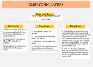
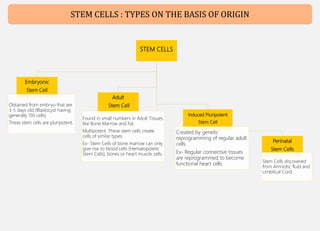
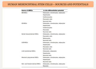
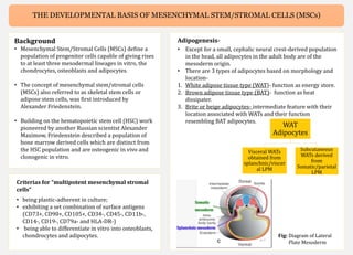
MESENCHYMAL STEM CELLS
By Swapnali Behera
CONTENT
1. Embryonic Layers (Classification)
2. Stem Cells
3. Classification of Stem Cells (On basis of Origin)
4. Human Mesenchymal Stem Cells - Current Trends And Future
Prospective
5. The Developmental Basis of Mesenchymal Stem/ Stromal Cells (MSCs)
6. Trilineage Differentiation of (MSCs) into Osteocytes, Adipocytes and
Chondrocytes
7. Mesenchymal Stem Cell Therapy For Severe Covid-19
EMBRYONIC LAYERS
Embryonic Layers
Parts of body which fall into each
germ layer
Ectoderm
1. Epidermis, hairs, nails, lens of
eye, sebaceous glands, Cornea,
Tooth Enamel, Epithelium of
mouth and nose.
2. Peripheral nervous system,
Adrenal Medulla, Facial
cartilage.
3. Brain, Spinal Cord, Posterior
pituitary, Motor Neurons,
Retina.
Mesoderm
1. Notochord, Kidneys and
gonads.
2. Circulatory system including
heart and spleen.
3.Wall of gut and wall of human
body.
4. Dentine of teeth, Genitourinary
system, Serous membranes,
Adipose tissue, Connective tissue.
Endoderm
1. Epithelial lining of digestive tract
except parts of mouth, pharynx and
terminal part of rectum; lining cells of
all glands which open to digestive
tract, epithelium of auditory tube and
tympanic cavity, the trachea, bronchi,
alveoli of lungs, bladder and part of
the urethra, follicle lining of thyroid
glands and thymus.
2. Form the Pharynx, esophagus,
stomach, small intestine, colon, liver,
pancreas, bladder, epithelial parts of
trachea, bronchi, lungs, thyroid and
parathyroid.
STEM CELLS : GENERAL THINGS
What are Stem Cells?
Stem cells are unspecialized cells of the
human body, having ability to differentiate
into any cell of an organism and having the
ability of self-renewal.
Potency
• No. of possible fates open to a cell.
• Decreases with age and decreass as
parts of organism become specialized
(Except for some Stem Cells)
Potency
Types
Totipotent
All fates are possible
Ex- Zygote
Pluripotent
Can develop into
any of the 3 linages
Multipotent
Gives rise to specific
tissue linages
Ex- Mesenchymal
stem cells
Oligopotent
Few fates possible.
More restricted
than Multipotent
cells.
Why study Stem cells?
1. Increases understanding of how diseases occur- by watching the
growth of stem cells into various organs.
2. Used as Regenerative Medicine- Diseases like Spinal Cord Injuries,
Type 1 diabetes, Amyotrophic Lateral Sclerosis, Alzheimer’s diseases
and lot more.
3. Test of Drugs for Safety and effectiveness.
Types of Stem Cells:
1. Embryonic Stem Cells:-
• Obtained from embryo that are 3-5 days old
(Blastocyst having generally 150 cells)
• Pluripotent.
2. Perinatal Stem Cells:-
• Stem Cells discovered from Amniotic fluid and
Umbilical Cord.
3. Adult Stem Cell:-
• Found in small numbers in Adult Tissues like Bone Marrow and Fat.
• Multipotent. These stem cells create cells of similar types.
• Ex- Stem Cells of bone marrow can only give rise to blood cells
(Hematopoietic Stem Cells), bones or heart muscle cells.
4. Induced Pluripotent Cells;-
• Created by genetic reprogramming of regular adult cells.
• Ex- Regular connective tissues are reprogrammed to become
functional heart cells.
STEM CELLS
Embryonic
Stem Cell
Obtained from embryo that are
3-5 days old (Blastocyst having
generally 150 cells)
These stem cells are pluripotent.
Adult
Stem Cell
Found in small numbers in Adult Tissues
like Bone Marrow and Fat.
Multipotent. These stem cells create
cells of similar types.
Ex- Stem Cells of bone marrow can only
give rise to blood cells (Hematopoietic
Stem Cells), bones or heart muscle cells.
Induced Pluripotent
Stem Cell
Created by genetic
reprogramming of regular adult
cells.
Ex- Regular connective tissues
are reprogrammed to become
functional heart cells.
Perinatal
Stem Cells
Stem Cells discovered
from Amniotic fluid and
Umbilical Cord.
STEM CELLS : TYPES ON THE BASIS OF ORIGIN
HUMAN MESENCHYMAL STEM CELLS -
CURRENT TRENDS AND FUTURE
PROSPECTIVE
Imran Ullah, Raghavendra Baregundi Subbarao and Gyu Jin Rho
Review Article
Article 1. Summary
HUMAN MESENCHYMAL STEM CELLS - CURRENT TRENDS AND FUTURE PROSPECTIVE
Why is it being studied so widely?
They are self-renewable, multipotent,
easily accessible and culturally expandable
in vitro with exceptional genomic stability
and few ethical issues, marking its
importance in cell therapy, regenerative
medicine and tissue repairmen.
Human MSCs
These are the non-haematopoietic,
multipotent stem cells with the capacity to
differentiate into mesodermal lineage such
as osteocytes, adipocytes and chondrocytes
as well ectodermal (neurocytes) and
endodermal lineages (hepatocytes).
Minimum criterias to define hMSCs-
1. These cells should exhibits plastic
adherence.
2. should possess specific set of cell surface
markers, i.e. cluster of differentiation
(CD)73, D90, CD105 and lack expression
of CD14, CD34, CD45 and human
leucocyte antigen-DR & have the ability to
differentiate in vitro into adipocyte,
chondrocyte and osteoblast
Sources to derive hMSCs-
Apart from fetal tissues, MSCs are also found in-
1. Efficient population of MSCs has been reported from bone marrow.
2. MSCs were also isolated from adipose tissue, amniotic fluid, amniotic
membrane, dental tissues, endometrium, limb bud, menstrual blood,
peripheral blood, placenta and fetal membrane, salivary gland, skin
and foreskin, sub-amniotic umbilical cord lining membrane, synovial
fluid and Wharton’s jelly.
Major challenges to hMSCs’ research-
One of the major challenges is to obtain adequate number of cells as
these cells were found to lose their potency during sub-culturing and at
higher passages. Reason? The senescence and aging of MSCs during in
vitro expansion due to the decrease in telomerase activity i.e. shortening
of the telomere length and morphological alterations.
In vitro differentiation potential of hMSCs
hMSCs
Ectoderm
Mesoderm
Endoderm
Linages it can divide into
SOURCES, METHOD OF ISOLATION, AND INITIAL CULTURING OF VARIOUS hMSCs
HUMAN MESENCHYMAL STEM CELLS – SOURCES AND POTENTIALS
HUMAN MESENCHYMAL STEM CELLS - CURRENT TRENDS AND FUTURE PROSPECTIVE
Immunomodulatory features
Due to low expression of MHC I and lack
expression of MHC class II along with co-
stimulatory molecules, like CD80, CD40 and
CD86, MSCs are unable to bring substantial
alloreactivity and these features protects
MSCs from natural killer (NK) cells lysis. The
MSCs therapy might alleviate disease
response by increasing the conversion from
Th2 (T helper cells) response to Th1 cellular
immune response through modulation of
interleukin (IL)-4 and interferon (IFN)-γ
levels in effector T-cells. By the secretion of
suppressors of T-cells development,
inhibitory factors i.e. leukaemia inhibitory
factor (LIF) and IFN-γ enhance
immunomodulatory properties of MSCs.
Moreover, it is observed that human BM-
MSCs were not recognized by NK cells, as
they expressed HLA-DR molecules
Homing of MSCs
• Homing is the term used when cells are delivered to the site of injury.
• Local delivery and homing of cells are found beneficial due to
interaction with the host tissues, accompanied by the secretion of
trophic factors.
• Factors like cells age, culturing conditions, cell passage number and the
delivery method, which influence the homing ability of MSCs to the
injured site.
• While culturing MSCs, Oxygen condition, availability of cytokines and
growth factors supplements in the culture media triggers important
factors which are helpful in the homing of MSCs.
Cryopreservation and Banking-
• For the selection of optimal cryopreservation media, uniform change in
temperature during freezing and thawing, employed freezing device and
long term storage in liquid nitrogen are considered.
• A serum-free media is substantial for the cryopreservation of MSC. Most
recently, human albumin and neuropeptide were used instead of FBS and
MSCs maintained their cell survival and proliferation potential in the
culture conditions.
• Additionally, cryoprotective agents (CPAs) are required for the
cryopreservation media to prevent any freezing damage to cells. A large
number of CPAs are available among which DMSO is the most common
CPAs used in cryopreservation of MSCs.
• But due to it’s toxic nature, patients develop mild complications like
nausea, vomiting, headache, hypertension, diarrhoea and hypotension
and also severe effects like cardiovascular and respiratory issues.
HUMAN MESENCHYMAL STEM CELLS - CURRENT TRENDS AND FUTURE PROSPECTIVE
MSCs in clinical trials
• Currently, there are 463 registered clinical trials in different
clinical phases (phase I, II etc.), evaluating the potential of
MSC-based cell therapy throughout the world.
• Among 463 registered trials, 264 trials are in open status
which is open for recruitment whereas 199 trials are closed;
out of which 106 studies are completed whereas the rest are
in active phases.
• Clinical trials conducted with MSCs showed very less
detrimental effects; however, few of them showed mild
adverse effects.
Cryopreservation and Banking-
• Most recently for tissue cryopreservation, a new
method was introduced using the mixture of 0.05
M glucose, 0.05 M sucrose and 1.5 M ethylene
glycol in phosphate buffer saline, shown
successful isolation and characterization of MSCs
after 3 months of cryopreservation of the tissue.
• For cryopreservation of MSCs, the second
important factor is the freezing temperature rate.
Mostly slow freezing at the rate of 1 ◦C/min is the
optimum rate for MSCs preservation
• current controlled rate freezers (CRFs) are
suitable for controlling temperature, maintaining
the rate of temperature during cryopreservation.
These CRFs can be programmed to find out the
exact temperature which the sample is
experiencing during freezing .
• Despite of these benefits, these CRFs lack the
uniformity of temperature to all vials during
large-scale banking of MSCs.
• Recently more advanced CRF, which provides
unidirectional flow of cryogen to each sample,
were created by Praxair Inc.
Fig: No. of diseases registered under MSCs based cell therapy
THE DEVELOPMENTAL BASIS OF
MESENCHYMAL STEM/STROMAL CELLS
(MSC)
Guojun Sheng
Sheng BMC Developmental Biology (2015)
Article 2. Summary
THE DEVELOPMENTAL BASIS OF MESENCHYMAL STEM/STROMAL CELLS (MSCs)
Background
• Mesenchymal Stem/Stromal Cells (MSCs) define a
population of progenitor cells capable of giving rises
to at least three mesodermal lineages in vitro, the
chondrocytes, osteoblasts and adipocytes.
• The concept of mesenchymal stem/stromal cells
(MSCs) also referred to as skeletal stem cells or
adipose stem cells, was first introduced by
Alexander Friedenstein.
• Building on the hematopoietic stem cell (HSC) work
pioneered by another Russian scientist Alexander
Maximow, Friedenstein described a population of
bone marrow derived cells which are distinct from
the HSC population and are osteogenic in vivo and
clonogenic in vitro.
Adipogenesis-
• Except for a small, cephalic neural crest-derived population
in the head, all adipocytes in the adult body are of the
mesoderm origin.
• There are 3 types of adipocytes based on morphology and
location-
1. White adipose tissue type (WAT)- function as energy store.
2. Brown adipose tissue type (BAT)- function as heat
dissipater.
3. Brite or beige adipocytes- intermediate feature with their
location associated with WATs and their function
resembling BAT adipocytes.
Criterias for “multipotent mesenchymal stromal
cells”
• being plastic-adherent in culture;
• exhibiting a set combination of surface antigens
(CD73+, CD90+, CD105+, CD34-, CD45-, CD11b-,
CD14-, CD19-, CD79a- and HLA-DR-)
• being able to differentiate in vitro into osteoblasts,
chondrocytes and adipocytes.
WAT
Adipocytes
Visceral WATs
obtained from
splanchnic/viscer
al LPM
Subcutaneous
WATs derived
from
Somatic/parietal
LPM
Fig: Diagram of Lateral
Plate Mesoderm
THE DEVELOPMENTAL BASIS OF MESENCHYMAL STEM/STROMAL CELLS (MSCS)
Chondrogenesis and osteogenesis
•These are two separate, but tightly-linked
skeletogenic processes.
• With the exception of the clavicle, all post-cranial
bones form through endochondrial ossification, i.e.,
secretion of bone-specific matrix proteins and
subsequent mineralization of this matrix take place
in a tissue architecture modeled by the
chondrocytes.
• Three mesoderm lineages in development, the axial,
paraxial and lateral plate, are capable of generating
skeletal elements.
• The axial mesoderm gives rise to the embryonic
notochord and the adult nucleus pulposus and
expresses many cartilage-specific markers (e.g.,
type II collagen and aggrecan).
• The paraxial/somitic mesoderm generates all axial
and associated skeletal elements (the vertebrae, ribs
and part of the shoulder girdle), whereas all distal
skeletal elements (bones in the limbs, the pelvic
girdle, the sternum and part of the shoulder girdle)
are derived from the somatic/parietal layer of the
LPM
Fig. Schematic diagram of mesoderm formation and patterning during
vertebrate early development.
A. Mesoderm precursors located in the primitive streak (PS) undergo
epithelial-to-mesenchymal transition (EMT) and migrate between the
ectoderm and endoderm germ layers to their final destinations in a spatially
and temporally coordinated manner (white stippled lines). B. Major
mesoderm lineages (axial, paraxial, intermediate, lateral plate and
extraembryonic) are laid out along the medio-lateral axis of the early
embryo.
Major lineages of the mesoderm germ layer
Prior to the onset of gastrulation which generates an embryo
with three germ layers, mesoderm precursors are specified
molecularly when they are still part of the epiblast. These
mesoderm precursor cells ingress from the epiblast to become
bona fide mesoderm cells through an epithelial to mesenchymal
transition (EMT) process, which takes place in an embryonic
structure called primitive streak. EMT of mesoderm precursors
at the primitive streak proceeds in a temporally and spatially
ordered manner.
THE DEVELOPMENTAL BASIS OF MESENCHYMAL STEM/STROMAL CELLS (MSCS)
The LPM and its somatic and splanchnic layers
•All mesoderm cells undergo at least one round MET
(mesenchymal to epithelial transition) after their
initial EMT, and many undergo several rounds of
subsequent EMT/MET processes before their final
differentiation.
•Nascent LPM cells then polarize to form two
epithelial layers, located-
1. adjacent to the endoderm- splanchnic/visceral
layer
2. adjacent to the ectoderm- somatic/parietal layer.
• The apical side of both epithelial layers faces the
enclosed internal space, the coelomic cavity.
• Cells from the splanchnic layer of the LPM
contribute to nearly the entire cardiovascular
system, including the cardiac and smooth muscles,
endothelial cells, pericytes and HSCs, and to the
mesothelial lining of visceral organs and visceral
adipocytes.
• The somatic layer of the LPM gives rise to the
dermis and hypodermis in the lateral and ventral
body wall, the chondrocytes and osteocytes in all
distal skeletal elements, the vast majority of
subcutaneous adipocytes including those in the
abdominal and gluteofemoral regions.
Fig. Schematic diagram of somatic LPM morphogenesis.
Epithelial-to-mesenchymal transition (EMT) of the epithelial
somatic LPM produces a homogenous mesenchymal cell
population located between the ectoderm and the remaining
LPM epithelium. These mesenchymal cells differentiate into
many mesoderm lineageslineages, including the adipocytes,
chondrocytes and osteoblasts. MSCs are hypothesized to exist
both in the naïve somatic LPM population and in a more
differentiated LPM tissue environment
THE DEVELOPMENTAL BASIS OF MESENCHYMAL STEM/STROMAL CELLS (MSCS)
Mesenchymal stem cells or mesenchymal
stromal cells
• Although most of the cell and tissue types which have
been associated with MSCs come ontologically from
the somatic LPM, many of them do not.
• For example, as target differentiation lineages, the
cardiac and most of the smooth muscles come from
the splanchnic LPM and the skeletal muscles are from
the somitic mesoderm.
• One may therefore argue that the MSCs are a sub-
population of, and are distinct from, a pan-
mesodermal stem cell population, the latter of which
can be viewed as the equivalent of primitive streak-
like mesoderm progenitors in vivo.
Conclusions
This article has outlined the ontological, molecular and
cellular evidence in support of the existence of MSCs in
vivo. The somatic LPM is the most important
mesoderm compartment for the cells and tissues
commonly associated with MSCs. The term
“mesenchymal stem cells” is preferred by this author
and a clear distinction should be made between
somatic LPM-derived MSCs and mesenchymal shaped
stem-like cells derived from other mesodermal
compartments.
Fig: Comparison of MSC-related phenomena in vivo
and in vitro. In vivo, progenitor populations that will
give rise to the adipocyte, chondrocyte and osteoblast
lineages pass through developmental phases that
progressively restrict their fate choices. This is
correlated with progressively limited differentiation
potentials of corresponding stem cell populations
cultured in vitro. ICM: inner cell mass; ESCs: embryonic
stem cells; Epi-SCs: epiblast stem cells; LPM: lateral
plate mesoderm; MSCs: mesenchymal stem cells.
Corresponding stem cell populations for the mesoderm
(pan-mesoderm stem cells) and LPM (pan-lateral plate
mesoderm stem cells) have not yet been reported
TRILINEAGE DIFFERENTIATION OF
MULTIPOTENT HUMAN MESENCHYMAL
STEM CELLS (MSC) INTO OSTEOCYTES,
ADIPOCYTES AND CHONDROCYTES
Merck
Article 3. Summary
Trilineage Differentiation of (MSCs) into Osteocytes, Adipocytes and Chondrocytes
Background
.Mesenchymal stem cells (MSCs) are
fibroblastoid multipotent adult stem cells
isolated from several human tissues.
• MSC originate from the perivascular niche,
a tight network present throughout the
vasculature of the whole body. These
perivascular cells lack endothelial and
hematopoietic markers, e.g. CD31, CD34
and CD45, but express CD146, PDGFR beta,
and alkaline phosphatase.
• MSC express the surface markers CD73,
CD90 and CD105 and stain negative for
CD14 or CD11b, CD34, CD45, CD79α or
CD19, and HLADR.
• The most common and reliable way to
identify a population of MSC is to verify
their multipotency.
• MSC can differentiate into adipocytes,
osteoblasts, myocytes, and chondrocytes in
vivo and in vitro. Transdifferentiation of
MSC into cells of nonmesenchymal origin,
such as hepatocytes, neurons and
pancreatic islet cells, has been observed in
vitro when specific culture conditions and
stimuli are applied.
Osteogenesis Differentiation Protocol
1. Coat the culture vessel. Coat a 6well tissue culture plate with 10 μg/mL human
fibronectin or bovine fibronectin.
2. Seed Mesenchymal Stem Cells. Plate MSCs at 1x10⁵ cells per well in the
fibronectincoated tissue culture plate using MSC Growth Medium 2 (C-28009). Work in
duplicate.
3. Allow Mesenchymal Stem Cells to grow. Allow the cells to reach 8090% confluency.
This will take 2448 hours.
4. Induce Mesenchymal Stem Cells. Induce one of the duplicate samples with MSC
Osteogenic Differentiation Medium (C-28013). Use MSC Growth Medium 2 for the
remaining well as a negative control.
5. Induce Mesenchymal Stem Cells. Incubate for 1214 days. Change the medium every
third day. Be careful not to disturb the cell monolayer.
6. Wash the cells. After differentiation steps are complete, remove the cells from the
incubator and carefully aspirate the medium. Gently wash the cells with Dulbecco’s
phosphate buffered saline (PBS) w/o Ca++/Mg++ (D8537).
7. Fix the cells. Carefully aspirate the PBS and add enough Saccomanno Fixation Solution
to cover the cellular monolayer. After at least 30 min gently aspirate the fixation solution
and wash the cells with distilled water.
8. Stain the cells. Immediately before use, pass the required amount of Alizarin Red S
staining solution (TMS008) through a 0.22 μm Millex PES filter. Carefully aspirate the
distilled water and add enough filtered Alizarin Red S staining solution to cover the
cellular monolayer. Incubate at room temperature in the dark for 1015 min. Monitor
staining progress for 10 min and stop the process when staining intensity is sufficient.
9. Wash the cells. Carefully aspirate the Alizarin Red S staining solution and wash the cell
monolayer four times with 1 mL distilled water. Carefully aspirate the distilled water
and add PBS.
10. Analyze the cells Analyze the sample immediately, as the dye may bleed upon
prolonged storage without embedding. Undifferentiated MSC (without extracellular
calcium deposits) are colorless/slightly purple, whereas MSC derived osteoblasts (with
extracellular calcium deposits) stain bright orangered
Trilineage Differentiation of (MSCs) into Osteocytes, Adipocytes and Chondrocytes
Adipogenesis Differentiation Protocol
1. Coat the culture vessel. Coat a 6well tissue culture plate with 10 μg/mL human fibronectin or bovine fibronectin
according to the instruction manual.
2. Seed the Mesenchymal Stem Cells. In a fibronectin coated 6well tissue culture plate, plate 1 x 10⁵ MSC per well using
MSC Growth Medium 2 (C-28009). Work in duplicate.
3. Allow the Mesenchymal Stem Cells grow. Allow the cells to reach 8090% confluency. This will take 2448 hours.
4. Induce the Mesenchymal Stem Cells. Induce one of the duplicate samples with MSC Adipogenic Differentiation Medium
2 (C-28016). Use MSC Growth Medium 2 for the remaining well as a negative control.
5. Differentiation of the induced Mesenchymal Stem Cells. Incubate for 1214 days. Change the medium every third day
taking care not to disturb the cell monolayer.
6. Wash the cells. After differentiation steps are complete, remove the cells from the incubator and carefully aspirate the
medium. Gently wash the cells with Dulbecco’s phosphate buffered saline (PBS) w/o Ca++/Mg++ (D8537).
7. Fix the cells. Carefully aspirate the PBS (D8537) and add enough Saccomanno Fixation Solution to cover the cell
monolayer. Incubate at room temperature for at least 30 min.
8. Wash the cells. Carefully aspirate the fixation buffer and wash the cell monolayer with distilled water. Gently aspirate
the water and add enough 60% isopropanol to cover the cell monolayer. Incubate at room temperature for 5 min.
9. Stain the cells. Carefully aspirate the 60% isopropanol and add enough OilRedO staining solution (O1391) to cover the
cell monolayer. Incubate at room temperature for 1015 min.
10.Wash the cells. Carefully aspirate the staining solution and wash the cell monolayer several times with distilled water
until the water is clear. Blot the vessel containing the stained cells upside down on a paper towel to remove as much
water as possible.
11.Analyze the cells. Cover with PBS and analyse the stained samples promptly as the dye tends to fade upon prolonged
light exposure. Intracellular lipid vesicles in mature adipocytes will be stained bright red.
Trilineage Differentiation of (MSCs) into Osteocytes, Adipocytes and Chondrocytes
Chondrogenesis Differentiation Protocol
1. Preparation of the Negative Control Medium. The negative control medium is Dulbecco’s Modified Eagle’s Medium
(DMEM, lowglucose) with 2 mM L-glutamine and 10% fetal bovine serum.
2. Seed Mesenchymal Stem Cells. Plate MSC at 2 x 10⁵ cells per well in a 96well Ubottom suspension culture plate using
the negative control medium. Work in duplicate.
3. MSCspheroid formation. Spheroids will spontaneously form within 2448 hours of incubation.
4. Induce MSCspheroids. Induce one of the duplicate samples with MSC Chondrogenic Differentiation Medium (C-28012).
Use the negative control medium for the remaining wells.
5. Differentiate induced MSC spheroids. Incubate for 21 days. Change the medium every third day taking care not to
aspirate the spheroids.
6. Wash the cartilage spheroids. After differentiation steps are complete, remove the cells from the incubator and
carefully aspirate the medium. Gently wash the spheroids with Dulbecco’s phosphatebuffered saline (PBS) w/o
Ca++/Mg++ (D8537).
7. Fixation of the cartilage spheroids. Carefully aspirate the PBS. Add enough Saccomanno Fixation Solution to cover the
cartilage spheroids. Incubate at room temperature for 3 hours.
8. Wash the cartilage spheroids. Carefully aspirate the fixative and wash the spheroids twice with distilled water.
9. Stain the cells. Immediately before use, pass the required amount of Alcian Blue staining solution (TMS010) through a
0.22 μm Millex PES filter. Carefully aspirate the distilled water and add enough filtered Alcian Blue staining solution to
generously cover the cartilage spheroids, as some evaporation will occur. Incubate in the dark for 45 minutes at room
temperature.
10.Wash the cells. Carefully aspirate the Alcian Blue staining solution and wash the cartilage spheroids with the destaining
solution for 10 min. Repeat the wash step twice. Carefully aspirate the destaining solution and add PBS.
11.Analyze the cartilage spheroids. Cartilage will be stained an intense dark blue, whereas other tissue will, at most, stain
light blue.
MESENCHYMAL STEM CELL
THERAPY FOR SEVERE COVID-19
Lei Shi1 , Lifeng Wang1 , Ruonan Xu1 , Chao Zhang 1 , Yunbo Xie1 , Kai Liu1 , Tiantian Li1 ,
Wei Hu1 , Cheng Zhen1 and Fu-Sheng Wang 1
Signal Transduction and Targeted Therapy
Article 4. Summary
MESENCHYMAL STEM CELL THERAPY FOR SEVERE COVID-19
Background
In severe cases, acute SARS-CoV-2 infection
leads to immune disorders and damage to
both the adaptive and innate immune
responses. Mesenchymal stem cells (MSCs)
serve as a therapeutic option may regulate
the over-activated inflammatory response
and promote recovery of lung damage.
The host’s innate and adaptive immune
responses, especially specific adaptive
immunity to SARS-CoV-2, play an essential
role in controlling viral infection. Excessive
inflammation and the cytokine storm cause
organ damage on being affected by severe
COVID-19. Therefore, aside from direct
antiviral treatment and supplemental oxygen
therapy for COVID-19 cases,
immunomodulatory therapeutic strategies
may potentially prevent disease progression
and rescue COVID-19 patients. Many
immunotherapeutic approaches have been
used for COVID-19, including glucocorticoid
therapy, convalescent plasma therapy, and
anti-interleukin (IL-6) receptor antibody
therapy. However, their side effects and
variable treatment efficacy are still under
study.
Use of MSCs
The safety and efficacy of mesenchymal stem cell (MSC) therapies have
been recently illustrated in clinical trials, such as immune-mediated
inflammatory diseases as systemic lupus erythematosus12 and graft-
versus-host disease (GVHD).
To date, more than 60 stem-cell clinical trials for COVID-19 therapy have
been registered at ClinicalTrials.gov.
Rationale of Stem Cell Therapy
Pathology and pathogenesis of COVID-19
• Bilateral diffuse alveolar injury was observed in the lungs, accompanied
by cellular fibrous mucinous exudates and ARDS. Interstitial
mononuclear inflammatory infiltration, dominated by lymphocytes, was
widespread in both lungs.
• During SARS-CoV-2 infection, the proportions of natural killer (NK) cells,
CD4 + T cells, and CD8 + T cells significantly decrease.
• The proportions of dendritic cell compartments are significantly
decreased, while the IFN response profiles are elevated. In addition, γδ T
cells, NK cells, and CD16 + monocytes are significantly activated.
Immunomodulation and therapeutic principles of MSCs
• MSCs have differentiational and regenerative properties and can secrete
hepatocyte growth factor, vascular endothelial growth factor, and
keratinocyte growth factor to promote the regeneration of type II
alveolar epithelial cells.
MESENCHYMAL STEM CELL THERAPY FOR SEVERE COVID-19
Rationale of Stem Cell Therapy
Immunomodulation and therapeutic principles of
MSCs
• MSCs can be attracted to sites of inflammation by
different chemokines and exert the potential to
modulate the functions of various immunocytes— such
as NK cells, dendritic cells, B cells, T cells, neutrophils,
and macrophages—through direct contact and
paracrine effects.
• MSC treatment has been observed to reduce pulmonary
lesions and inhibit the inflammatory response induced
by influenza virus infection.
• Thus, MSCs may offer a therapeutic option for patients
with severe or critical COVID-19, potentially
contributing to recovery from lung damage, suppressing
the over-activated inflammatory response, and
influencing the progression of pulmonary fibrosis.
Fig: Proposed mechanisms for MSC action in patients with
severe COVID-19. At first, SARS-CoV-2 primarily occupies the
respiratory tract including the lung; the infiltration of
immune cells (neutrophils, monocytes/macrophages, NK,
CD4 + T, CD8 + T, Th17, and B cells) increases; then cytokine
storms (including IFN-α, IL-1, IL-6 and TNF-α) occur. Hyaline
membrane formation, the release of cellular fibromyxoid
exudates, and pneumocyte desquamation are also observed.
After stem-cell infusion, the number of infiltrated immune
cells decreases significantly, and the damaged lung tissue is
repaired. MSCs play a role in regeneration and immune
regulation.
MESENCHYMAL STEM CELL THERAPY FOR SEVERE COVID-19
CLINICAL TRIALS OF MSC THERAPIES FOR COVID-
19
Phase-1 trials
• Zhao et al. - Intravenous administration of MSCs could
improve the clinical outcome of COVID-19 patients.
• Li et al.- Trial using menstrual blood-derived MSCs (MB-
MSCs) for severe patients, which indicated that MSC
transplantation might serve as an alternative option for
treating COVID-19, particularly in critical patients.
• Umbilical cord MSC (UC-MSC) infusion in moderate or
severe COVID-19 patients is safe.
• Mirakaj et al. - Patients who underwent MSC treatment,
compared against control patients, experienced
significantly lower lung damage (Murray score) upon
discharge, had a higher survival rate to discharge, and
had better recovery of pulmonary functions.
• Wu et al.- Lung fibrotic lesions were decreased by
transfusion of hESCIMRCs in patients with pulmonary
fibrosis.
Phase-2 trials
• In Wuhan, China, 101 patients with severe COVID-19
were recruited and assigned randomly at a 2:1 ratio to
receive UC-MSCs or a placebo, respectively. Compared
with the placebo, UC-MSC transfusion exerted a
improvement in lung lesion volume from baseline to day
28.
• Lanzoni et al. performed another double-blind,
randomized, controlled trial and found that UC-MSC
infusions significantly decreased cytokine levels at day 6
and improved survival in COVID-19 patients with ARDS.
CURRENT CHALLENGES
• Need to confirm its efficacy in controlling pulmonary
fibrosis by means of a multi-cohort, randomized,
controlled trial with a long-term follow-up.
• Standardize MSC products and their clinical application
protocols.
• Key areas to optimize include the type of MSCs to be
transfused, whether the cells are fresh or frozen (and their
viability before infusion), the administration regimen
(including dosage, interval, and number of cycles), the
route of administration, and delivery of MSCs within a
specific phase of the COVID-19 disease.
• MSC therapies provide a promising and challenging
opportunity for patients with COVID-19. Phase-3 trials are
still necessary for further evaluation of the efficacy and
associated mechanisms of MSC treatment.
Mesenchymal Stem Cells.pptx

Mesenchymal Stem Cells.pptx

  • 1.
  • 2.
    CONTENT 1. Embryonic Layers(Classification) 2. Stem Cells 3. Classification of Stem Cells (On basis of Origin) 4. Human Mesenchymal Stem Cells - Current Trends And Future Prospective 5. The Developmental Basis of Mesenchymal Stem/ Stromal Cells (MSCs) 6. Trilineage Differentiation of (MSCs) into Osteocytes, Adipocytes and Chondrocytes 7. Mesenchymal Stem Cell Therapy For Severe Covid-19
  • 3.
    EMBRYONIC LAYERS Embryonic Layers Partsof body which fall into each germ layer Ectoderm 1. Epidermis, hairs, nails, lens of eye, sebaceous glands, Cornea, Tooth Enamel, Epithelium of mouth and nose. 2. Peripheral nervous system, Adrenal Medulla, Facial cartilage. 3. Brain, Spinal Cord, Posterior pituitary, Motor Neurons, Retina. Mesoderm 1. Notochord, Kidneys and gonads. 2. Circulatory system including heart and spleen. 3.Wall of gut and wall of human body. 4. Dentine of teeth, Genitourinary system, Serous membranes, Adipose tissue, Connective tissue. Endoderm 1. Epithelial lining of digestive tract except parts of mouth, pharynx and terminal part of rectum; lining cells of all glands which open to digestive tract, epithelium of auditory tube and tympanic cavity, the trachea, bronchi, alveoli of lungs, bladder and part of the urethra, follicle lining of thyroid glands and thymus. 2. Form the Pharynx, esophagus, stomach, small intestine, colon, liver, pancreas, bladder, epithelial parts of trachea, bronchi, lungs, thyroid and parathyroid.
  • 4.
    STEM CELLS :GENERAL THINGS What are Stem Cells? Stem cells are unspecialized cells of the human body, having ability to differentiate into any cell of an organism and having the ability of self-renewal. Potency • No. of possible fates open to a cell. • Decreases with age and decreass as parts of organism become specialized (Except for some Stem Cells) Potency Types Totipotent All fates are possible Ex- Zygote Pluripotent Can develop into any of the 3 linages Multipotent Gives rise to specific tissue linages Ex- Mesenchymal stem cells Oligopotent Few fates possible. More restricted than Multipotent cells. Why study Stem cells? 1. Increases understanding of how diseases occur- by watching the growth of stem cells into various organs. 2. Used as Regenerative Medicine- Diseases like Spinal Cord Injuries, Type 1 diabetes, Amyotrophic Lateral Sclerosis, Alzheimer’s diseases and lot more. 3. Test of Drugs for Safety and effectiveness. Types of Stem Cells: 1. Embryonic Stem Cells:- • Obtained from embryo that are 3-5 days old (Blastocyst having generally 150 cells) • Pluripotent. 2. Perinatal Stem Cells:- • Stem Cells discovered from Amniotic fluid and Umbilical Cord. 3. Adult Stem Cell:- • Found in small numbers in Adult Tissues like Bone Marrow and Fat. • Multipotent. These stem cells create cells of similar types. • Ex- Stem Cells of bone marrow can only give rise to blood cells (Hematopoietic Stem Cells), bones or heart muscle cells. 4. Induced Pluripotent Cells;- • Created by genetic reprogramming of regular adult cells. • Ex- Regular connective tissues are reprogrammed to become functional heart cells.
  • 5.
    STEM CELLS Embryonic Stem Cell Obtainedfrom embryo that are 3-5 days old (Blastocyst having generally 150 cells) These stem cells are pluripotent. Adult Stem Cell Found in small numbers in Adult Tissues like Bone Marrow and Fat. Multipotent. These stem cells create cells of similar types. Ex- Stem Cells of bone marrow can only give rise to blood cells (Hematopoietic Stem Cells), bones or heart muscle cells. Induced Pluripotent Stem Cell Created by genetic reprogramming of regular adult cells. Ex- Regular connective tissues are reprogrammed to become functional heart cells. Perinatal Stem Cells Stem Cells discovered from Amniotic fluid and Umbilical Cord. STEM CELLS : TYPES ON THE BASIS OF ORIGIN
  • 6.
    HUMAN MESENCHYMAL STEMCELLS - CURRENT TRENDS AND FUTURE PROSPECTIVE Imran Ullah, Raghavendra Baregundi Subbarao and Gyu Jin Rho Review Article Article 1. Summary
  • 7.
    HUMAN MESENCHYMAL STEMCELLS - CURRENT TRENDS AND FUTURE PROSPECTIVE Why is it being studied so widely? They are self-renewable, multipotent, easily accessible and culturally expandable in vitro with exceptional genomic stability and few ethical issues, marking its importance in cell therapy, regenerative medicine and tissue repairmen. Human MSCs These are the non-haematopoietic, multipotent stem cells with the capacity to differentiate into mesodermal lineage such as osteocytes, adipocytes and chondrocytes as well ectodermal (neurocytes) and endodermal lineages (hepatocytes). Minimum criterias to define hMSCs- 1. These cells should exhibits plastic adherence. 2. should possess specific set of cell surface markers, i.e. cluster of differentiation (CD)73, D90, CD105 and lack expression of CD14, CD34, CD45 and human leucocyte antigen-DR & have the ability to differentiate in vitro into adipocyte, chondrocyte and osteoblast Sources to derive hMSCs- Apart from fetal tissues, MSCs are also found in- 1. Efficient population of MSCs has been reported from bone marrow. 2. MSCs were also isolated from adipose tissue, amniotic fluid, amniotic membrane, dental tissues, endometrium, limb bud, menstrual blood, peripheral blood, placenta and fetal membrane, salivary gland, skin and foreskin, sub-amniotic umbilical cord lining membrane, synovial fluid and Wharton’s jelly. Major challenges to hMSCs’ research- One of the major challenges is to obtain adequate number of cells as these cells were found to lose their potency during sub-culturing and at higher passages. Reason? The senescence and aging of MSCs during in vitro expansion due to the decrease in telomerase activity i.e. shortening of the telomere length and morphological alterations. In vitro differentiation potential of hMSCs hMSCs Ectoderm Mesoderm Endoderm Linages it can divide into
  • 8.
    SOURCES, METHOD OFISOLATION, AND INITIAL CULTURING OF VARIOUS hMSCs
  • 9.
    HUMAN MESENCHYMAL STEMCELLS – SOURCES AND POTENTIALS
  • 10.
    HUMAN MESENCHYMAL STEMCELLS - CURRENT TRENDS AND FUTURE PROSPECTIVE Immunomodulatory features Due to low expression of MHC I and lack expression of MHC class II along with co- stimulatory molecules, like CD80, CD40 and CD86, MSCs are unable to bring substantial alloreactivity and these features protects MSCs from natural killer (NK) cells lysis. The MSCs therapy might alleviate disease response by increasing the conversion from Th2 (T helper cells) response to Th1 cellular immune response through modulation of interleukin (IL)-4 and interferon (IFN)-γ levels in effector T-cells. By the secretion of suppressors of T-cells development, inhibitory factors i.e. leukaemia inhibitory factor (LIF) and IFN-γ enhance immunomodulatory properties of MSCs. Moreover, it is observed that human BM- MSCs were not recognized by NK cells, as they expressed HLA-DR molecules Homing of MSCs • Homing is the term used when cells are delivered to the site of injury. • Local delivery and homing of cells are found beneficial due to interaction with the host tissues, accompanied by the secretion of trophic factors. • Factors like cells age, culturing conditions, cell passage number and the delivery method, which influence the homing ability of MSCs to the injured site. • While culturing MSCs, Oxygen condition, availability of cytokines and growth factors supplements in the culture media triggers important factors which are helpful in the homing of MSCs. Cryopreservation and Banking- • For the selection of optimal cryopreservation media, uniform change in temperature during freezing and thawing, employed freezing device and long term storage in liquid nitrogen are considered. • A serum-free media is substantial for the cryopreservation of MSC. Most recently, human albumin and neuropeptide were used instead of FBS and MSCs maintained their cell survival and proliferation potential in the culture conditions. • Additionally, cryoprotective agents (CPAs) are required for the cryopreservation media to prevent any freezing damage to cells. A large number of CPAs are available among which DMSO is the most common CPAs used in cryopreservation of MSCs. • But due to it’s toxic nature, patients develop mild complications like nausea, vomiting, headache, hypertension, diarrhoea and hypotension and also severe effects like cardiovascular and respiratory issues.
  • 11.
    HUMAN MESENCHYMAL STEMCELLS - CURRENT TRENDS AND FUTURE PROSPECTIVE MSCs in clinical trials • Currently, there are 463 registered clinical trials in different clinical phases (phase I, II etc.), evaluating the potential of MSC-based cell therapy throughout the world. • Among 463 registered trials, 264 trials are in open status which is open for recruitment whereas 199 trials are closed; out of which 106 studies are completed whereas the rest are in active phases. • Clinical trials conducted with MSCs showed very less detrimental effects; however, few of them showed mild adverse effects. Cryopreservation and Banking- • Most recently for tissue cryopreservation, a new method was introduced using the mixture of 0.05 M glucose, 0.05 M sucrose and 1.5 M ethylene glycol in phosphate buffer saline, shown successful isolation and characterization of MSCs after 3 months of cryopreservation of the tissue. • For cryopreservation of MSCs, the second important factor is the freezing temperature rate. Mostly slow freezing at the rate of 1 ◦C/min is the optimum rate for MSCs preservation • current controlled rate freezers (CRFs) are suitable for controlling temperature, maintaining the rate of temperature during cryopreservation. These CRFs can be programmed to find out the exact temperature which the sample is experiencing during freezing . • Despite of these benefits, these CRFs lack the uniformity of temperature to all vials during large-scale banking of MSCs. • Recently more advanced CRF, which provides unidirectional flow of cryogen to each sample, were created by Praxair Inc. Fig: No. of diseases registered under MSCs based cell therapy
  • 12.
    THE DEVELOPMENTAL BASISOF MESENCHYMAL STEM/STROMAL CELLS (MSC) Guojun Sheng Sheng BMC Developmental Biology (2015) Article 2. Summary
  • 13.
    THE DEVELOPMENTAL BASISOF MESENCHYMAL STEM/STROMAL CELLS (MSCs) Background • Mesenchymal Stem/Stromal Cells (MSCs) define a population of progenitor cells capable of giving rises to at least three mesodermal lineages in vitro, the chondrocytes, osteoblasts and adipocytes. • The concept of mesenchymal stem/stromal cells (MSCs) also referred to as skeletal stem cells or adipose stem cells, was first introduced by Alexander Friedenstein. • Building on the hematopoietic stem cell (HSC) work pioneered by another Russian scientist Alexander Maximow, Friedenstein described a population of bone marrow derived cells which are distinct from the HSC population and are osteogenic in vivo and clonogenic in vitro. Adipogenesis- • Except for a small, cephalic neural crest-derived population in the head, all adipocytes in the adult body are of the mesoderm origin. • There are 3 types of adipocytes based on morphology and location- 1. White adipose tissue type (WAT)- function as energy store. 2. Brown adipose tissue type (BAT)- function as heat dissipater. 3. Brite or beige adipocytes- intermediate feature with their location associated with WATs and their function resembling BAT adipocytes. Criterias for “multipotent mesenchymal stromal cells” • being plastic-adherent in culture; • exhibiting a set combination of surface antigens (CD73+, CD90+, CD105+, CD34-, CD45-, CD11b-, CD14-, CD19-, CD79a- and HLA-DR-) • being able to differentiate in vitro into osteoblasts, chondrocytes and adipocytes. WAT Adipocytes Visceral WATs obtained from splanchnic/viscer al LPM Subcutaneous WATs derived from Somatic/parietal LPM Fig: Diagram of Lateral Plate Mesoderm
  • 14.
    THE DEVELOPMENTAL BASISOF MESENCHYMAL STEM/STROMAL CELLS (MSCS) Chondrogenesis and osteogenesis •These are two separate, but tightly-linked skeletogenic processes. • With the exception of the clavicle, all post-cranial bones form through endochondrial ossification, i.e., secretion of bone-specific matrix proteins and subsequent mineralization of this matrix take place in a tissue architecture modeled by the chondrocytes. • Three mesoderm lineages in development, the axial, paraxial and lateral plate, are capable of generating skeletal elements. • The axial mesoderm gives rise to the embryonic notochord and the adult nucleus pulposus and expresses many cartilage-specific markers (e.g., type II collagen and aggrecan). • The paraxial/somitic mesoderm generates all axial and associated skeletal elements (the vertebrae, ribs and part of the shoulder girdle), whereas all distal skeletal elements (bones in the limbs, the pelvic girdle, the sternum and part of the shoulder girdle) are derived from the somatic/parietal layer of the LPM Fig. Schematic diagram of mesoderm formation and patterning during vertebrate early development. A. Mesoderm precursors located in the primitive streak (PS) undergo epithelial-to-mesenchymal transition (EMT) and migrate between the ectoderm and endoderm germ layers to their final destinations in a spatially and temporally coordinated manner (white stippled lines). B. Major mesoderm lineages (axial, paraxial, intermediate, lateral plate and extraembryonic) are laid out along the medio-lateral axis of the early embryo. Major lineages of the mesoderm germ layer Prior to the onset of gastrulation which generates an embryo with three germ layers, mesoderm precursors are specified molecularly when they are still part of the epiblast. These mesoderm precursor cells ingress from the epiblast to become bona fide mesoderm cells through an epithelial to mesenchymal transition (EMT) process, which takes place in an embryonic structure called primitive streak. EMT of mesoderm precursors at the primitive streak proceeds in a temporally and spatially ordered manner.
  • 15.
    THE DEVELOPMENTAL BASISOF MESENCHYMAL STEM/STROMAL CELLS (MSCS) The LPM and its somatic and splanchnic layers •All mesoderm cells undergo at least one round MET (mesenchymal to epithelial transition) after their initial EMT, and many undergo several rounds of subsequent EMT/MET processes before their final differentiation. •Nascent LPM cells then polarize to form two epithelial layers, located- 1. adjacent to the endoderm- splanchnic/visceral layer 2. adjacent to the ectoderm- somatic/parietal layer. • The apical side of both epithelial layers faces the enclosed internal space, the coelomic cavity. • Cells from the splanchnic layer of the LPM contribute to nearly the entire cardiovascular system, including the cardiac and smooth muscles, endothelial cells, pericytes and HSCs, and to the mesothelial lining of visceral organs and visceral adipocytes. • The somatic layer of the LPM gives rise to the dermis and hypodermis in the lateral and ventral body wall, the chondrocytes and osteocytes in all distal skeletal elements, the vast majority of subcutaneous adipocytes including those in the abdominal and gluteofemoral regions. Fig. Schematic diagram of somatic LPM morphogenesis. Epithelial-to-mesenchymal transition (EMT) of the epithelial somatic LPM produces a homogenous mesenchymal cell population located between the ectoderm and the remaining LPM epithelium. These mesenchymal cells differentiate into many mesoderm lineageslineages, including the adipocytes, chondrocytes and osteoblasts. MSCs are hypothesized to exist both in the naïve somatic LPM population and in a more differentiated LPM tissue environment
  • 16.
    THE DEVELOPMENTAL BASISOF MESENCHYMAL STEM/STROMAL CELLS (MSCS) Mesenchymal stem cells or mesenchymal stromal cells • Although most of the cell and tissue types which have been associated with MSCs come ontologically from the somatic LPM, many of them do not. • For example, as target differentiation lineages, the cardiac and most of the smooth muscles come from the splanchnic LPM and the skeletal muscles are from the somitic mesoderm. • One may therefore argue that the MSCs are a sub- population of, and are distinct from, a pan- mesodermal stem cell population, the latter of which can be viewed as the equivalent of primitive streak- like mesoderm progenitors in vivo. Conclusions This article has outlined the ontological, molecular and cellular evidence in support of the existence of MSCs in vivo. The somatic LPM is the most important mesoderm compartment for the cells and tissues commonly associated with MSCs. The term “mesenchymal stem cells” is preferred by this author and a clear distinction should be made between somatic LPM-derived MSCs and mesenchymal shaped stem-like cells derived from other mesodermal compartments. Fig: Comparison of MSC-related phenomena in vivo and in vitro. In vivo, progenitor populations that will give rise to the adipocyte, chondrocyte and osteoblast lineages pass through developmental phases that progressively restrict their fate choices. This is correlated with progressively limited differentiation potentials of corresponding stem cell populations cultured in vitro. ICM: inner cell mass; ESCs: embryonic stem cells; Epi-SCs: epiblast stem cells; LPM: lateral plate mesoderm; MSCs: mesenchymal stem cells. Corresponding stem cell populations for the mesoderm (pan-mesoderm stem cells) and LPM (pan-lateral plate mesoderm stem cells) have not yet been reported
  • 17.
    TRILINEAGE DIFFERENTIATION OF MULTIPOTENTHUMAN MESENCHYMAL STEM CELLS (MSC) INTO OSTEOCYTES, ADIPOCYTES AND CHONDROCYTES Merck Article 3. Summary
  • 18.
    Trilineage Differentiation of(MSCs) into Osteocytes, Adipocytes and Chondrocytes Background .Mesenchymal stem cells (MSCs) are fibroblastoid multipotent adult stem cells isolated from several human tissues. • MSC originate from the perivascular niche, a tight network present throughout the vasculature of the whole body. These perivascular cells lack endothelial and hematopoietic markers, e.g. CD31, CD34 and CD45, but express CD146, PDGFR beta, and alkaline phosphatase. • MSC express the surface markers CD73, CD90 and CD105 and stain negative for CD14 or CD11b, CD34, CD45, CD79α or CD19, and HLADR. • The most common and reliable way to identify a population of MSC is to verify their multipotency. • MSC can differentiate into adipocytes, osteoblasts, myocytes, and chondrocytes in vivo and in vitro. Transdifferentiation of MSC into cells of nonmesenchymal origin, such as hepatocytes, neurons and pancreatic islet cells, has been observed in vitro when specific culture conditions and stimuli are applied. Osteogenesis Differentiation Protocol 1. Coat the culture vessel. Coat a 6well tissue culture plate with 10 μg/mL human fibronectin or bovine fibronectin. 2. Seed Mesenchymal Stem Cells. Plate MSCs at 1x10⁵ cells per well in the fibronectincoated tissue culture plate using MSC Growth Medium 2 (C-28009). Work in duplicate. 3. Allow Mesenchymal Stem Cells to grow. Allow the cells to reach 8090% confluency. This will take 2448 hours. 4. Induce Mesenchymal Stem Cells. Induce one of the duplicate samples with MSC Osteogenic Differentiation Medium (C-28013). Use MSC Growth Medium 2 for the remaining well as a negative control. 5. Induce Mesenchymal Stem Cells. Incubate for 1214 days. Change the medium every third day. Be careful not to disturb the cell monolayer. 6. Wash the cells. After differentiation steps are complete, remove the cells from the incubator and carefully aspirate the medium. Gently wash the cells with Dulbecco’s phosphate buffered saline (PBS) w/o Ca++/Mg++ (D8537). 7. Fix the cells. Carefully aspirate the PBS and add enough Saccomanno Fixation Solution to cover the cellular monolayer. After at least 30 min gently aspirate the fixation solution and wash the cells with distilled water. 8. Stain the cells. Immediately before use, pass the required amount of Alizarin Red S staining solution (TMS008) through a 0.22 μm Millex PES filter. Carefully aspirate the distilled water and add enough filtered Alizarin Red S staining solution to cover the cellular monolayer. Incubate at room temperature in the dark for 1015 min. Monitor staining progress for 10 min and stop the process when staining intensity is sufficient. 9. Wash the cells. Carefully aspirate the Alizarin Red S staining solution and wash the cell monolayer four times with 1 mL distilled water. Carefully aspirate the distilled water and add PBS. 10. Analyze the cells Analyze the sample immediately, as the dye may bleed upon prolonged storage without embedding. Undifferentiated MSC (without extracellular calcium deposits) are colorless/slightly purple, whereas MSC derived osteoblasts (with extracellular calcium deposits) stain bright orangered
  • 19.
    Trilineage Differentiation of(MSCs) into Osteocytes, Adipocytes and Chondrocytes Adipogenesis Differentiation Protocol 1. Coat the culture vessel. Coat a 6well tissue culture plate with 10 μg/mL human fibronectin or bovine fibronectin according to the instruction manual. 2. Seed the Mesenchymal Stem Cells. In a fibronectin coated 6well tissue culture plate, plate 1 x 10⁵ MSC per well using MSC Growth Medium 2 (C-28009). Work in duplicate. 3. Allow the Mesenchymal Stem Cells grow. Allow the cells to reach 8090% confluency. This will take 2448 hours. 4. Induce the Mesenchymal Stem Cells. Induce one of the duplicate samples with MSC Adipogenic Differentiation Medium 2 (C-28016). Use MSC Growth Medium 2 for the remaining well as a negative control. 5. Differentiation of the induced Mesenchymal Stem Cells. Incubate for 1214 days. Change the medium every third day taking care not to disturb the cell monolayer. 6. Wash the cells. After differentiation steps are complete, remove the cells from the incubator and carefully aspirate the medium. Gently wash the cells with Dulbecco’s phosphate buffered saline (PBS) w/o Ca++/Mg++ (D8537). 7. Fix the cells. Carefully aspirate the PBS (D8537) and add enough Saccomanno Fixation Solution to cover the cell monolayer. Incubate at room temperature for at least 30 min. 8. Wash the cells. Carefully aspirate the fixation buffer and wash the cell monolayer with distilled water. Gently aspirate the water and add enough 60% isopropanol to cover the cell monolayer. Incubate at room temperature for 5 min. 9. Stain the cells. Carefully aspirate the 60% isopropanol and add enough OilRedO staining solution (O1391) to cover the cell monolayer. Incubate at room temperature for 1015 min. 10.Wash the cells. Carefully aspirate the staining solution and wash the cell monolayer several times with distilled water until the water is clear. Blot the vessel containing the stained cells upside down on a paper towel to remove as much water as possible. 11.Analyze the cells. Cover with PBS and analyse the stained samples promptly as the dye tends to fade upon prolonged light exposure. Intracellular lipid vesicles in mature adipocytes will be stained bright red.
  • 20.
    Trilineage Differentiation of(MSCs) into Osteocytes, Adipocytes and Chondrocytes Chondrogenesis Differentiation Protocol 1. Preparation of the Negative Control Medium. The negative control medium is Dulbecco’s Modified Eagle’s Medium (DMEM, lowglucose) with 2 mM L-glutamine and 10% fetal bovine serum. 2. Seed Mesenchymal Stem Cells. Plate MSC at 2 x 10⁵ cells per well in a 96well Ubottom suspension culture plate using the negative control medium. Work in duplicate. 3. MSCspheroid formation. Spheroids will spontaneously form within 2448 hours of incubation. 4. Induce MSCspheroids. Induce one of the duplicate samples with MSC Chondrogenic Differentiation Medium (C-28012). Use the negative control medium for the remaining wells. 5. Differentiate induced MSC spheroids. Incubate for 21 days. Change the medium every third day taking care not to aspirate the spheroids. 6. Wash the cartilage spheroids. After differentiation steps are complete, remove the cells from the incubator and carefully aspirate the medium. Gently wash the spheroids with Dulbecco’s phosphatebuffered saline (PBS) w/o Ca++/Mg++ (D8537). 7. Fixation of the cartilage spheroids. Carefully aspirate the PBS. Add enough Saccomanno Fixation Solution to cover the cartilage spheroids. Incubate at room temperature for 3 hours. 8. Wash the cartilage spheroids. Carefully aspirate the fixative and wash the spheroids twice with distilled water. 9. Stain the cells. Immediately before use, pass the required amount of Alcian Blue staining solution (TMS010) through a 0.22 μm Millex PES filter. Carefully aspirate the distilled water and add enough filtered Alcian Blue staining solution to generously cover the cartilage spheroids, as some evaporation will occur. Incubate in the dark for 45 minutes at room temperature. 10.Wash the cells. Carefully aspirate the Alcian Blue staining solution and wash the cartilage spheroids with the destaining solution for 10 min. Repeat the wash step twice. Carefully aspirate the destaining solution and add PBS. 11.Analyze the cartilage spheroids. Cartilage will be stained an intense dark blue, whereas other tissue will, at most, stain light blue.
  • 21.
    MESENCHYMAL STEM CELL THERAPYFOR SEVERE COVID-19 Lei Shi1 , Lifeng Wang1 , Ruonan Xu1 , Chao Zhang 1 , Yunbo Xie1 , Kai Liu1 , Tiantian Li1 , Wei Hu1 , Cheng Zhen1 and Fu-Sheng Wang 1 Signal Transduction and Targeted Therapy Article 4. Summary
  • 22.
    MESENCHYMAL STEM CELLTHERAPY FOR SEVERE COVID-19 Background In severe cases, acute SARS-CoV-2 infection leads to immune disorders and damage to both the adaptive and innate immune responses. Mesenchymal stem cells (MSCs) serve as a therapeutic option may regulate the over-activated inflammatory response and promote recovery of lung damage. The host’s innate and adaptive immune responses, especially specific adaptive immunity to SARS-CoV-2, play an essential role in controlling viral infection. Excessive inflammation and the cytokine storm cause organ damage on being affected by severe COVID-19. Therefore, aside from direct antiviral treatment and supplemental oxygen therapy for COVID-19 cases, immunomodulatory therapeutic strategies may potentially prevent disease progression and rescue COVID-19 patients. Many immunotherapeutic approaches have been used for COVID-19, including glucocorticoid therapy, convalescent plasma therapy, and anti-interleukin (IL-6) receptor antibody therapy. However, their side effects and variable treatment efficacy are still under study. Use of MSCs The safety and efficacy of mesenchymal stem cell (MSC) therapies have been recently illustrated in clinical trials, such as immune-mediated inflammatory diseases as systemic lupus erythematosus12 and graft- versus-host disease (GVHD). To date, more than 60 stem-cell clinical trials for COVID-19 therapy have been registered at ClinicalTrials.gov. Rationale of Stem Cell Therapy Pathology and pathogenesis of COVID-19 • Bilateral diffuse alveolar injury was observed in the lungs, accompanied by cellular fibrous mucinous exudates and ARDS. Interstitial mononuclear inflammatory infiltration, dominated by lymphocytes, was widespread in both lungs. • During SARS-CoV-2 infection, the proportions of natural killer (NK) cells, CD4 + T cells, and CD8 + T cells significantly decrease. • The proportions of dendritic cell compartments are significantly decreased, while the IFN response profiles are elevated. In addition, γδ T cells, NK cells, and CD16 + monocytes are significantly activated. Immunomodulation and therapeutic principles of MSCs • MSCs have differentiational and regenerative properties and can secrete hepatocyte growth factor, vascular endothelial growth factor, and keratinocyte growth factor to promote the regeneration of type II alveolar epithelial cells.
  • 23.
    MESENCHYMAL STEM CELLTHERAPY FOR SEVERE COVID-19 Rationale of Stem Cell Therapy Immunomodulation and therapeutic principles of MSCs • MSCs can be attracted to sites of inflammation by different chemokines and exert the potential to modulate the functions of various immunocytes— such as NK cells, dendritic cells, B cells, T cells, neutrophils, and macrophages—through direct contact and paracrine effects. • MSC treatment has been observed to reduce pulmonary lesions and inhibit the inflammatory response induced by influenza virus infection. • Thus, MSCs may offer a therapeutic option for patients with severe or critical COVID-19, potentially contributing to recovery from lung damage, suppressing the over-activated inflammatory response, and influencing the progression of pulmonary fibrosis. Fig: Proposed mechanisms for MSC action in patients with severe COVID-19. At first, SARS-CoV-2 primarily occupies the respiratory tract including the lung; the infiltration of immune cells (neutrophils, monocytes/macrophages, NK, CD4 + T, CD8 + T, Th17, and B cells) increases; then cytokine storms (including IFN-α, IL-1, IL-6 and TNF-α) occur. Hyaline membrane formation, the release of cellular fibromyxoid exudates, and pneumocyte desquamation are also observed. After stem-cell infusion, the number of infiltrated immune cells decreases significantly, and the damaged lung tissue is repaired. MSCs play a role in regeneration and immune regulation.
  • 24.
    MESENCHYMAL STEM CELLTHERAPY FOR SEVERE COVID-19 CLINICAL TRIALS OF MSC THERAPIES FOR COVID- 19 Phase-1 trials • Zhao et al. - Intravenous administration of MSCs could improve the clinical outcome of COVID-19 patients. • Li et al.- Trial using menstrual blood-derived MSCs (MB- MSCs) for severe patients, which indicated that MSC transplantation might serve as an alternative option for treating COVID-19, particularly in critical patients. • Umbilical cord MSC (UC-MSC) infusion in moderate or severe COVID-19 patients is safe. • Mirakaj et al. - Patients who underwent MSC treatment, compared against control patients, experienced significantly lower lung damage (Murray score) upon discharge, had a higher survival rate to discharge, and had better recovery of pulmonary functions. • Wu et al.- Lung fibrotic lesions were decreased by transfusion of hESCIMRCs in patients with pulmonary fibrosis. Phase-2 trials • In Wuhan, China, 101 patients with severe COVID-19 were recruited and assigned randomly at a 2:1 ratio to receive UC-MSCs or a placebo, respectively. Compared with the placebo, UC-MSC transfusion exerted a improvement in lung lesion volume from baseline to day 28. • Lanzoni et al. performed another double-blind, randomized, controlled trial and found that UC-MSC infusions significantly decreased cytokine levels at day 6 and improved survival in COVID-19 patients with ARDS. CURRENT CHALLENGES • Need to confirm its efficacy in controlling pulmonary fibrosis by means of a multi-cohort, randomized, controlled trial with a long-term follow-up. • Standardize MSC products and their clinical application protocols. • Key areas to optimize include the type of MSCs to be transfused, whether the cells are fresh or frozen (and their viability before infusion), the administration regimen (including dosage, interval, and number of cycles), the route of administration, and delivery of MSCs within a specific phase of the COVID-19 disease. • MSC therapies provide a promising and challenging opportunity for patients with COVID-19. Phase-3 trials are still necessary for further evaluation of the efficacy and associated mechanisms of MSC treatment.