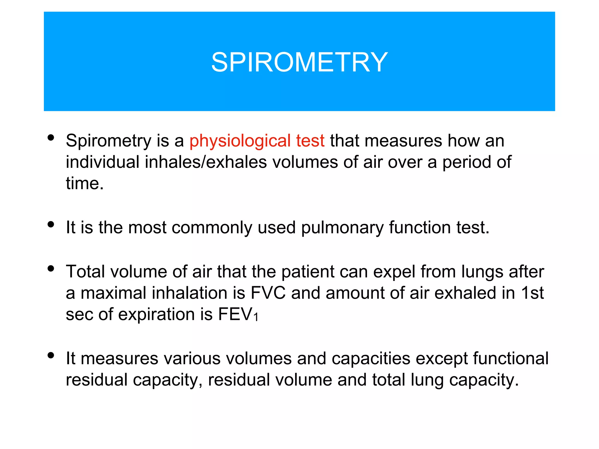
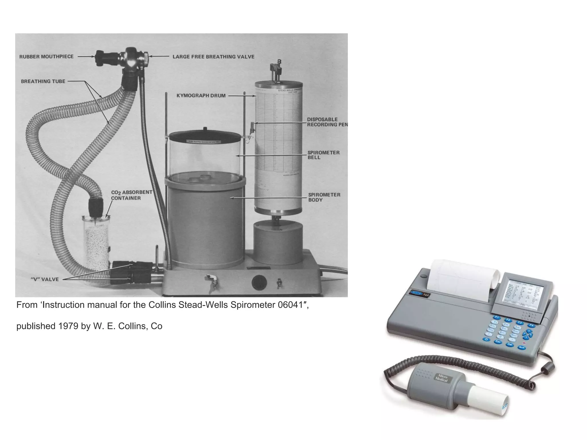
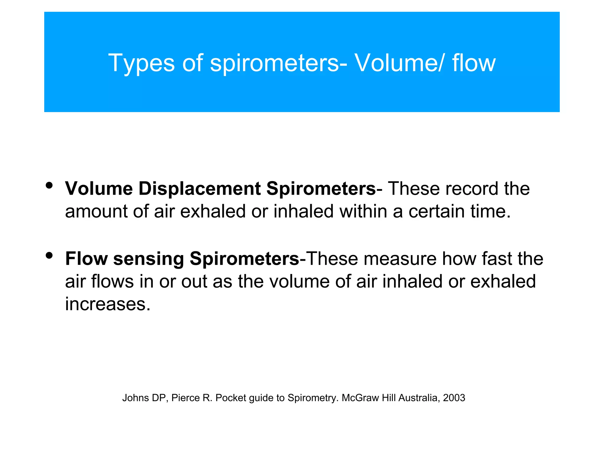
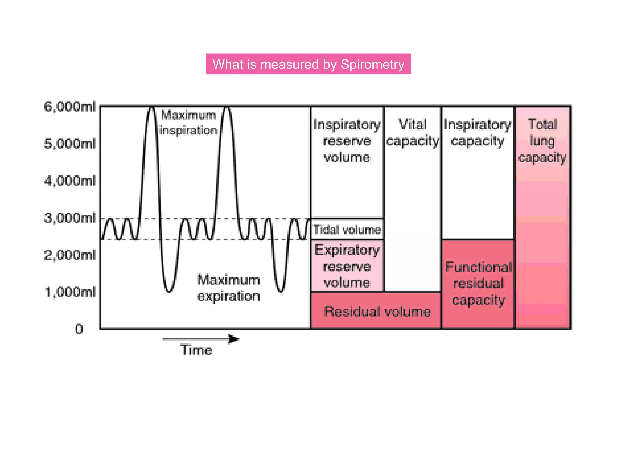
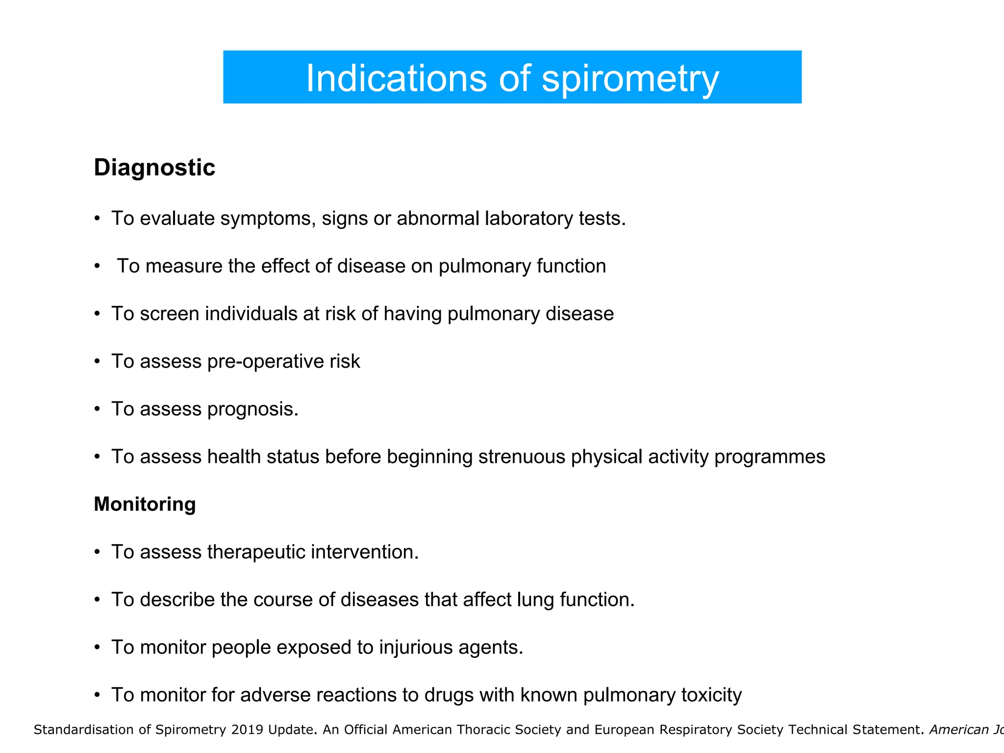
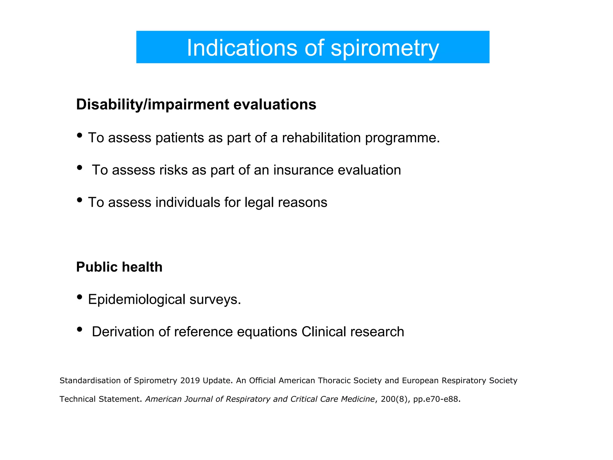
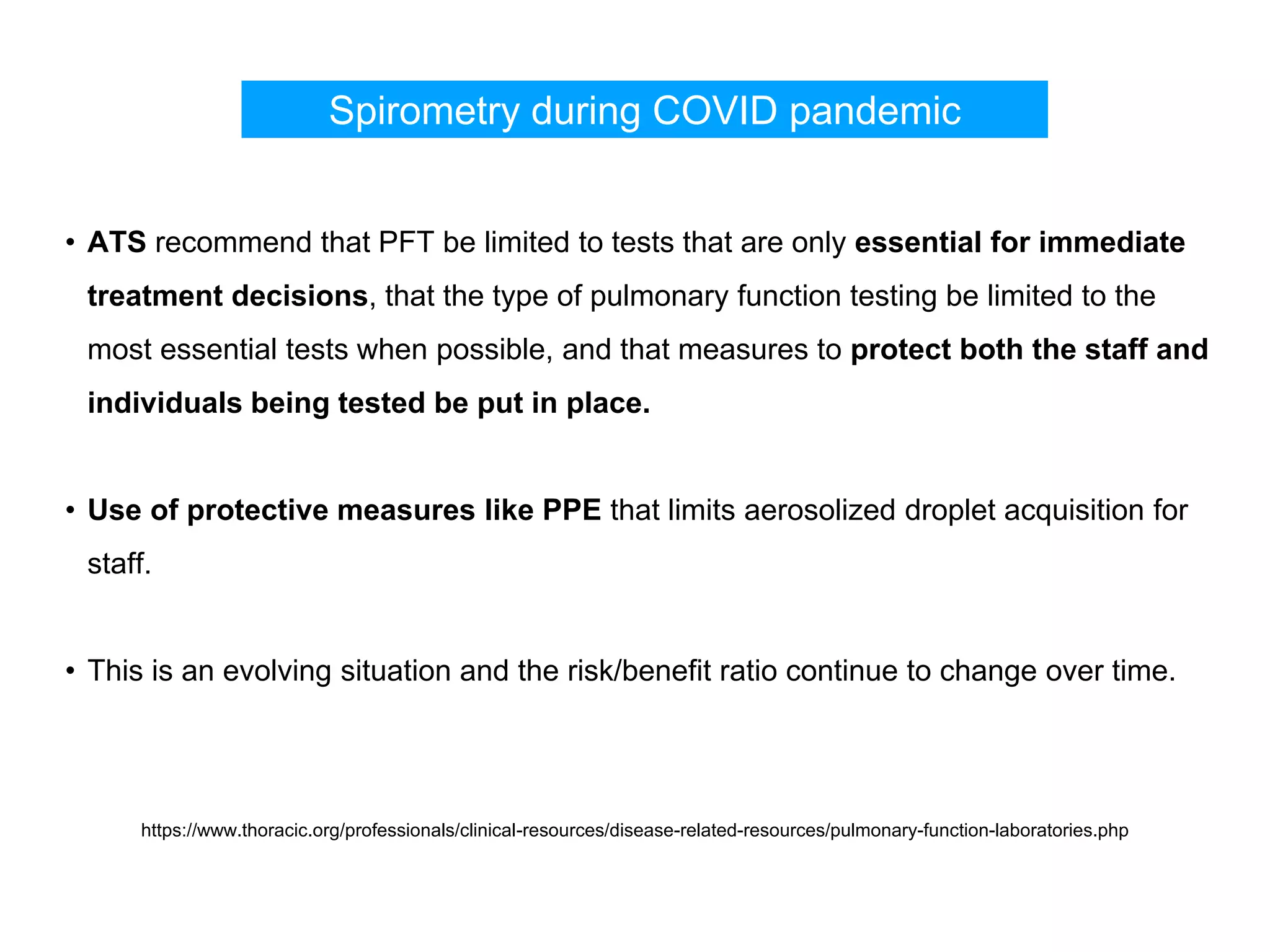
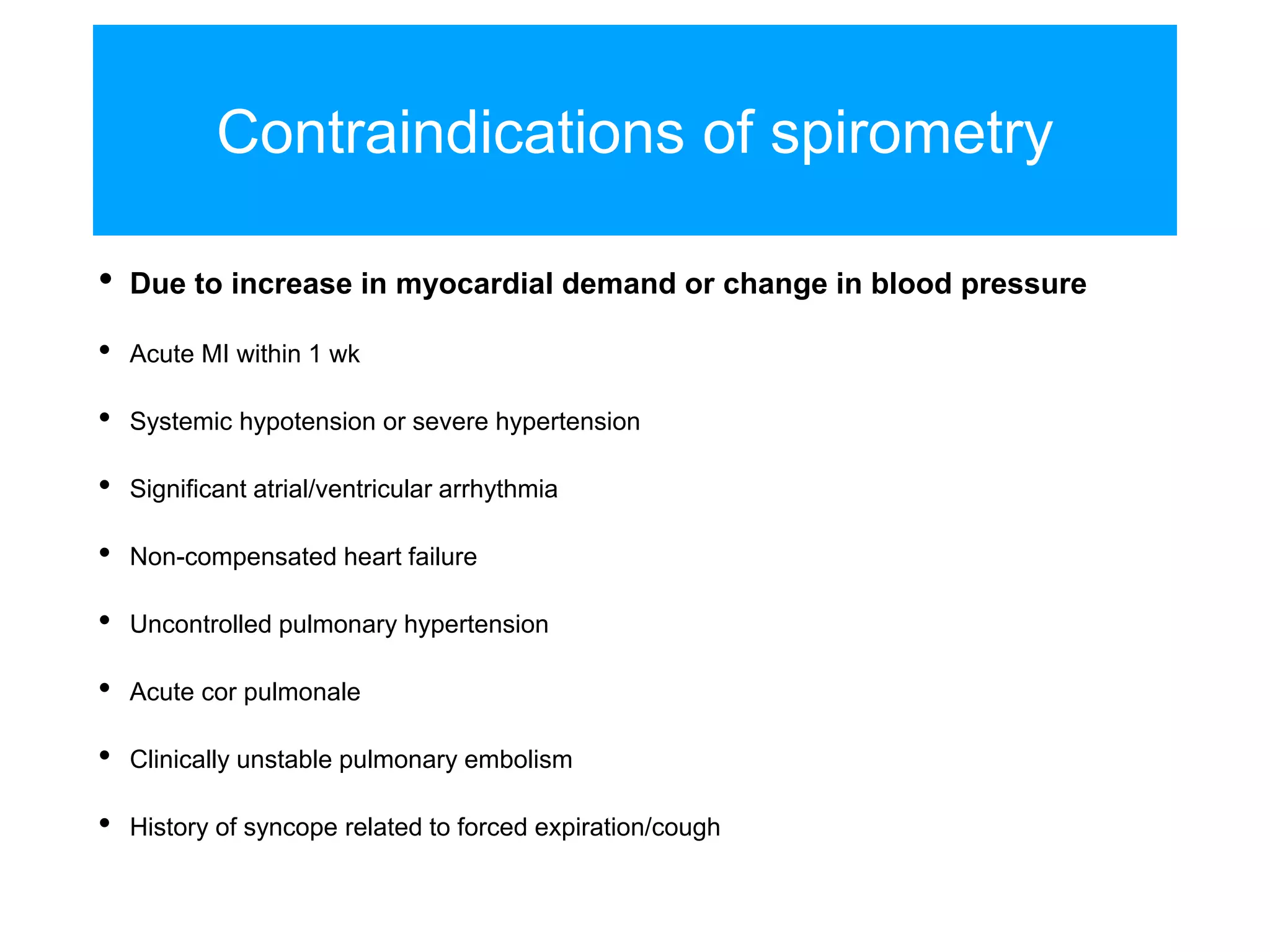
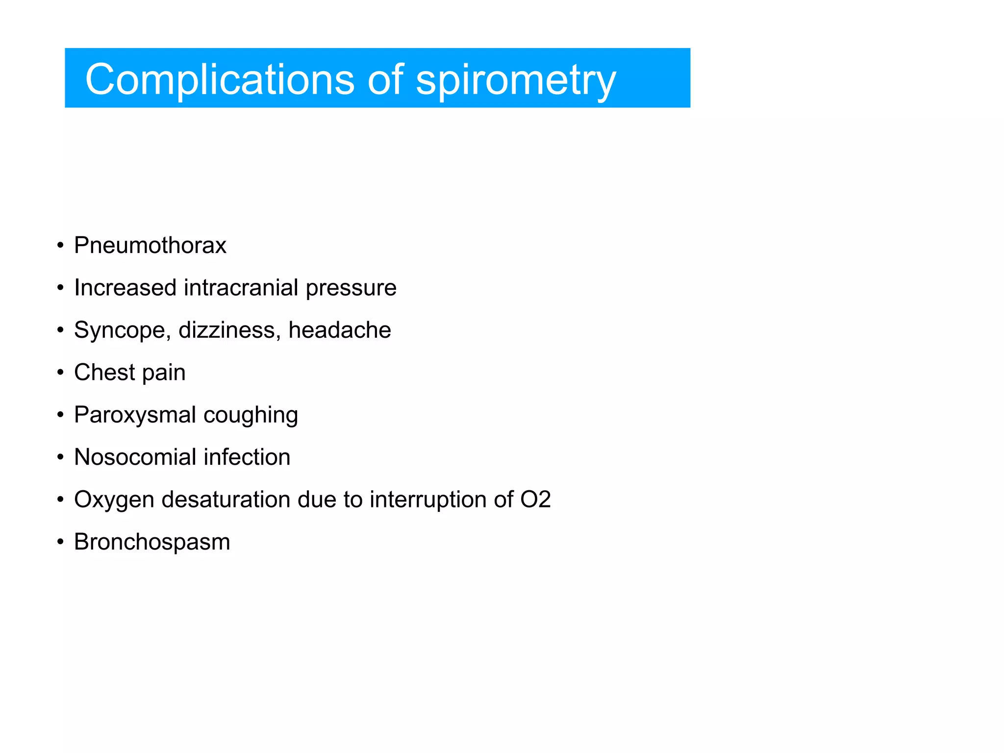
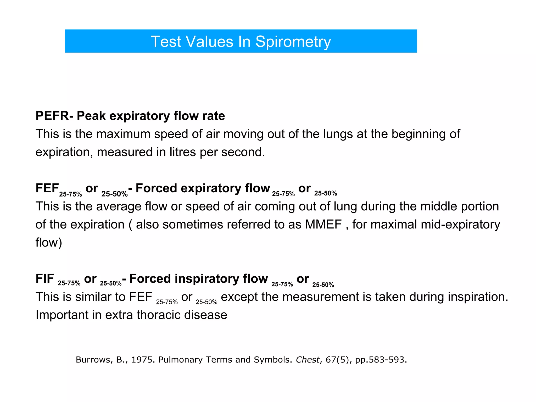
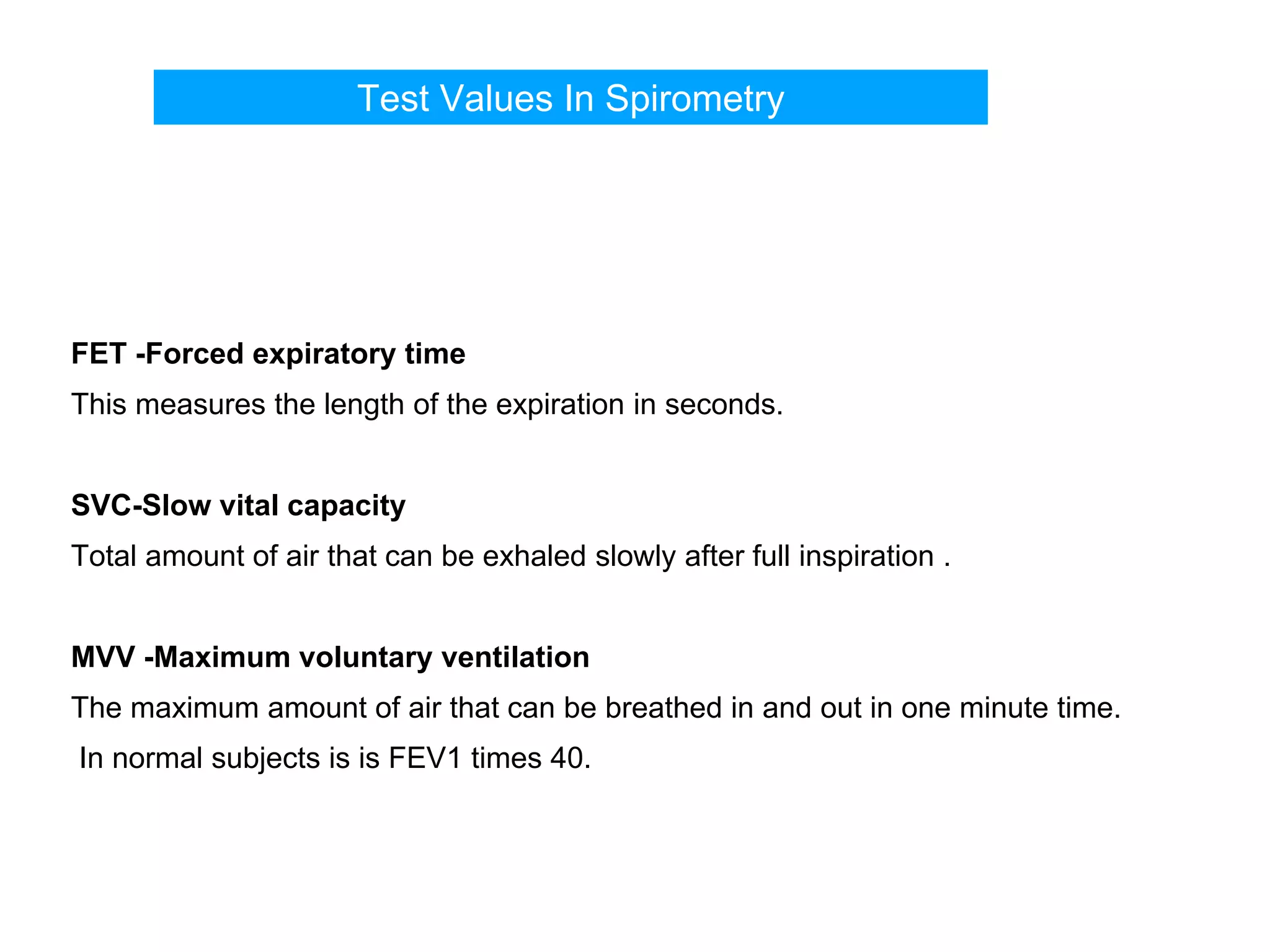
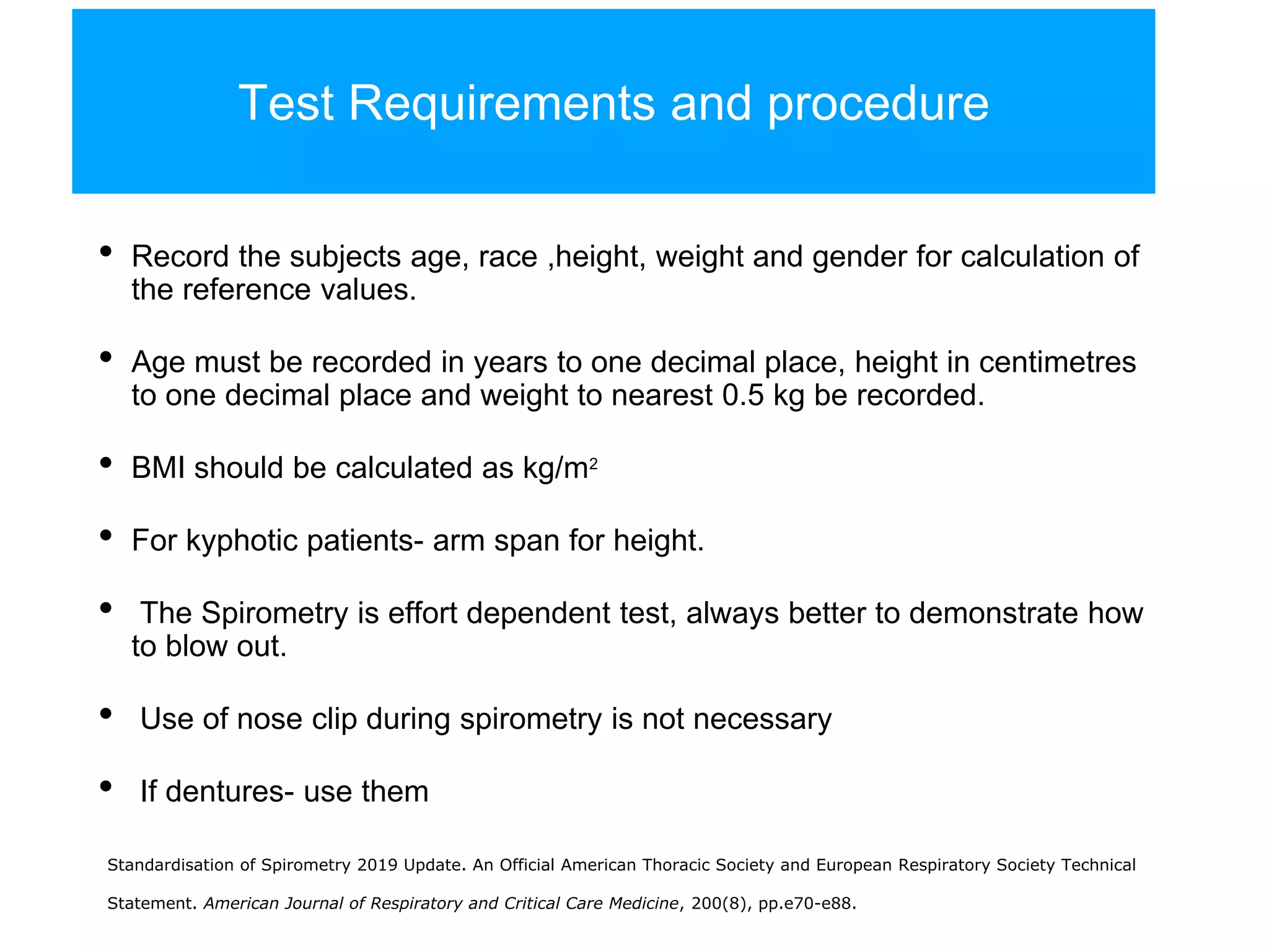
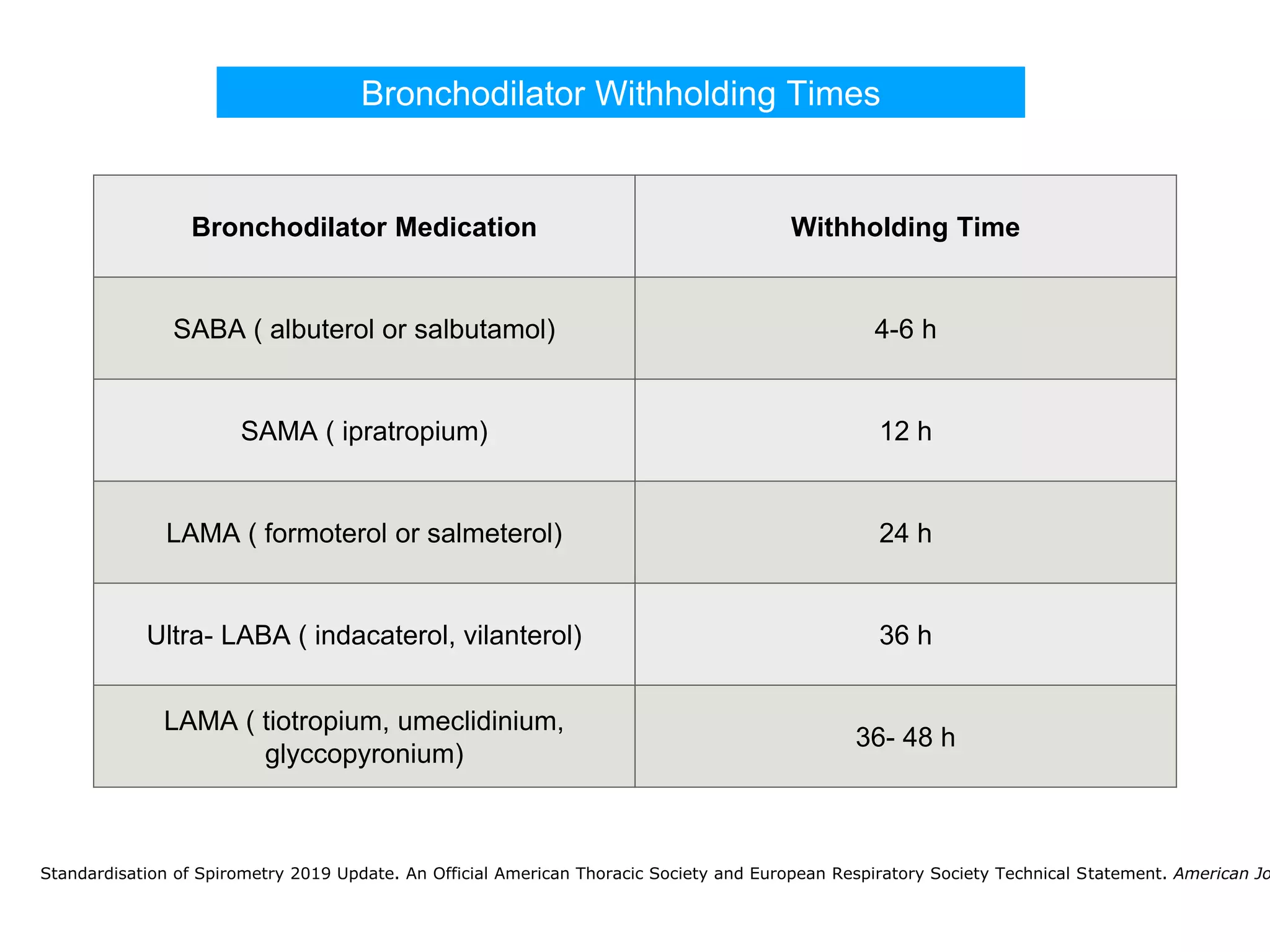
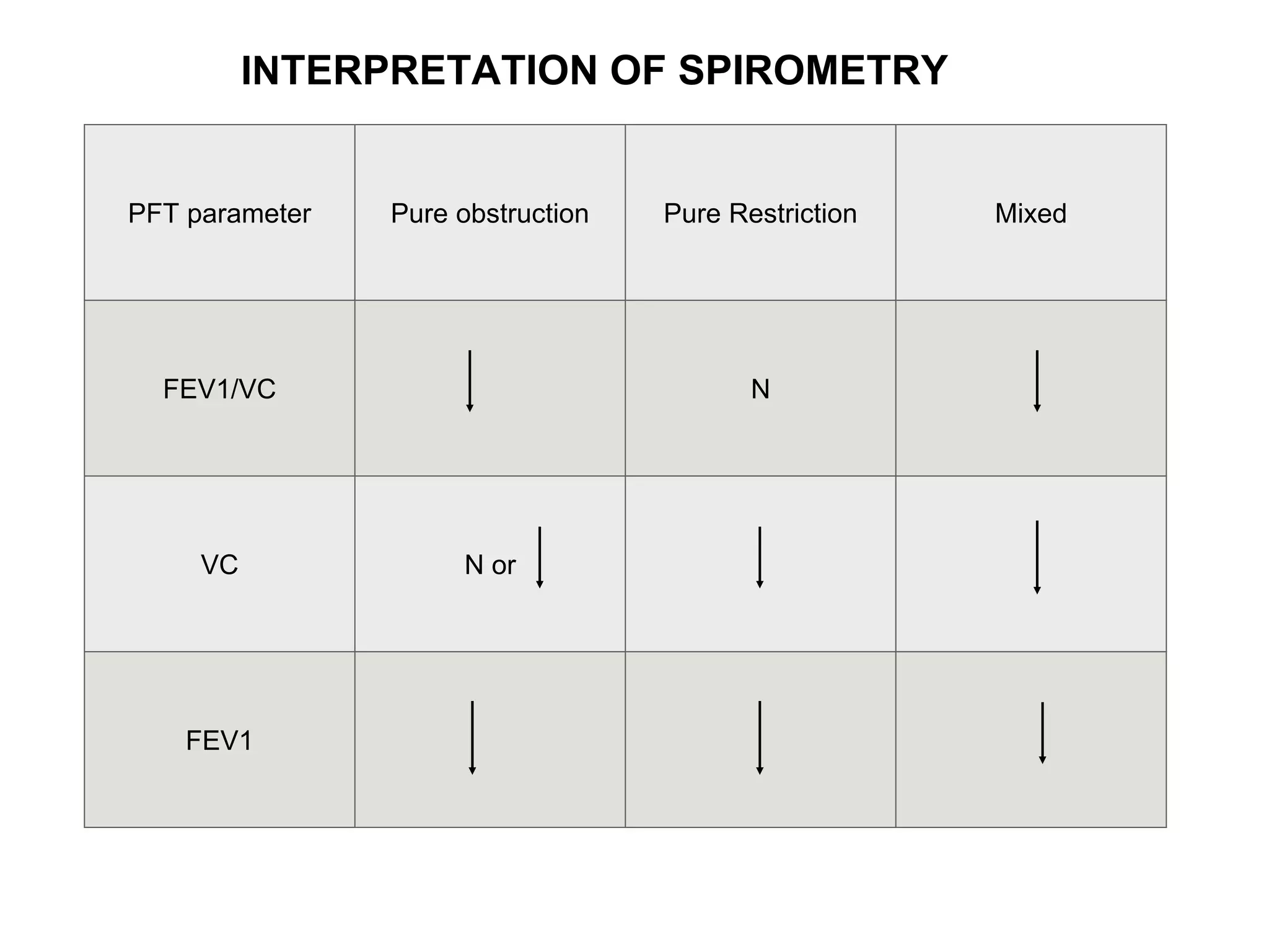
This presentation describes the indications, contraindications, methods of performing spirometry. It explains the interpretation of spirometry with examples.
This document provides information about pulmonary function tests, specifically spirometry. It describes how spirometry is performed and what measurements are taken, including forced vital capacity (FVC), forced expiratory volume in 1 second (FEV1), and ratios like FEV1/FVC. Normal values vary based on factors like age, height, sex, and race. Obstructive and restrictive lung patterns are identified based on these measurements. Spirometry is used to diagnose and monitor lung diseases.
Spirometry is a test that measures lung function by having the patient forcefully exhale air into a mouthpiece. It was originally developed in the 1840s and has since been improved with electronic devices. It is used to diagnose asthma and COPD by measuring airflow obstruction. The test involves withholding some medications beforehand, having the patient blow hard into the device multiple times, and may show a normal pattern or patterns indicating obstruction, restriction, or both. Reversibility testing with a bronchodilator helps distinguish asthma from COPD. Spirometry provides important information about lung health and disease.
Pulmonary function tests provide objective measurements of lung function through various tests. Spirometry is the most basic and widely used test that measures volumes of air inhaled and exhaled over time through a spirometer. It can detect obstructive or restrictive lung diseases patterns based on evaluations of parameters like FEV1, FVC, FEV1/FVC ratio, and flow-volume loops. Other tests measure lung volumes, diffusion capacity, and assess ventilation/perfusion ratios to further characterize lung abnormalities. Together, pulmonary function tests provide quantifiable data to support diagnoses suggested by symptoms and physical exams.
Pulmonary function tests are used to evaluate the respiratory system by measuring lung volumes, gas exchange, and other functions. They have several indications, including investigating symptoms of pulmonary disease, monitoring known lung diseases, and preoperative evaluation. The tests can be categorized as measuring mechanical lung function, gas exchange, or cardiopulmonary interaction. Common tests include spirometry, lung volume measurements, diffusion capacity tests, and exercise tests.
This document discusses bronchial hyperresponsiveness and bronchial provocation tests. It begins by defining asthma as a chronic inflammatory airway disease characterized by variable airflow obstruction and hyperresponsiveness to triggers. Bronchial hyperresponsiveness is an abnormal increase in airflow limitation following exposure to a stimulus and can be quantified using bronchial provocation tests. Several types of direct and indirect stimuli are described for use in bronchial provocation tests, with methacholine challenge being the most commonly used direct stimulus test due to its safety and sensitivity. The document outlines the procedures, interpretations, and indications for various bronchial provocation tests.
Spirometry is a common test used to measure lung function by analyzing breathing patterns during a forced breath maneuver. It measures the total air volume exhaled (forced vital capacity or FVC) and the rate of exhaling during the first second (forced expiratory volume in one second or FEV1). These values are used to diagnose respiratory conditions, monitor treatment effectiveness, and detect airway obstruction. A spirometer is a device that records expiratory flow and volume over time to produce graphs of lung function parameters like FVC, FEV1, and their ratio (FEV1/FVC).
1. The document discusses DLCO (diffusing capacity of the lungs for carbon monoxide), which measures the efficiency of the lungs in transporting oxygen across the alveolar capillary membrane.
2. It describes the single breath hold method for measuring DLCO, which involves inhaling a gas mixture containing carbon monoxide and exhaling into a collection device to measure gas concentrations.
3. DLCO can be lowered in conditions that decrease the surface area for diffusion like emphysema, or increase the thickness of the alveolar capillary membrane like interstitial lung diseases.
This document provides an overview of pulmonary function tests (PFTs). It discusses various PFT measurements including spirometry, lung volumes, diffusing capacity, and airway resistance. Static and dynamic spirometry are used to measure volumes like FVC, FEV1, and ratios like FEV1/FVC. Lung volumes are typically measured using body plethysmography and provide values such as TLC, FRC, and RV. PFTs are used to diagnose obstructive and restrictive lung diseases and evaluate therapeutic interventions.
Pulmonary function tests (PFTs) evaluate the different components of the respiratory system including the airways, lungs, blood vessels and chest wall muscles. Spirometry is the most common PFT and measures how much air the patient can inhale and exhale. It evaluates values like the forced vital capacity (FVC), forced expiratory volume in 1 second (FEV1), and the FEV1/FVC ratio. Obstructive patterns show reduced FEV1 and FVC with a low FEV1/FVC ratio while restrictive patterns have reduced FVC but a normal or increased FEV1/FVC ratio. PFTs are useful for diagnosing conditions like asthma, COPD, and inter
This document discusses peak expiratory flow rate (PEFR), including its purpose, measurement steps, and significance. PEFR is measured using a peak flow monitor to assess, diagnose, manage, and determine prognosis of lung diseases like asthma. It involves preparing the device, standing up straight, taking a deep breath, and blowing hard into the mouthpiece to measure airflow in L/min. Normal PEFR values vary based on age, sex, and height but are generally above 500 L/min for adult males and 400 L/min for adult females. PEFR tests allow patterns in airflow to be monitored over time to help adjust medications or identify environmental triggers.
Lung volumes and capacities can be measured using spirometry. There are four lung volumes - tidal volume, inspiratory reserve volume, expiratory reserve volume, and residual volume. There are also five lung capacities, which are combinations of the lung volumes - inspiratory capacity, expiratory capacity, vital capacity, functional residual capacity, and total lung capacity. Spirometry is used to measure the volumes exhaled during a forced vital capacity maneuver, including the forced expiratory volume in 1 second (FEV1) and ratios like FEV1/FVC, to distinguish between obstructive and restrictive lung diseases.
This document provides an overview of cardiopulmonary exercise testing (CPET) including:
1. CPET assesses the integrated exercise responses of multiple body systems.
2. It describes the equipment, protocols, measurements, and safety considerations for CPET.
3. The document explains how CPET can evaluate exercise limitation and provide diagnostic information for conditions like cardiomyopathy by measuring how the cardiopulmonary systems respond during exercise.
This document discusses pulmonary function tests (PFTs), including their goals, uses, limitations, procedures, and interpretations. PFTs are used to assess lung function before surgeries and characterize any pulmonary dysfunction. Key information obtained from PFTs includes measurements of forced vital capacity (FVC), forced expiratory volume in 1 second (FEV1), their ratio (FEV1/FVC), and peak expiratory flow rate. Interpretations of these values can indicate restrictive or obstructive lung disease. The document outlines how PFTs are performed using portable devices or clinic spirometers and flow-volume loops.
Body plethysmography is a technique used to measure lung volumes like intrathoracic gas volume (TGV) and airway resistance. It involves having the patient breathe in an enclosed chamber while measuring changes in pressure and volume. Specific airway resistance (sRaw) is determined from the relationship between respiratory flow and volume shifts in the chamber. Intrathoracic gas volume (ITGV) can also be measured by having the patient breathe against a shutter to create a closed system where changes in pressure and volume can estimate ITGV based on Boyle's law. Clinical applications include evaluating effects of pulmonary disorders on lung volumes like functional residual capacity (FRC) and residual volume (RV).
Pulmonary function tests (PFTs), such as spirometry, measure how well the lungs function by analyzing air flow. Spirometry yields measurements like FEV1, FVC, and their ratio (FEV1/FVC), which can help distinguish between obstructive and restrictive lung diseases. Obstructive diseases like asthma result in low FEV1/FVC due to airway narrowing, while restrictive diseases lower all lung volumes from reduced compliance. PFTs aid in diagnosis, monitoring treatment, and assessing pre-operative risk.
Simple Interpretation of Pulmonary Function testsGamal Agmy
This document discusses spirometry testing for obstructive lung diseases such as COPD and asthma. It provides information on the different measures assessed during spirometry including FEV1, FVC, and FEF25-75. It describes how to interpret the results and what values indicate restrictive versus obstructive lung disease. Guidelines for pre- and post-bronchodilator spirometry are covered as well as how to assess for reversibility and bronchial hyperresponsiveness. The importance of spirometry for diagnosing and monitoring lung diseases like COPD and asthma is emphasized.
Pulmonary Function Testing-Simplified description...!Sharmin Susiwala
Pulmonary function tests evaluate lung function through measurements of lung volumes, capacities, flows, and gas exchange. Spirometry is the most common pulmonary function test and measures volumes of air inhaled and exhaled over time through the use of a spirometer. Key spirometry metrics include forced vital capacity, forced expiratory volume in 1 second, and peak expiratory flow rate. Arterial blood gases analyze blood pH, oxygen, and carbon dioxide levels to assess lung function and respiratory disease. Together, pulmonary function tests provide diagnostic and monitoring tools for conditions like asthma, COPD, and lung impairments.
Pulmonary function tests provide objective assessments of respiratory symptoms and can help diagnose respiratory diseases. Spirometry is the most widely used test and measures volumes like FVC and FEV1. Restrictive diseases reduce total lung capacity while obstructive diseases cause decreased FEV1/FVC ratio. Other tests include lung volume measurements using plethysmography or gas dilution, diffusing capacity to assess gas exchange, and methacholine challenge for asthma diagnosis. Bedside tests can also help evaluate respiratory function.
- Administered questionnaires
- Performed skin prick tests to common aeroallergens
- Collected blood samples for total IgE & specific IgE
FENO measurement:
- Using NIOX MINO ( Aerocrine AB, Solna, Sweden)
- According to ATS/ERS guidelines
JACI. 2011; 127 ( 5) : 1165-72.e5.
Allergic sensitization:
- Positive SPT ( wheal diameter ≥ 3 mm) to at least one allergen
- Or specific IgE ≥ 0.35 kU/L to at least one allergen
Asthma:
What are the pulmonary function tests used?
What are the indications?
What are the contraindications?
How to perform each and prepare patients?
How to interpret and reach a diagnosis?
How to clean and calibrate devices?
The document discusses interpretation of spirometry data. It provides normal values for measures like FEV1, FVC and FEF25-75. An obstructive pattern is defined as a decreased FEV1/FVC ratio below 0.7, with decreased FEV1 and possibly decreased FVC. A restrictive pattern shows a normal or mildly reduced FEV1 but reduced FVC. Acceptability and repeatability criteria for spirometry tests are outlined.
Pulmonary function tests (PFTs) objectively assess lung function using tests such as spirometry, lung volume measurements, and diffusing capacity measurements. PFTs can predict, diagnose, and monitor pulmonary dysfunction. They can distinguish between obstructive and restrictive lung diseases and determine disease severity and treatment responses. PFTs also assess surgical risk and postoperative pulmonary complications. Simple bedside tests include breath counts, cough strength tests, and peak flow measurements. Laboratory PFTs precisely measure volumes, flows, gas exchange, and generate flow-volume loops. Proper interpretation of PFTs involves comparing values to predicted normals.
This document provides an overview of pulmonary function tests, with a focus on spirometry. It discusses the anatomy of the lungs and airways. Various lung volumes and capacities are defined, including tidal volume, inspiratory reserve volume, and total lung capacity. Spirometry is described as a test that measures airflow into and out of the lungs. Forced vital capacity, FEV1, and FEF25-75 are key spirometric measurements. The document reviews obstructive, restrictive, and mixed lung disease patterns based on spirometry results. Clinical applications and case examples are also briefly mentioned.
Pulmonary function tests (PFTs) measure how well the lungs work by testing how much air a person can breathe in and out, as well as the lungs' ability to bring oxygen to the body. PFTs can help diagnose several respiratory conditions and diseases. Before the tests, patients should avoid medications, large meals, caffeine, smoking, and strenuous exercise. During the tests, patients breathe into a machine to test lung volume and capacity through spirometry, plethysmography, and diffusion capacity tests.
The document discusses pulmonary function tests (PFTs), specifically spirometry. It provides details on lung anatomy and physiology, the purpose of PFTs in evaluating lung function and disease, how spirometry is performed and interpreted, and what values it measures such as FEV1, FVC, and their ratios. PFTs including spirometry are useful diagnostic tools to evaluate symptoms, monitor treatment effectiveness, and distinguish obstructive from restrictive lung diseases.
1) Cardiopulmonary exercise testing (CPET) provides a non-invasive evaluation of integrated exercise responses of the pulmonary, cardiovascular, and muscular systems through measurements taken during exercise.
2) CPET is useful for evaluating exercise intolerance, cardiovascular or respiratory disease, pre-operative risk assessment, and exercise prescription. It involves measurements of oxygen uptake, carbon dioxide production, and ventilatory parameters during a symptom-limited exercise test.
3) Interpretation of CPET results involves comparing measured parameters like peak oxygen uptake, anaerobic threshold, and ventilatory efficiency to reference values to determine if results are normal or abnormal.
Spirometry is a test that measures lung function by having the patient forcefully exhale after taking a deep breath. It measures volumes like forced vital capacity (FVC) and forced expiratory volume in 1 second (FEV1). Spirometry can detect both restrictive and obstructive lung diseases. The test involves having the patient blow into a mouthpiece attached to a spirometer, which produces volume-time and flow-volume curves. Key factors like technique, acceptability criteria, reference values, and bronchodilator responsiveness testing are important considerations when performing and interpreting spirometry results.
Pulmonary function test in children (spirometer)Azad Haleem
Spirometry is a noninvasive pulmonary function test that measures lung volumes and airflow. It is useful for diagnosing and monitoring respiratory diseases like asthma in children who are able to perform the test properly. Key measurements include forced vital capacity (FVC), forced expiratory volume in 1 second (FEV1), and ratios like FEV1/FVC. Obstructive patterns show reduced airflow while restrictive patterns show reduced lung volumes. Spirometry provides objective data to guide treatment and monitor response to medications like bronchodilators.
Pulmonary function testing (spirometry ) Dr Emad efat
Pulmonary function tests (PFTs) such as spirometry help diagnose and monitor respiratory diseases. Spirometry measures expiratory volumes and flow rates through forced inhalation and exhalation into a mouthpiece. For a test to be valid, exhalation must be smooth, continuous for 6 seconds, and meet reproducibility criteria of two largest FVC and FEV1 values within 0.2 L of each other. PFTs can detect restrictive and obstructive lung diseases and assess response to treatment.
Spirometry is a simple, inexpensive pulmonary function test used to evaluate respiratory diseases. It measures how much air a person can inhale and exhale. Sir John Hutchinson invented the first spirometer to measure vital capacity. Spirometry can identify obstructive and restrictive lung diseases, establish reversibility in asthma, screen smokers for risk, and monitor treatment effectiveness. It involves maximal inhalation and exhalation into a mouthpiece to measure volumes like forced vital capacity and flows like forced expiratory volume in one second. Graphs of the results can indicate normal lung function or patterns suggestive of various respiratory conditions.
This document discusses pulmonary function tests (PFTs), which objectively measure lung function and are useful for diagnosing and monitoring respiratory disease. It describes various PFTs including spirometry, peak expiratory flow rate tests, body plethysmography, diffusion capacity tests, bronchoprovocation challenge tests, and six-minute walk tests. Spirometry specifically measures volumes of air inhaled and exhaled, while peak flow tests measure maximum exhaled airflow and diffusion capacity tests evaluate gas exchange in the lungs. Together these PFTs provide clinicians with quantitative data on lung volumes, capacities, flows, and gas exchange to aid in respiratory disease assessment.
5-PFT- Dr BassamFFFFFFFFFFF Al- Selwey.pdfMosaHasen
The document outlines a presentation on pulmonary function tests, specifically spirometry, discussing the history, equipment, procedures, normal values, and interpretation for obstructive, restrictive, and mixed lung disease patterns. It also covers indications, contraindications, and practical techniques for performing spirometry tests.
Pulmonary function tests (PFTs) measure how well the lungs work by assessing lung volumes, air flow, and gas exchange. PFTs can help diagnose various respiratory diseases by detecting abnormalities. The tests involve spirometry to measure volumes of air inhaled and exhaled during normal and forced breathing maneuvers. Obstructive diseases reduce air flow, shown by a decreased FEV1 and FEV1/FVC ratio. Restrictive diseases limit lung expansion, shown by a reduced FVC but normal or increased FEV1/FVC ratio. PFTs are useful for evaluating respiratory system function and the effects of treatment.
Pulmonary function tests (PFTs) measure how well the lungs work by assessing lung volumes, air flow, and gas exchange. PFTs can help diagnose various respiratory diseases by detecting abnormalities. The tests involve spirometry to measure volumes of air inhaled and exhaled during normal and forced breathing maneuvers. Obstructive diseases reduce air flow, shown by a decreased FEV1 and FEV1/FVC ratio. Restrictive diseases limit lung expansion, shown by a reduced FVC but normal or increased FEV1/FVC ratio. PFTs are useful for evaluating respiratory function and monitoring treatment effects.
Pulmonary function tests (PFTs) evaluate aspects of lung function using standardized equipment. Common PFTs include spirometry, which measures volumes of air inhaled and exhaled over time; diffusing capacity of the lung for carbon monoxide (DLCO), which measures gas exchange; and body plethysmography, which measures lung volumes. Spirometry was invented in the 1800s and measures parameters such as forced vital capacity (FVC), forced expiratory volume in one second (FEV1), and their ratio (FEV1/FVC). Normal values vary based on age, height, sex, and ethnicity. PFTs can detect obstructive and restrictive lung diseases based on patterns of airflow
This document provides information on pulmonary function testing and spirometry. It defines key lung volumes and capacities that are measured, such as FVC, FEV1, FRC, RV. Normal values for various pulmonary function tests are provided. Spirometry is described as the most common pulmonary function test used to measure breath volume and flow. The document outlines the technique for spirometry and how to interpret the results to determine if a restrictive or obstructive ventilatory pattern is present. Limitations of spirometry and contraindications to its use are also discussed.
PULMONARY FUNCTION TESTS PLAY A VERY IMPORTANT ROLE IN ESTIMATING THE FUNCTION OF LUNGS ESPECIALLY IN ASTHAMA AND COPD, One of the frequent reasons patients see their primary care physicians is for the symptom of dyspnea. Among the objective tests to quantify this symptom is the pulmonary function test
Spirometry is a common pulmonary function test that measures breath volume and flow. It can be used to detect lung disease, monitor occupational exposures, and assess medication effects. This document provides an overview of how to properly perform and interpret spirometry tests, including ensuring patient preparation and positioning, using correct technique, assessing test quality, and comparing results to reference values to determine if lungs are normal or abnormal. Quality is important for obtaining accurate and reproducible results.
This document defines asthma and discusses its diagnosis and management. Key points:
- Asthma is a chronic inflammatory lung disease characterized by variable airflow obstruction and respiratory symptoms.
- Diagnosis is based on symptoms and evidence of variable lung function and airflow limitation through spirometry testing. Peak expiratory flow monitoring can help monitor the condition.
- Treatment involves long-term control medications like inhaled corticosteroids and quick-relief bronchodilators. The goals are to control symptoms, minimize future risk of exacerbations, and maintain normal lung function and activity levels.
Spirometry measures lung volumes and airflow. Modern spirometers use flow sensors to measure tidal volume, inspiratory reserve volume, expiratory reserve volume, residual volume, vital capacity, and total lung capacity. Key metrics include FEV1, FVC, and their ratio. Spirometry is used to diagnose and monitor lung diseases, assess surgical risk, and evaluate therapeutic interventions. Proper technique requires full inspiration and expiration over 6 seconds. Results are interpreted by comparing values to predicted norms and assessing for reversibility of obstruction.
Pulmonary Function Tests-Nursing Maseno.pptxakoeljames8543
This document discusses various pulmonary function tests (PFTs) including spirometry. PFTs measure how well the lungs work by assessing lung volumes, airway function, and gas exchange. Spirometry specifically measures airflow and lung capacity. It involves taking a deep breath and then forcibly exhaling for 6 seconds into a spirometer. Key measurements include FEV1, FVC, and their ratio (FEV1%), which can help identify obstructive or restrictive lung diseases. Abnormal PFT results are below 80% of predicted values and indicate the severity of lung impairment. PFTs are useful diagnostic tools that also monitor treatment effectiveness.
Pulmonary function tests measure how well the lungs work by assessing how much air they can hold and exhale. There are various lung volumes and capacities that are measured, including tidal volume, inspiratory reserve volume, and total lung capacity. Spirometry is the main test used, which measures exhaled air volumes over time. It can help diagnose obstructive lung diseases like asthma which show decreased FEV1/FVC ratio, versus restrictive diseases which show increased FEV1/FVC ratio. Other tests include diffusing capacity to measure gas exchange, body plethysmography to measure total lung capacity, and inhalational challenges to identify allergens.
Computer in pharmaceutical research and development-Mpharm(Pharmaceutics)MuskanShingari
Statistics- Statistics is the science of collecting, organizing, presenting, analyzing and interpreting numerical data to assist in making more effective decisions.
A statistics is a measure which is used to estimate the population parameter
Parameters-It is used to describe the properties of an entire population.
Examples-Measures of central tendency Dispersion, Variance, Standard Deviation (SD), Absolute Error, Mean Absolute Error (MAE), Eigen Value
Giloy in Ayurveda - Classical Categorization and SynonymsPlanet Ayurveda
Giloy, also known as Guduchi or Amrita in classical Ayurvedic texts, is a revered herb renowned for its myriad health benefits. It is categorized as a Rasayana, meaning it has rejuvenating properties that enhance vitality and longevity. Giloy is celebrated for its ability to boost the immune system, detoxify the body, and promote overall wellness. Its anti-inflammatory, antipyretic, and antioxidant properties make it a staple in managing conditions like fever, diabetes, and stress. The versatility and efficacy of Giloy in supporting health naturally highlight its importance in Ayurveda. At Planet Ayurveda, we provide a comprehensive range of health services and 100% herbal supplements that harness the power of natural ingredients like Giloy. Our products are globally available and affordable, ensuring that everyone can benefit from the ancient wisdom of Ayurveda. If you or your loved ones are dealing with health issues, contact Planet Ayurveda at 01725214040 to book an online video consultation with our professional doctors. Let us help you achieve optimal health and wellness naturally.
Know the difference between Endodontics and Orthodontics.Gokuldas Hospital
Your smile is beautiful.
Let’s be honest. Maintaining that beautiful smile is not an easy task. It is more than brushing and flossing. Sometimes, you might encounter dental issues that need special dental care. These issues can range anywhere from misalignment of the jaw to pain in the root of teeth.
STUDIES IN SUPPORT OF SPECIAL POPULATIONS: GERIATRICS E7shruti jagirdar
Unit 4: MRA 103T Regulatory affairs
This guideline is directed principally toward new Molecular Entities that are
likely to have significant use in the elderly, either because the disease intended
to be treated is characteristically a disease of aging ( e.g., Alzheimer's disease) or
because the population to be treated is known to include substantial numbers of
geriatric patients (e.g., hypertension).
Breast cancer: Post menopausal endocrine therapyDr. Sumit KUMAR
Breast cancer in postmenopausal women with hormone receptor-positive (HR+) status is a common and complex condition that necessitates a multifaceted approach to management. HR+ breast cancer means that the cancer cells grow in response to hormones such as estrogen and progesterone. This subtype is prevalent among postmenopausal women and typically exhibits a more indolent course compared to other forms of breast cancer, which allows for a variety of treatment options.
Diagnosis and Staging
The diagnosis of HR+ breast cancer begins with clinical evaluation, imaging, and biopsy. Imaging modalities such as mammography, ultrasound, and MRI help in assessing the extent of the disease. Histopathological examination and immunohistochemical staining of the biopsy sample confirm the diagnosis and hormone receptor status by identifying the presence of estrogen receptors (ER) and progesterone receptors (PR) on the tumor cells.
Staging involves determining the size of the tumor (T), the involvement of regional lymph nodes (N), and the presence of distant metastasis (M). The American Joint Committee on Cancer (AJCC) staging system is commonly used. Accurate staging is critical as it guides treatment decisions.
Treatment Options
Endocrine Therapy
Endocrine therapy is the cornerstone of treatment for HR+ breast cancer in postmenopausal women. The primary goal is to reduce the levels of estrogen or block its effects on cancer cells. Commonly used agents include:
Selective Estrogen Receptor Modulators (SERMs): Tamoxifen is a SERM that binds to estrogen receptors, blocking estrogen from stimulating breast cancer cells. It is effective but may have side effects such as increased risk of endometrial cancer and thromboembolic events.
Aromatase Inhibitors (AIs): These drugs, including anastrozole, letrozole, and exemestane, lower estrogen levels by inhibiting the aromatase enzyme, which converts androgens to estrogen in peripheral tissues. AIs are generally preferred in postmenopausal women due to their efficacy and safety profile compared to tamoxifen.
Selective Estrogen Receptor Downregulators (SERDs): Fulvestrant is a SERD that degrades estrogen receptors and is used in cases where resistance to other endocrine therapies develops.
Combination Therapies
Combining endocrine therapy with other treatments enhances efficacy. Examples include:
Endocrine Therapy with CDK4/6 Inhibitors: Palbociclib, ribociclib, and abemaciclib are CDK4/6 inhibitors that, when combined with endocrine therapy, significantly improve progression-free survival in advanced HR+ breast cancer.
Endocrine Therapy with mTOR Inhibitors: Everolimus, an mTOR inhibitor, can be added to endocrine therapy for patients who have developed resistance to aromatase inhibitors.
Chemotherapy
Chemotherapy is generally reserved for patients with high-risk features, such as large tumor size, high-grade histology, or extensive lymph node involvement. Regimens often include anthracyclines and taxanes.
How to Control Your Asthma Tips by gokuldas hospital.Gokuldas Hospital
Respiratory issues like asthma are the most sensitive issue that is affecting millions worldwide. It hampers the daily activities leaving the body tired and breathless.
The key to a good grip on asthma is proper knowledge and management strategies. Understanding the patient-specific symptoms and carving out an effective treatment likewise is the best way to keep asthma under control.
Travel Clinic Cardiff: Health Advice for International TravelersNX Healthcare
Travel Clinic Cardiff offers comprehensive travel health services, including vaccinations, travel advice, and preventive care for international travelers. Our expert team ensures you are well-prepared and protected for your journey, providing personalized consultations tailored to your destination. Conveniently located in Cardiff, we help you travel with confidence and peace of mind. Visit us: www.nxhealthcare.co.uk
These lecture slides, by Dr Sidra Arshad, offer a simplified look into the mechanisms involved in the regulation of respiration:
Learning objectives:
1. Describe the organisation of respiratory center
2. Describe the nervous control of inspiration and respiratory rhythm
3. Describe the functions of the dorsal and respiratory groups of neurons
4. Describe the influences of the Pneumotaxic and Apneustic centers
5. Explain the role of Hering-Breur inflation reflex in regulation of inspiration
6. Explain the role of central chemoreceptors in regulation of respiration
7. Explain the role of peripheral chemoreceptors in regulation of respiration
8. Explain the regulation of respiration during exercise
9. Integrate the respiratory regulatory mechanisms
10. Describe the Cheyne-Stokes breathing
Study Resources:
1. Chapter 42, Guyton and Hall Textbook of Medical Physiology, 14th edition
2. Chapter 36, Ganong’s Review of Medical Physiology, 26th edition
3. Chapter 13, Human Physiology by Lauralee Sherwood, 9th edition
- Video recording of this lecture in English language: https://youtu.be/Pt1nA32sdHQ
- Video recording of this lecture in Arabic language: https://youtu.be/uFdc9F0rlP0
- Link to download the book free: https://nephrotube.blogspot.com/p/nephrotube-nephrology-books.html
- Link to NephroTube website: www.NephroTube.com
- Link to NephroTube social media accounts: https://nephrotube.blogspot.com/p/join-nephrotube-on-social-media.html
PGx Analysis in VarSeq: A User’s PerspectiveGolden Helix
Since our release of the PGx capabilities in VarSeq, we’ve had a few months to gather some insights from various use cases. Some users approach PGx workflows by means of array genotyping or what seems to be a growing trend of adding the star allele calling to the existing NGS pipeline for whole genome data. Luckily, both approaches are supported with the VarSeq software platform. The genotyping method being used will also dictate what the scope of the tertiary analysis will be. For example, are your PGx reports a standalone pipeline or would your lab’s goal be to handle a dual-purpose workflow and report on PGx + Diagnostic findings.
The purpose of this webcast is to:
Discuss and demonstrate the approaches with array and NGS genotyping methods for star allele calling to prep for downstream analysis.
Following genotyping, explore alternative tertiary workflow concepts in VarSeq to handle PGx reporting.
Moreover, we will include insights users will need to consider when validating their PGx workflow for all possible star alleles and options you have for automating your PGx analysis for large number of samples. Please join us for a session dedicated to the application of star allele genotyping and subsequent PGx workflows in our VarSeq software.
Discover the benefits of homeopathic medicine for irregular periods with our guide on 5 common remedies. Learn how these natural treatments can help regulate menstrual cycles and improve overall menstrual health.
Visit Us: https://drdeepikashomeopathy.com/service/irregular-periods-treatment/
2. SPIROMETRY
• Spirometry is a physiological test that measures how an
individual inhales/exhales volumes of air over a period of
time.
• It is the most commonly used pulmonary function test.
• Total volume of air that the patient can expel from lungs after
a maximal inhalation is FVC and amount of air exhaled in 1st
sec of expiration is FEV1
• It measures various volumes and capacities except functional
residual capacity, residual volume and total lung capacity.
3. HISTORY
• In mid 1800s, John Hutchinson developed a simple
spirometer based on water seal principle that measured the
maximum volume of air which can move in and out of lung,
vital capacity.
• Around 1950, Gansler attached a microswitch to a water seal
spirometer to time the vital capacity. He observed that healthy
people can exhale than 80% of their vital capacity in first
second and almost all the vital capacity in 3 seconds.
• In late 1950s, Robert Hyatt and others began using flow
volume display to measure lung function.
Sprigs EA. The history of spirometry. Br J Dis Chest 1978; 72: 165-180
4. From ‘Instruction manual for the Collins Stead-Wells Spirometer 06041″,
published 1979 by W. E. Collins, Co
5. Types of spirometers- Volume/ flow
• Volume Displacement Spirometers- These record the
amount of air exhaled or inhaled within a certain time.
• Flow sensing Spirometers-These measure how fast the
air flows in or out as the volume of air inhaled or exhaled
increases.
Johns DP, Pierce R. Pocket guide to Spirometry. McGraw Hill Australia, 2003
7. Diagnostic
• To evaluate symptoms, signs or abnormal laboratory tests.
• To measure the effect of disease on pulmonary function
• To screen individuals at risk of having pulmonary disease
• To assess pre-operative risk
• To assess prognosis.
• To assess health status before beginning strenuous physical activity programmes
Monitoring
• To assess therapeutic intervention.
• To describe the course of diseases that affect lung function.
• To monitor people exposed to injurious agents.
• To monitor for adverse reactions to drugs with known pulmonary toxicity
Indications of spirometry
Standardisation of Spirometry 2019 Update. An Official American Thoracic Society and European Respiratory Society Technical Statement. American Jo
8. Disability/impairment evaluations
• To assess patients as part of a rehabilitation programme.
• To assess risks as part of an insurance evaluation
• To assess individuals for legal reasons
Public health
• Epidemiological surveys.
• Derivation of reference equations Clinical research
Indications of spirometry
Standardisation of Spirometry 2019 Update. An Official American Thoracic Society and European Respiratory Society
Technical Statement. American Journal of Respiratory and Critical Care Medicine, 200(8), pp.e70-e88.
9. Spirometry during COVID pandemic
• ATS recommend that PFT be limited to tests that are only essential for immediate
treatment decisions, that the type of pulmonary function testing be limited to the
most essential tests when possible, and that measures to protect both the staff and
individuals being tested be put in place.
• Use of protective measures like PPE that limits aerosolized droplet acquisition for
staff.
• This is an evolving situation and the risk/benefit ratio continue to change over time.
https://www.thoracic.org/professionals/clinical-resources/disease-related-resources/pulmonary-function-laboratories.php
10. Bedside tests
• Sabrase breath holding test
• Single breath count
• Forced expiratory time
• Debono’s whistle blowing test
• Wright’s peak flow meter
• Wright’s respirometer
11. Contraindications of spirometry
• Due to increase in myocardial demand or change in blood pressure
• Acute MI within 1 wk
• Systemic hypotension or severe hypertension
• Significant atrial/ventricular arrhythmia
• Non-compensated heart failure
• Uncontrolled pulmonary hypertension
• Acute cor pulmonale
• Clinically unstable pulmonary embolism
• History of syncope related to forced expiration/cough
12. Due to increase in intra-cranial/ intra-ocular pressure
Cerebral aneurysm
Brain surgery within 4 wk
Recent concussion with continuing symptoms
Eye surgery within 1 wk
Due to increase in sinus and middle and middle ear pressure
Sinus or middle ear surgery or infection within 1 wk
Due to increase in intrathoracic and intraabdominal pressure
Presence of pneumothorax
Thoracic/abdominal surgery within 4 wk
Late term pregnancy
Infection control issues
Active or suspected transmissible respiratory or systemic infection like TB
Standardisation of Spirometry 2019 Update. An Official American Thoracic Society and European Respiratory Society Technical
Statement. American Journal of Respiratory and Critical Care Medicine, 200(8), pp.e70-e88.
13. • Pneumothorax
• Increased intracranial pressure
• Syncope, dizziness, headache
• Chest pain
• Paroxysmal coughing
• Nosocomial infection
• Oxygen desaturation due to interruption of O2
• Bronchospasm
Complications of spirometry
14. FVC -Forced vital capacity
This is the total amount of air that one can forcibly blow out after full inspiration, measured in
liters
FEV1- Forced expiratory volume in one second
This is the amount of air that one can forcibly blow out in one second, measured in liters.
Along with FVC it is considered one of the primary indicators of lung function
FEV1/FVC
The proportion of the total volume of air that can be expired in the first second of expiration.
Test Values In Spirometry
Burrows, B., 1975. Pulmonary Terms and Symbols. Chest, 67(5), pp.583-593.
15. PEFR- Peak expiratory flow rate
This is the maximum speed of air moving out of the lungs at the beginning of
expiration, measured in litres per second.
FEF25-75%
or 25-50%
- Forced expiratory flow 25-75%
or 25-50%
This is the average flow or speed of air coming out of lung during the middle portion
of the expiration ( also sometimes referred to as MMEF , for maximal mid-expiratory
flow)
FIF 25-75% or 25-50%- Forced inspiratory flow 25-75%
or 25-50%
This is similar to FEF 25-75% or 25-50% except the measurement is taken during inspiration.
Important in extra thoracic disease
Test Values In Spirometry
Burrows, B., 1975. Pulmonary Terms and Symbols. Chest, 67(5), pp.583-593.
16. FET -Forced expiratory time
This measures the length of the expiration in seconds.
SVC-Slow vital capacity
Total amount of air that can be exhaled slowly after full inspiration .
MVV -Maximum voluntary ventilation
The maximum amount of air that can be breathed in and out in one minute time.
In normal subjects is is FEV1 times 40.
Test Values In Spirometry
17. Test Requirements and procedure
• Record the subjects age, race ,height, weight and gender for calculation of
the reference values.
• Age must be recorded in years to one decimal place, height in centimetres
to one decimal place and weight to nearest 0.5 kg be recorded.
• BMI should be calculated as kg/m2
• For kyphotic patients- arm span for height.
• The Spirometry is effort dependent test, always better to demonstrate how
to blow out.
• Use of nose clip during spirometry is not necessary
• If dentures- use them
Standardisation of Spirometry 2019 Update. An Official American Thoracic Society and European Respiratory Society Technical
Statement. American Journal of Respiratory and Critical Care Medicine, 200(8), pp.e70-e88.
18. Calibration of Spirometers
• For correct and reproducible results
• Verify calibration frequently
• Syringes- 1L and 3L
19. Patients should be advised to avoid the following prior to testing:
• Smoking and/or vaping and/or water pipe within 1 hour before testing
• Consuming intoxicants within 8 hours before testing.
• Performing vigorous exercise within 1 hr before testing.
• Wearing clothing that substantially restricts full chest and abdominal
expansion.
Pre Test preparation
Standardisation of Spirometry 2019 Update. An Official American Thoracic Society and European Respiratory Society Technical
Statement. American Journal of Respiratory and Critical Care Medicine, 200(8), pp.e70-e88.
20. Bronchodilator Medication Withholding Time
SABA ( albuterol or salbutamol) 4-6 h
SAMA ( ipratropium) 12 h
LAMA ( formoterol or salmeterol) 24 h
Ultra- LABA ( indacaterol, vilanterol) 36 h
LAMA ( tiotropium, umeclidinium,
glyccopyronium)
36- 48 h
Bronchodilator Withholding Times
Standardisation of Spirometry 2019 Update. An Official American Thoracic Society and European Respiratory Society Technical Statement. American Jo
21. exhalation
Inhalation
Spirometry requires a coordinated maximum effort. The three steps are:
• Step 1: Coach the patient to take as deep a breath as possible
• Step 2: Loudly prompt the patient to Blast out the air into the
spirometer
• Step 3: Encourage the patient to continue exhaling for maximum of 15 seconds.
• Step 4 : Inspiration at maximal flow back to maximum lung volume.
EXHALATION
INHALATION
Phase 1
Inhale
Phase 2
Blast
Phase 3
Keep going
23. Flow Volume Loop- Inspiratory / Expiratory limbs
• Flow volume curve provides a graphic illustration of a
patient’s spirometry efforts.
• Flow is plotted against volume to display a continuous loop
from inspiration to expiration.
• A normal flow volume loop has a rapid peak expiratory
flow rate (termed as ‘peak of the curve’). The expiratory
flow rate then falls and the tracing moves downward to
meet the volume axis. It is termed ‘the slope of the curve.
• The inspiratory portion of the loop is a deep curve plotted
on the negative portion of the flow axis. It indicates upper
airway disease.
• The overall shape of the flow volume loop is important in
interpreting spirometry results.
Peak
Slope
24. • Blunt peak (Sand mound): Such appearance
indicates inadequate effort and the test needs to
be repeated (Fig. 1).
• Notch: A notch in the initial part indicates a
cough or hesitant start. After the initial flow, the
first peak appears and then the glottis is closed,
leading to notch. Flow restarts making a second
peak, test should be repeated ( Fig. 2)
• Delayed peak: Sometimes, the curve starts from
zero, but the peak is delayed. This pattern
indicates defective start and the test should be
repeated (Fig. 3).
• Flat peak: Reduced flow rate along with
expiratory plateau indicates intrathoracic
obstruction (Fig. 4).
Abnormal Patterns in Peak
Fig.1 Fig.2
Fig. 3 Fig.4
25. • Steep Curve: In restrictive lung diseases. curve is steep
and straight ( Fig. 1)
• Rat tail appearance: characteristics of obstructive
airways, airflow starts with a sharp peak, but flow rapidly
declines due to airway collapse resulting in shift of upward
concavity proximally and a long plateau. (Fig.2)
• Notches on slope: Sometimes, the descending slope has
undulations and these are because of cough. Notches in the
proximal part indicate a need for repetition of the test, since
it can give a falsely reduced FEV1. Coughing in the later
part of the slope does not affect the results (Fig. 3).
• Abrupt termination of the slope: Instead of a slow and
smooth pattern, the tracings abruptly fall on the volume axis
after the peak. During the test, this pattern appears when the
patients stops expiration before complete exhalation.
Therefore, the test should be repeated. In such situations,
the spirometric parameters will show a typical restrictive
defect with FEV1/FVC ratio as high as 100% (Fig. 4). This
is commonly seen in children.
Abnormal patterns in slope
Fig.1 Fig.2
Fig.3
Fig.4
26. • The volume versus time curve is an alternative
way of plotting spirometric results.and is another
useful illustration of patient’s performance.
• It shows the amount of air expired from the lungs
as a function to time.
• The normal volume time curve has a rapid up
slope and approaches a plateau soon after
exhalation.
• The maximum volume attained represents the
forced vital capacity (FVC), while the volume
attained after one second represents the forced
expiratory volume (FEV1).
Volume Time curve
27. • Steep ascent: Restrictive defects. The
duration of expiration is reduced.
• Shallow ascent: In airflow obstruction, instead
of being steep, the slope is shallow due to a low
flow rate. The duration of expiration is
prolonged.
• Ledges on the slope: Because of coughing,
the ascending slope shows small edges. If these
appear in the first second, the test should be
repeated. Cough in the later part does not affect
the results.
Abnormal Patterns
28. Standardisation of Spirometry 2019 Update. An Official American Thoracic Society and European Respiratory Society Technical Statement. American Jo
29. Back-extraploted volume(BEV). Time 0 is found by drawing a line with a slope
equal to peak flow on V-T curve and setting Time 0 to the point where this line
intersects the time axis. The BEV is equal to volume of gas exhaled before time 0
30.
31. • Repeatability criteria (applied to acceptable FVC and FEV1 values)
• Age >6 yr: The difference between the two largest FVC values must be ≤0.150
L, and the difference between the two largest FEV1 values must be ≤0.150 L
• Age ≤6 yr: The difference between the two largest FVC values must be ≤0.100
L or 10% of the highest value, whichever is greater, and the difference between
the two largest FEV1 values must be ≤0.100 L or 10% of the highest value,
whichever is greater.
32. Acceptability
criteria
Within the manoeuvre Between the manoeuvre
Free from artefacts
• Cough
• Glottic closure
• Early termination
• Submaximal effort
• Leaks
Having good start
Satisfactory exhalation
• Two largest value of FVC must be
within 0.15 L of each other.
• Two largest value of FEV1 must be
within 0.15 L of each other.
If FVC is < 1 L then it is 100 ml
33. • Both ATS and ERS recommend the use of LLN, to delineate between healthy
and suspected disease.
• These are set at the fifth percentile (equivalent to a z- score of -1.645) so that
95% of healthy population fall within the normal range and lowest 5% would be
false positive.
• The true LLN is age and/or height dependent and therefore will occur at varying
percent values in different individuals.
• The fixed values used (e.g. 80% predicted for, 0.70 for FEV1/FVC) are estimates
based on middle aged adults, and therefore erroneous clinical decisions based
on these fixed cut offs are likely to occur in children and in older shorter adults.
Using Reference Data in Interpretation of Results
Culver et al, 2017. Recommendations for a Standardized Pulmonary Function Report. An Official American Thoracic Society
Technical Statement. American Journal of Respiratory and Critical Care Medicine, 196(11), pp.1463-1472.
34. Algorithm for Spirometry Interpretation
Check FEV1/FVC
Normal
Reduced
Check FVC (VC)
Normal Reduced
Obstructive lung disease
VC
Low
Normal Restrictive
Lung disease
Additional testing
(lung volume)
Normal
Combined
obstructive and
restrictive disease
Pure obstructive
disease
Bronchodilator
test
Obstruction
reversible
Probably
asthma
Obstruction
not reversible
Probably
COPD
35. Pellegrino, R., 2005. Interpretative strategies for lung function tests. European Respiratory Journal, 26(5), pp.948-968.
36. PFT parameter Pure obstruction Pure Restriction Mixed
FEV1/VC N
VC N or
FEV1
INTERPRETATION OF SPIROMETRY
37. Bronchodilator reversibility test
• Bronchodilator reversibility (BDR) testing should be performed at baseline in all subjects
suspected or found to have airflow obstruction . However, in subsequent serial testing in
such subjects, BDR test is usually not required.
• BDR test should be performed between 15 and 20 min after administering salbutamol
(four puffs of 100 μg) or equivalent doses of levo salbutamol (4 puffs of 50 μg).
• If use of salbutamol is contraindicated, ipratropium (8 puffs of 20 μg) may be used as an
alternative with spirometry performed after 30 min.
• The bronchodilator should be delivered with a metered dose inhaler (MDI) device, ideally
with a spacer, using correct technique .
• Alternative preparations such as nebulisation or dry powder inhaler may be used in
subjects who are unable to take MDIs.
38. What criteria should be used to define bronchodilator reversibility?
An increase in FEV1 and/or FVC of 200 mL and 12% of the baseline should be
used as the criterion for defining BDR
The least improvement is seen when baseline values are very low ( often due
to diffuse mucous plugging ) or very high( relatively normal bronchial tone).
39. Severity of Obstructive lung
defect
• When FEV1/FVC is below LLN or < predicted normal.
• Based on FEV% predicted normal.
Mild 70 to < LLN
Moderate 60-69
Moderately severe 50-59
Severe 35-49
Very severe <35
Pellegrino, R., 2005. Interpretative strategies for lung function tests. European Respiratory Journal, 26(5), pp.948-968.
40. Assessment of severity of obstruction
in COPD ( GOLD guidelines)
• Post- bronchodilator spirometry FEV1/FVC < 0.7
• Severity assessed by FEV as % predicted normal
GOLD Stage
Degree of
obstruction
FEV1%
1 Mild > 80% predicted
2 Moderate 50- <80%
3 Severe 30- <50%
4 Very severe <30%
41. Severity of Restrictive lung
defect
• Most reliable is on basis of TLC measurement.
• Based on VC% of predicted normal severity of restriction
can be graded.
Degree of restriction
VC% of predicted
normal
MILD > 70 to LLN
MODERATE 50-69
SEVERE 35-49
VERY SEVERE <35
Pellegrino, R., 2005. Interpretative strategies for lung function tests. European Respiratory Journal, 26(5), pp.948-968
48. Diseases associated with Mixed Ventillatory Defects
• COPD
• Bronchiectasis
• Sarcoidosis
• Hypersensitivity Pneumonitis
• LAM
• CPFE
• OSA
• Both obstructive and restrictive disease
49. Spirometry in Central/ Upper Airways Lesions
• PEF may be severly affected.
• FEV1/PEF > 8 ml/L/min indicate need for inspiratory and expiratory
F/V loop evaluation.
• Extra thoracic lesions : MIF 50% is reduced.
• Intra thoracic lesions : MEF 50% is reduced.
60. RV/TLC % predicted Severity of Air trapping
Severity of Restriction
TLC % predicted
80 to 120 % pred Normal 80-120 %
130 to 140 % pred Mild >70 %
140 to 170 % pred Moderate 60- 69 %
Above 170 % pred Severe < 60 %
Severity Assessment on Lung Volume