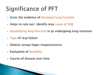
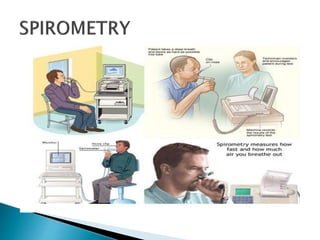
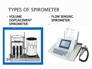
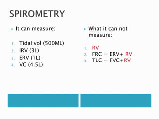
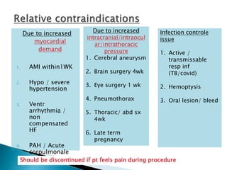
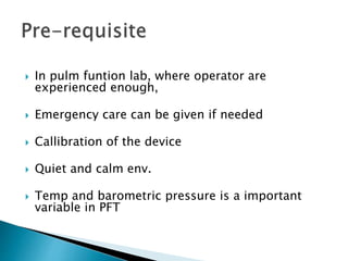
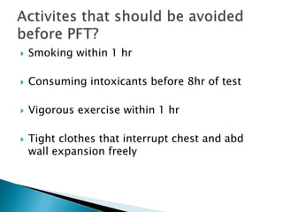
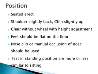
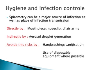
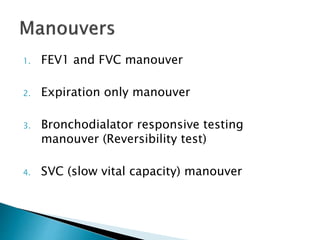
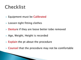
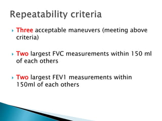
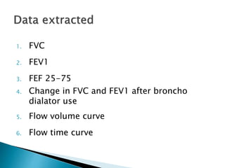
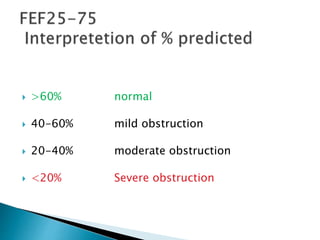
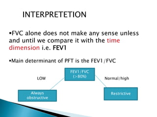
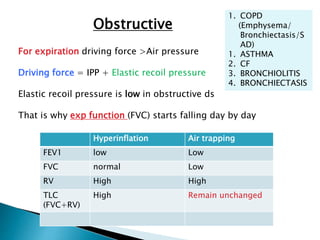
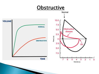
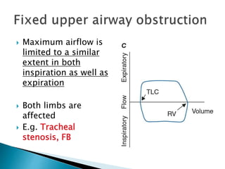
PULMONARY FUNCTION TESTS PLAY A VERY IMPORTANT ROLE IN ESTIMATING THE FUNCTION OF LUNGS ESPECIALLY IN ASTHAMA AND COPD, One of the frequent reasons patients see their primary care physicians is for the symptom of dyspnea. Among the objective tests to quantify this symptom is the pulmonary function test
Pulmonary function tests (PFTs) are noninvasive tests that show how well the lungs are working. The tests measure lung volume, capacity, rates of flow, and gas exchange. This information can help your healthcare provider diagnose and decide the treatment of certain lung disorders.
Various types of Pulmonary function tests, physiology , how to do spirometry, how to interpret, precautions while doing it, newer pfts : described in this ppt.
PULMONARY FUNCTION TESTS - LAB DATA INTERPRETATIONLincyAsha
PULMONARY FUNCTION TESTS
LAB DATA INTERPRETATION
CLINICAL PHARMACY PRACTICE
M.PHARMACY
PHARMACY PRACTICE
1ST YEAR
Pulmonary function tests are a series of tests performed to examine a patient’s respiratory system and identify the severity of pulmonary impairment.
These tests are performed to measure a patient’s lung volume, capacity, flow rate and gas exchange.
This allows medical professionals to obtain an accurate diagnosis and determine the best course of medical intervention for the patient.
In general there are two types of lung disorders that these tests can be used to assess
Obstructive lung diseases
Restrictive lung diseases
1.OBSTRUCTIVE LUNG DISEASES
It include conditions that make it difficult to exhale air out of the lungs
This results in shortness of breath that occurs from narrowing and constriction of the airways and causes the patient to have decreased flow rates. Eg. COPD, Asthma
2.RESTRICTIVE LUNG DISEASES
It include conditions that make it difficult to fully fill the lungs with air during inhalation.
When the lungs aren’t fully able to expand it causes the patient to have decreased lung volumes. Eg. Pulmonary fibrosis, interstitial lung disease
Pulmonary function tests would be indicated for the following:
On healthy patients as part of a routine physical exam
Evaluate signs and symptoms of lung disease
Diagnosis of certain medical conditions
Measure current stage of disease and evaluate its progress
Assess how a patient is responding to different treatments
Determine patient’s condition before surgery to assess the risk of respiratory complications
Screen people who are at risk of pulmonary disease
Determine how much a patient’s airways have narrowed due to disorders
In certain types of work environments to assess the health of employees.
Additionally PFTs may be indicated for the following
Chronic lung conditions
Restrictive airway problems
Asthma
COPD
Shortness of breath
Impairment or disability
Early morning wheezing
Chest muscle weakness
Lung cancer
Respiratory infections
STATIC LUNG VOLUMES
Lung volume is the amount of air breathed by an individual under a specific condition.
1.Tidal Volume (TV)
It is the volume of air inspired or expired during normal breathing at rest.
2.Inspiratory Reserve Volume (IRV)
It is the volume of air inspired with maximum effort over and above the normal tidal volume.
3.Expiratory Reserve Volume (ERV)
It is the volume of air expired forcefully after a normal respiration.
4.Residual Volume (RV)
It is the volume of air remaining in the lungs after a forceful expiration
STATIC LUNG CAPACITIES
1.Inspiratory capacity (IC)
It is the amount of air a person can inspire forcefully after a normal respiration.
IC = TV+IRV
2.Functional Residual Capacity (FRC)
It is the amount of air that remains in the lungs at the end of normal respiration.
FRC = ERV+RV
3.Vital Capacity (VC)
It is the maximum volume of air exhaled forcefully from the lungs after a maximum inspiration.
4.Total Lung Capacity
Title: Sense of Smell
Presenter: Dr. Faiza, Assistant Professor of Physiology
Qualifications:
MBBS (Best Graduate, AIMC Lahore)
FCPS Physiology
ICMT, CHPE, DHPE (STMU)
MPH (GC University, Faisalabad)
MBA (Virtual University of Pakistan)
Learning Objectives:
Describe the primary categories of smells and the concept of odor blindness.
Explain the structure and location of the olfactory membrane and mucosa, including the types and roles of cells involved in olfaction.
Describe the pathway and mechanisms of olfactory signal transmission from the olfactory receptors to the brain.
Illustrate the biochemical cascade triggered by odorant binding to olfactory receptors, including the role of G-proteins and second messengers in generating an action potential.
Identify different types of olfactory disorders such as anosmia, hyposmia, hyperosmia, and dysosmia, including their potential causes.
Key Topics:
Olfactory Genes:
3% of the human genome accounts for olfactory genes.
400 genes for odorant receptors.
Olfactory Membrane:
Located in the superior part of the nasal cavity.
Medially: Folds downward along the superior septum.
Laterally: Folds over the superior turbinate and upper surface of the middle turbinate.
Total surface area: 5-10 square centimeters.
Olfactory Mucosa:
Olfactory Cells: Bipolar nerve cells derived from the CNS (100 million), with 4-25 olfactory cilia per cell.
Sustentacular Cells: Produce mucus and maintain ionic and molecular environment.
Basal Cells: Replace worn-out olfactory cells with an average lifespan of 1-2 months.
Bowman’s Gland: Secretes mucus.
Stimulation of Olfactory Cells:
Odorant dissolves in mucus and attaches to receptors on olfactory cilia.
Involves a cascade effect through G-proteins and second messengers, leading to depolarization and action potential generation in the olfactory nerve.
Quality of a Good Odorant:
Small (3-20 Carbon atoms), volatile, water-soluble, and lipid-soluble.
Facilitated by odorant-binding proteins in mucus.
Membrane Potential and Action Potential:
Resting membrane potential: -55mV.
Action potential frequency in the olfactory nerve increases with odorant strength.
Adaptation Towards the Sense of Smell:
Rapid adaptation within the first second, with further slow adaptation.
Psychological adaptation greater than receptor adaptation, involving feedback inhibition from the central nervous system.
Primary Sensations of Smell:
Camphoraceous, Musky, Floral, Pepperminty, Ethereal, Pungent, Putrid.
Odor Detection Threshold:
Examples: Hydrogen sulfide (0.0005 ppm), Methyl-mercaptan (0.002 ppm).
Some toxic substances are odorless at lethal concentrations.
Characteristics of Smell:
Odor blindness for single substances due to lack of appropriate receptor protein.
Behavioral and emotional influences of smell.
Transmission of Olfactory Signals:
From olfactory cells to glomeruli in the olfactory bulb, involving lateral inhibition.
Primitive, less old, and new olfactory systems with different path
Basavarajeeyam is an important text for ayurvedic physician belonging to andhra pradehs. It is a popular compendium in various parts of our country as well as in andhra pradesh. The content of the text was presented in sanskrit and telugu language (Bilingual). One of the most famous book in ayurvedic pharmaceutics and therapeutics. This book contains 25 chapters called as prakaranas. Many rasaoushadis were explained, pioneer of dhatu druti, nadi pareeksha, mutra pareeksha etc. Belongs to the period of 15-16 century. New diseases like upadamsha, phiranga rogas are explained.
NVBDCP.pptx Nation vector borne disease control programSapna Thakur
NVBDCP was launched in 2003-2004 . Vector-Borne Disease: Disease that results from an infection transmitted to humans and other animals by blood-feeding arthropods, such as mosquitoes, ticks, and fleas. Examples of vector-borne diseases include Dengue fever, West Nile Virus, Lyme disease, and malaria.
TEST BANK for Operations Management, 14th Edition by William J. Stevenson, Ve...kevinkariuki227
TEST BANK for Operations Management, 14th Edition by William J. Stevenson, Verified Chapters 1 - 19, Complete Newest Version.pdf
Recomendações da OMS sobre cuidados maternos e neonatais para uma experiência pós-natal positiva.
Em consonância com os ODS – Objetivos do Desenvolvimento Sustentável e a Estratégia Global para a Saúde das Mulheres, Crianças e Adolescentes, e aplicando uma abordagem baseada nos direitos humanos, os esforços de cuidados pós-natais devem expandir-se para além da cobertura e da simples sobrevivência, de modo a incluir cuidados de qualidade.
Estas diretrizes visam melhorar a qualidade dos cuidados pós-natais essenciais e de rotina prestados às mulheres e aos recém-nascidos, com o objetivo final de melhorar a saúde e o bem-estar materno e neonatal.
Uma “experiência pós-natal positiva” é um resultado importante para todas as mulheres que dão à luz e para os seus recém-nascidos, estabelecendo as bases para a melhoria da saúde e do bem-estar a curto e longo prazo. Uma experiência pós-natal positiva é definida como aquela em que as mulheres, pessoas que gestam, os recém-nascidos, os casais, os pais, os cuidadores e as famílias recebem informação consistente, garantia e apoio de profissionais de saúde motivados; e onde um sistema de saúde flexível e com recursos reconheça as necessidades das mulheres e dos bebês e respeite o seu contexto cultural.
Estas diretrizes consolidadas apresentam algumas recomendações novas e já bem fundamentadas sobre cuidados pós-natais de rotina para mulheres e neonatos que recebem cuidados no pós-parto em unidades de saúde ou na comunidade, independentemente dos recursos disponíveis.
É fornecido um conjunto abrangente de recomendações para cuidados durante o período puerperal, com ênfase nos cuidados essenciais que todas as mulheres e recém-nascidos devem receber, e com a devida atenção à qualidade dos cuidados; isto é, a entrega e a experiência do cuidado recebido. Estas diretrizes atualizam e ampliam as recomendações da OMS de 2014 sobre cuidados pós-natais da mãe e do recém-nascido e complementam as atuais diretrizes da OMS sobre a gestão de complicações pós-natais.
O estabelecimento da amamentação e o manejo das principais intercorrências é contemplada.
Recomendamos muito.
Vamos discutir essas recomendações no nosso curso de pós-graduação em Aleitamento no Instituto Ciclos.
Esta publicação só está disponível em inglês até o momento.
Prof. Marcus Renato de Carvalho
www.agostodourado.com
Ozempic: Preoperative Management of Patients on GLP-1 Receptor Agonists Saeid Safari
Preoperative Management of Patients on GLP-1 Receptor Agonists like Ozempic and Semiglutide
ASA GUIDELINE
NYSORA Guideline
2 Case Reports of Gastric Ultrasound
263778731218 Abortion Clinic /Pills In Harare ,sisternakatoto
263778731218 Abortion Clinic /Pills In Harare ,ABORTION WOMEN’S CLINIC +27730423979 IN women clinic we believe that every woman should be able to make choices in her pregnancy. Our job is to provide compassionate care, safety,affordable and confidential services. That’s why we have won the trust from all generations of women all over the world. we use non surgical method(Abortion pills) to terminate…Dr.LISA +27730423979women Clinic is committed to providing the highest quality of obstetrical and gynecological care to women of all ages. Our dedicated staff aim to treat each patient and her health concerns with compassion and respect.Our dedicated group ABORTION WOMEN’S CLINIC +27730423979 IN women clinic we believe that every woman should be able to make choices in her pregnancy. Our job is to provide compassionate care, safety,affordable and confidential services. That’s why we have won the trust from all generations of women all over the world. we use non surgical method(Abortion pills) to terminate…Dr.LISA +27730423979women Clinic is committed to providing the highest quality of obstetrical and gynecological care to women of all ages. Our dedicated staff aim to treat each patient and her health concerns with compassion and respect.Our dedicated group of receptionists, nurses, and physicians have worked together as a teamof receptionists, nurses, and physicians have worked together as a team wwww.lisywomensclinic.co.za/
Title: Sense of Taste
Presenter: Dr. Faiza, Assistant Professor of Physiology
Qualifications:
MBBS (Best Graduate, AIMC Lahore)
FCPS Physiology
ICMT, CHPE, DHPE (STMU)
MPH (GC University, Faisalabad)
MBA (Virtual University of Pakistan)
Learning Objectives:
Describe the structure and function of taste buds.
Describe the relationship between the taste threshold and taste index of common substances.
Explain the chemical basis and signal transduction of taste perception for each type of primary taste sensation.
Recognize different abnormalities of taste perception and their causes.
Key Topics:
Significance of Taste Sensation:
Differentiation between pleasant and harmful food
Influence on behavior
Selection of food based on metabolic needs
Receptors of Taste:
Taste buds on the tongue
Influence of sense of smell, texture of food, and pain stimulation (e.g., by pepper)
Primary and Secondary Taste Sensations:
Primary taste sensations: Sweet, Sour, Salty, Bitter, Umami
Chemical basis and signal transduction mechanisms for each taste
Taste Threshold and Index:
Taste threshold values for Sweet (sucrose), Salty (NaCl), Sour (HCl), and Bitter (Quinine)
Taste index relationship: Inversely proportional to taste threshold
Taste Blindness:
Inability to taste certain substances, particularly thiourea compounds
Example: Phenylthiocarbamide
Structure and Function of Taste Buds:
Composition: Epithelial cells, Sustentacular/Supporting cells, Taste cells, Basal cells
Features: Taste pores, Taste hairs/microvilli, and Taste nerve fibers
Location of Taste Buds:
Found in papillae of the tongue (Fungiform, Circumvallate, Foliate)
Also present on the palate, tonsillar pillars, epiglottis, and proximal esophagus
Mechanism of Taste Stimulation:
Interaction of taste substances with receptors on microvilli
Signal transduction pathways for Umami, Sweet, Bitter, Sour, and Salty tastes
Taste Sensitivity and Adaptation:
Decrease in sensitivity with age
Rapid adaptation of taste sensation
Role of Saliva in Taste:
Dissolution of tastants to reach receptors
Washing away the stimulus
Taste Preferences and Aversions:
Mechanisms behind taste preference and aversion
Influence of receptors and neural pathways
Impact of Sensory Nerve Damage:
Degeneration of taste buds if the sensory nerve fiber is cut
Abnormalities of Taste Detection:
Conditions: Ageusia, Hypogeusia, Dysgeusia (parageusia)
Causes: Nerve damage, neurological disorders, infections, poor oral hygiene, adverse drug effects, deficiencies, aging, tobacco use, altered neurotransmitter levels
Neurotransmitters and Taste Threshold:
Effects of serotonin (5-HT) and norepinephrine (NE) on taste sensitivity
Supertasters:
25% of the population with heightened sensitivity to taste, especially bitterness
Increased number of fungiform papillae
Ethanol (CH3CH2OH), or beverage alcohol, is a two-carbon alcohol
that is rapidly distributed in the body and brain. Ethanol alters many
neurochemical systems and has rewarding and addictive properties. It
is the oldest recreational drug and likely contributes to more morbidity,
mortality, and public health costs than all illicit drugs combined. The
5th edition of the Diagnostic and Statistical Manual of Mental Disorders
(DSM-5) integrates alcohol abuse and alcohol dependence into a single
disorder called alcohol use disorder (AUD), with mild, moderate,
and severe subclassifications (American Psychiatric Association, 2013).
In the DSM-5, all types of substance abuse and dependence have been
combined into a single substance use disorder (SUD) on a continuum
from mild to severe. A diagnosis of AUD requires that at least two of
the 11 DSM-5 behaviors be present within a 12-month period (mild
AUD: 2–3 criteria; moderate AUD: 4–5 criteria; severe AUD: 6–11 criteria).
The four main behavioral effects of AUD are impaired control over
drinking, negative social consequences, risky use, and altered physiological
effects (tolerance, withdrawal). This chapter presents an overview
of the prevalence and harmful consequences of AUD in the U.S.,
the systemic nature of the disease, neurocircuitry and stages of AUD,
comorbidities, fetal alcohol spectrum disorders, genetic risk factors, and
pharmacotherapies for AUD.
micro teaching on communication m.sc nursing.pdfAnurag Sharma
Microteaching is a unique model of practice teaching. It is a viable instrument for the. desired change in the teaching behavior or the behavior potential which, in specified types of real. classroom situations, tends to facilitate the achievement of specified types of objectives.
2. Its a generic term used to indicate a
battery of test or manouvers
performed using standardised
equipment to evaluate function of
lung.
3. 1. Tests for ventilatory functions:
Evaluate lung volumes and capacities:
- Spirometry
- Body Plethysmography
- Gas dilution method
( FRC & RV detection)
Evaluate hypersensitivity of airway
- Bronco provocation test
4. 2.Tests for gas exchange:
DLCO , ABG, Oxymetry and Capnography.
3. Other Tests:
Tests for lung compliance.
Test for resistance and impedence: Impulse oscillometry.
Assessment of regional lung functions.
Assessment of respiratory muscle strength.
Breath condensate.
5. Gives the evidence of deranged lung function
Helps to rule out/ identify resp cause of SOB
Quantifying lung function in pt undergoing lung resection
Type of resp failure
Detects airway hyper responsiveness
Evaluation of disability
Course of disease over time
14. It can measure:
1. Tidal vol (500ML)
2. IRV (3L)
3. ERV (1L)
4. VC (4.5L)
What it can not
measure:
1. RV
2. FRC = ERV+ RV
3. TLC = FVC+RV
15. 1.Diagnostic:
A.To evaluate symptoms, signs, and
abnormal lab tests
• Symptoms: dyspnea wheezing, orthopnea, cough, phlegm
production, chest pain
• Signs: diminished breath sound, over inflation, expiratory
slowing, cyanosis, chest deformity, unexplained crackles
• Abnormal lab test: hypoxemia, hypercapnia, abnormal chest
radiograph
B. To measure the effect of disease on
pulmonary function
16. C. To screen individuals at risk of having
pulmonary disease
Smokers
Occupational exposure
D. To assess preop risk
prognosis( lung transplant)
health ststus before begining of a
strenous physical activity program
17. 2. Monitoring
To assess therapeutic intervention
a) Bronchodilator therapy
b) Steroid therapy (Asthma, ILD)
c) Antibiotics in cystic fibrosis
To describe the course of disease that affect lung
function
Pulmonary disease (obstructive airway disiese,ILD)
Cardiac disease (CHF)
NM disorders (GB syndrome)
18. To monitor people exposed to injurious agent
To monitor adverse reaction to drug with known
pulmonary toxicity
3.To identify Flow Volume Loop patterns
4. Disability/Impairment evaluation
To assess patients as part of rehabilitation
program
To assess risk as a part of an insurance
evaluation
5. Public Health: For clinical research
19. Due to increased
myocardial
demand
1. AMI within1WK
2. Hypo / severe
hypertension
3. Ventr
arrhythmia /
non
compensated
HF
4. PAH / Acute
corpulmonale
Due to increased
intracranial/intraocul
ar/intrathoracic
pressure
1. Cerebral aneurysm
2. Brain surgery 4wk
3. Eye surgery 1 wk
4. Pneumothorax
5. Thoracic/ abd sx
4wk
6. Late term
pregnancy
Infection controle
issue
1. Active /
transmissable
resp inf
(TB/covid)
2. Hemoptysis
3. Oral lesion/ bleed
Should be discontinued if pt feels pain during procedure
20. In pulm funtion lab, where operator are
experienced enough,
Emergency care can be given if needed
Callibration of the device
Quiet and calm env.
Temp and barometric pressure is a important
variable in PFT
21. Smoking within 1 hr
Consuming intoxicants before 8hr of test
Vigorous exercise within 1 hr
Tight clothes that interrupt chest and abd
wall expansion freely
22. Seated erect
Shoulder slightly back, Chin slightly up
Chair without wheel with height adjustment
Feet should be flat on the floor
Nose clip or manual occlusion of nose
should be used
Test in standing position are more or less
similar to sitting
23. Spirometry can be a major source of infection as
well as place of infection transmission
Directly by : Mouthpiece, noseclip, chair arms
Indirectly by : Aerosol droplet generation
Avoide this risks by : Handwashing/sanitisation
Use of disposable
equipment where possible
26. Equipment must be Calibrated
Loosen tight fitting clothes
Denture if they are loose better tobe removed
Age, Weight, Height is recorded
Explain the pt about the procedure
Counsel that the procedure may not be comfortable
27. Maximum
inspiration
•Start at flow zero
•Inspire as deeply as possible
•No pause
•Wait till the inspiration is complete ( eye brow
becomes widened, head starts quivering)
Blast of
expiration
• Don’t just blow, blow as much and as forcefully
possible
Continued
expiration
At least 6 sec is acceptable
3 sec for <10 yrs
Wait for the plateau phase in display
Ask for the next step
Maximum
inspiration
after forced
expiration
•To return to TLC and complete the flow volume
curve
•This will cross check whether the
pt began exp from full
inspiration or not
FEV1/FVC MANEUVER
28. Expiration only manouver
(Done for children only)
Inspire maximum lung vol within 2 s
Insert mouth piece
Innitiate max expiration
Remove mouth piece at end of forced expiration
29. Bronchodialator responsive test
Degree of improvement of air flow in response to bronco
dilator
Can differentiate Asthma from other COPD
But neither asthma nor COPD is diagnosed on
spirometry
bronchodilator Dose FEV1 before and
after
Salbutamol 200-400mcg
via large spacer
15 min
Terbutaline 500 mcg via
turbohaler
15 min
Ipratropium 160 mcg via
spacer
45 min
30. The test can be concluded
when both ACCEPTABILITY and
REPEATABILITY criteria are met
To ensure the REPRODUCIBILITY of
the test
Requires 5 to maximum 8 attempt
31. Free from artefact ( Cough / Early glottis closure)
Good start
Free from leaks
Extrapolation back from the PEFR gives a
theoretical start time (should be within 5% of FVC
or within 150 ml)
Acceptable exhalation
Adults :at least 6 sec of exhalation and plateue
Children <10yrs : at least 3 sec of exhalation
32. Three acceptable maneuvers (meeting above
criteria)
Two largest FVC measurements within 150 ml
of each others
Two largest FEV1 measurements within
150ml of each others
33. 1. FVC
2. FEV1
3. FEF 25-75
4. Change in FVC and FEV1 after broncho
dialator use
5. Flow volume curve
6. Flow time curve
36. Total volume of air that
can be exhaled forcefully
from TLC
The majority of FVC can
be exhaled in<3 seconds
in normal
Often prolonged in
Obstructive lung disease
Measured in liters
37. 80-120% = Normal
70-79% = Mild
reduction
50-69% = Moderate
reduction
<50% = Severe
reduction
38. Volume of air forcefully
expired from full
inspiration (TLC) in first
second
Normally 75-80% of
FVC is exhaled in first
second
Thus FEV1/FVC can be
utilised to characterise
lung disease
39. Mean forced
expiratory flow
during middle half of
FVC
May reflect effort
independent
expiration And the
status of the small
air way
40.
41. >60% normal
40-60% mild obstruction
20-40% moderate obstruction
<20% Severe obstruction
42. INTERPRETETION
FVC alone does not make any sense unless
and until we compare it with the time
dimension i.e. FEV1
Main determinant of PFT is the FEV1/FVC
FEV1/FVC
(>80%)
LOW Normal/high
Always
obstructive
Restrictive
43. Obstructive
1. COPD
(Emphysema/
Bronchiectasis/S
AD)
1. ASTHMA
2. CF
3. BRONCHIOLITIS
4. BRONCHIECTASIS
For expiration driving force >Air pressure
Driving force = IPP + Elastic recoil pressure
Elastic recoil pressure is low in obstructive ds
That is why exp function (FVC) starts falling day by day
Hyperinflation Air trapping
FEV1 low Low
FVC normal Low
RV High High
TLC
(FVC+RV)
High Remain unchanged
45. Q. Can obstr lung dis have normal FEV1/FVC ?
Yes, in Small air way disease.
Here we diagnose SAD by FEF 25-75 /MMEFR/MEAN FORCED
EXPIRATORY FLOW RATE
It is the average flow rate of lung in middle 50% of
the FVC manouver
It is the slope of the line
46. Reversibility test by SABA
If FEV1 > 12%
and
FVC > 200ml
It indicates
Bronchial
Asthma
49. Here lung’s inspiratory function is affected
So IRV is decreased
FVC= IRV + TV +ERV = FVC
FEV1 remains normal more or less
FEV1/FVC remains normal/high
53. Intra
parenchymal
NM disorder Chest wall
deformity
DLCO LOW NORMAL
TLC LOW LOW NORMAL
RV LOW NORMAL
RV/TLC HIGH NORMAL
KCO
(DLCO/VOL)
NORMAL HIGH
54. It uses a small amount of CO to measure gas
exchange across the alveolar membrane
during a 10 sec breath hold.
CO in exhaled air is analysed to determine
the quantity of CO crossing the membrane
55. FACTORS INCREASD DLCO DECREASED
DLCO
Thickness of alveolar
membrane
ILD
smoking
Altered
volume/surface area
ratio
Emphysema
Hb available Polycythemia Anaemia
Pregnancy
Blood coming to
capilleris
Pulmonary Hge
Asthma
Left to right shunt
Exercise
Pulmonary vascular
disease
56. Diffusion limited gas
Affinity of Hb for CO is >200 times
Partial pressure of CO in pulmonary
capillaries rises very slowly
KCO (Diffusion coefficient)= DLCO/
Lung Volume
Normal in ILD
Raised in Extra parencymal disease
57. Intra
parenchymal
NM disorder Chest wall
deformity
DLCO LOW NORMAL
TLC LOW LOW NORMAL
RV LOW NORMAL
RV/TLC HIGH NORMAL
KCO
(DLCO/VOL)
NORMAL HIGH
58. Different flow vol loops
NORMAL
Scooped
pattern
Fixed airway
obstruction
Extra thoracic
variable obstruction
Intra thoracic
variable obstr
60. Narrowing is maximal
in Expiration
As lesion is intra
thoracic
Intra thoracic pressure
is maximum in
expiration and lower
than air
Thus expiratory limb is
flattened
e.g. Tracheomalacia
61. Obstruction worsens
in inspiration
As negative pressure
narrows trachea
Thus Inspiratory limb
flattens
E.g extrinsic
compression from
Goiter, LN
62. Maximum airflow is
limited to a similar
extent in both
inspiration as well as
expiration
Both limbs are
affected
E.g. Tracheal
stenosis, FB
63. Most common cause is poor patient technique
Sub optimal inspiration
Sub maximal expiratory effort
Delay in forced expiration
Shortened expiratory time
Air leak around the mouth piece
Poor posture = leaning forward
Subjects must be observed and encouraged through the
procedure
66. Highly dependent on patient compliance and
effort
Thus FEV1 and FVC may be underestimated
Not useful for <4years/unconcious/sedated
Can not measure RV,FRC,TLC