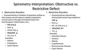
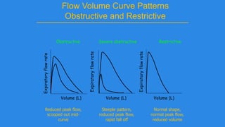
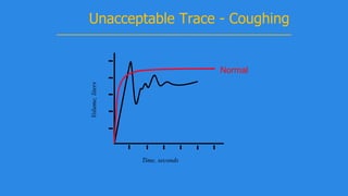
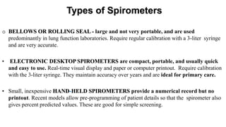
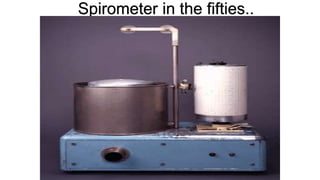
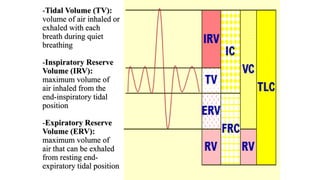
DR. GIRISH JAIN
Dept. of Pulmonary Medicine,
Mahatma Gandhi Medical College , Jaipur
• Pulmonary function tests is a term used to indicate studies or
maneuvers that performed using standardized equipment to
measure lung function.
• Evaluates one or more aspects of the respiratory system
• – Respiratory mechanics
• – Lung parenchymal function/ Gas exchange
• – Cardiopulmonary interaction
INTRODUCTION
PFT INCLUDES:-
1. SPIROMETRY
2. DLCO
3. BODY PLETHYSMOGRAPHY
4. ABG
SPIROMETRY
Definition
Spirometry is a method of assessing lung function by
measuring the volume of air that patient can expel
from lungs after a maximal inspiration.
o physiological test that measures how an individual
inhales or exhales volumes of air as a function of
time.
• Measures VC, FVC, FEV1, PEFR.
• CAN’T MEASURE – FRC, RV, TLC.
John Hutchinson 1811-1861 A.D.
• Born - 1811
• Medical education in London
• M.R.C.S in 1836; M.D. in 1848
• Assistant Physician to the Hospital,
Brompton in 1850
• Invented the spirometer and the science
of spirometry
• Related vital capacity to height and
presented his work to Statistical Society and
Royal Medical and Surgical Society
1846, “On the capacity of the lungs” reported
on 2130 subjects in Medico-surgical
Transactions 29:137, London
Silhouette of Hutchinson Performing Spirometry
From
Chest,
2002
Types of Spirometers
o BELLOWS OR ROLLING SEAL - large and not very portable, and are used
predominantly in lung function laboratories. Require regular calibration with a 3-liter syringe
and are very accurate.
• ELECTRONIC DESKTOP SPIROMETERS are compact, portable, and usually quick
and easy to use. Real-time visual display and paper or computer printout. Require calibration
with the 3-liter syringe. They maintain accuracy over years and are ideal for primary care.
• Small, inexpensive HAND-HELD SPIROMETERS provide a numerical record but no
printout. Recent models allow pre-programming of patient details so that the spirometer also
gives percent predicted values. These are good for simple screening.
Spirometers
• Volume – displacement
 Water seal spirometer
 Dry rolling seal spirometer
 Bellows spirometer
• Flow sensing device
• Rotating vanes (Turbine)
• Pressure differential flow sensing spirometers
• Heated wire flow sensors
• Pitot tube flow sensors
Spirometer in the fifties..
Volume Measuring Spirometer
Flow Measuring Spirometer
NOW-Hand-held spirometer
Many types are available..
Spirobank
MicroLoop
SpiroPro
Simplicity
Datospir 70 SpiroStar
Pulmonary Function Parameters
• Total lung capacity (TLC)
• Vital capacity (VC)
• Inspiratory capacity (IC)
• Functional residual capacity (FRC)
• Inspiratory reserve volume (IRV)
• Expiratory reserve volume (ERV)
• Tidal volume (TV)
• Residual volume (RV)
-Tidal Volume (TV):
volume of air inhaled or
exhaled with each
breath during quiet
breathing
-Inspiratory Reserve
Volume (IRV):
maximum volume of
air inhaled from the
end-inspiratory tidal
position
-Expiratory Reserve
Volume (ERV):
maximum volume of
air that can be exhaled
from resting end-
expiratory tidal position
*Residual Volume (RV):
->Volume of air remaining in lungs after
maximium exhalation
->Indirectly measured (FRC-ERV) not by
spirometry
*Total Lung Capacity (TLC): Sum of all
volume compartments or volume of air in
lungs after maximum inspiration
*Vital Capacity (VC): TLC minus RV or
maximum volume of air exhaled from
maximal inspiratory level
*Inspiratory Capacity (IC): Sum of IRV and
TV or the maximum volume of air that can be
inhaled from the end-expiratory tidal position
Functional Residual
Capacity (FRC):
Sum of RV and ERV
or the volume of air
in the lungs at end-
expiratory tidal
position
Measured with
multiple-breath
closed-circuit
helium dilution,
multiple-breath
open-circuit nitrogen
washout, or body
plethysmography
(not by spirometry)
Indications for spirometry:-
• Diagnostic
• To evaluate symptoms, signs, or abnormal laboratory tests
• -Symptoms: dyspnea, wheezing, orthopnea, cough, chest pain
• -Signs: diminished breath sounds, overinflation,cyanosis, chest deformity, unexplained crepts
• -Abnormal laboratory tests: hypoxemia, hypercapnia, polycythemia,
• abnormal chest radiographs
• To measure the effect of disease on pulmonary function
• To screen individuals at risk of having pulmonary diseases
• -Smokers
• -Individuals in occupations with exposures to injurious substances
• -Some routine physical examinations
• To assess preoperative risk
• To assess prognosis (lung transplant, etc.)
• To assess health status before enrollment in strenuous physical activity programs
• Monitoring
• To assess therapeutic interventions
• -bronchodilator therapy
• -Steroid treatment for asthma, interstitial lung disease, etc.
• -Management of congestive heart failure
• To describe the course of diseases affecting lung function
• -Pulmonary diseases
• Obstructive airways diseases
• Interstitial lung diseases
• -Cardiac diseases
• Congestive heart failure
• -Neuromuscular diseases
• Guillain-Barre Syndrome
• To monitor persons in occupations with exposure to injurious agents
• To monitor for adverse reactions to drugs with known pulmonary toxicity
Contraindications
• Patients within 1 month of a myocardial infarction/Unstable cardiovascular status.
• Significant Chest or abdominal pain of any cause.
• Oral or facial pain may exacerbated by a mouthpiece.
• Stress incontinence, hernia.
• Dementia or confused state.
• Very ill or dyspnoeic patient.
• Hemoptysis of unknown origin.
• Pneumothorax, pulmonary embolism, severe infection.
• Thoracic/abdominal/cerebral aneurysms.
• Recent eye surgery/surgery of thorax or abdomen.
• Presence of acute disease process that may interfere with performance (e.g. Nausea,
vomiting, ac severe asthma}.
• history of stroke or severe head injury.
Complications of Spirometry
Nosocomial infections.
Syncope, dizziness, light headedness.
Chest pain, muscle cramps.
Paroxymal coughing.
Bronchospasm.
Oxygen desaturation due to interruption of oxygen
therapy.
Performing spirometry
 Spirometry can be performed at the bedside, physician’s
consulting room or a laboratory and must be recorded.
 Sitting is considered safe in order to prevent falling due
to syncope. However, in obese and pregnant subjects, the
standing position may be preferred. In children[more
than 4 yrs] seated position is preferred.
Preliminaries to Patient Testing
• Physical assessment
• Pulmonary history
• Instructions prior to testing
– Drink / drugs / chocolate / smoke
• Physical measurement
– Age, sex, height, weight
Subject should be asked to avoid :
Smoking 24 hours
Drinks containing caffeine
/ theobromines
12 hours
Chocolates 8 hours
Alcohol 4 hours
Heavy meal 2 hours
Exercise 30 minutes
Bronchodilators &
Bronchoconstrictors
 Short-acting bronchodilators, Anticholinergics- within previous
6 hours.
 Long-acting bronchodilators- within previous 12 hours.
 Slow release theophyllines- within previous 24 hours.
Precaution: while testing reversibility
*Patients should not use Bronchodilator in preceding hours- May cause
false negative test
-Short acting beta-2 agonist -6hours
-Long acting/oral beta 2 agonist-12 hours
-Slow release theophyllines- within previous 24 hours.
Patient Instructions
• Good seal around mouthpiece
• Take a few normal breaths in and out.
• Take a deep breath in, and then blow out as hard and as fast
as possible.
Remove nose ring & loose denture if any.
Test procedure
Three distinct phases to the FVC manoeuvre:
1) maximal inspiration;
2) ‘‘blast’’ of exhalation;
3) continued complete exhalation to the end of test (EOT).
Technician should demonstrate appropriate technique
1. Maximal Inspiration;
Inhale rapidly and completely from functional residual capacity (FRC),
the breathing tube should be inserted into the subject’s mouth , making
sure the lips are sealed around the mouthpiece and that the tongue
does not occlude it, and then t FVC manoeuvre should be begun
without hesitation.
-Preceding inspiration should be fast and any pause at full inspiration
should be minimal (i.e. only for 1–2 s).
- inhalation should full before beginning forced exhalation
‘2.‘Blast’’ of exhalation
The subject should prompted to ‘‘blast,’’ not just ‘‘blow,’’ air from their
lungs, and then subject should encouraged to fully exhale.
3.Continued complete exhalation to the end of test (EOT).
Throughout manoeuvre, enthusiastic coaching of the subject using
appropriate body language and phrases, such as ‘‘keep going’’, is
required.
If patient feels ‘‘dizzy’’manoeuvre should stopped, since syncope could
followed after procedure.
If testing undertaken on standing or in another position, this must be
documented on report.
Flow Volume Curve
Expiratory
flow rate
L/sec
Volume (L)
FVC
Maximum
expiratory flow
(PEF)
Inspiratory
flow rate
L/sec
RVTLC
Restrictive pattern
Normal
Obstructive pattern
Fixed obstruction:
constant airflow limitation on inspiration and expiration
so loop are flattened and look like rectangle -such as in
tumor, tracheal stenosis
Variable extrathoracic obstruction:
limitation of inspiratory flow, flattened inspiratory loop
expiratory flow is unimpaired—such as in vocal cord
dysfunction, unilateral vocal cord paralysis
Variable intrathoracic obstruction:
flattening of expiratory limb; as in malignancy or
tracheomalacia
Upper Airway Obstruction
Graph Showing FEV1 and FVC
1 2 3 4 5 6
1
2
3
4Volume,liters
Time, seconds
FVC5
1
FEV1 = 4L
FVC = 5L
FEV1/FVC = 0.8
Spirogram Patterns
• Normal
• Obstructive
• Restrictive
• Mixed Obstructive and Restrictive
Predicted Normal Values
 Age
 Height
 Sex
 Ethnic Origin
Affected by:
Standard Spirometric Indicies
• FEV1 - Forced expiratory volume in one second:
The volume of air expired in the first second of
the blow
• FVC - Forced vital capacity:
The total volume of air that can be forcibly
exhaled in one breath
• FEV1/FVC ratio:
The fraction of air exhaled in the first second
relative to the total volume exhaled
FVC
• Forced vital capacity
(FVC):
– Total volume of air that can
be exhaled forcefully from
TLC
– The majority of FVC can be
exhaled in <3 seconds in
normal people, but often is
much more prolonged in
obstructive diseases
– Measured in liters (L)
FVC
• Interpretation of % predicted:
– 80-120% Normal
– 70-79% Mild reduction
– 50%-69% Moderate reduction
– <50% Severe reduction
FVC
FEV1
• Forced expiratory volume
in 1 second: (FEV1)
– Volume of air forcefully
expired from full inflation
(TLC) in the first second
– Measured in liters (L)
– Normal people can exhale
more than 75-80% of their
FVC in the first second;
thus the FEV1/FVC can
be utilized to characterize
lung disease
FEV1
• Interpretation of % predicted:
– >75% Normal
– 60%-75% Mild obstruction
– 50-59% Moderate obstruction
– <49% Severe obstruction
FEV1 FVC
FEV1/FVC ratio:
Called as Tiffeneau-pinelli index
The fraction of air exhaled in the first second
relative to the total volume exhaled
80 or higher - Normal
79 or lower - Abnormal
FEF25-75
• Forced expiratory flow 25-
75% (FEF25-75)
– Mean forced expiratory flow
during middle half of FVC
– Measured in L/sec
– May reflect status of the
small airways
– Highly variable
– Depends heavily on FVC
FEF25-75
»Interpretation of % predicted:
»>80% Normal
»60-79% Mild obstruction
»40-59% Moderate obstruction
»<40% Severe obstruction
MAX. VOLUNTARY VENTILATION
Largest volume that can be breath per minute by
voluntary effort , as hard & as fast as possible.
N – 150-175 l/min.
Estimate of max. Ventilation available to meet increased
physiological demand.
Measured for 12 secs – extrapolated for 1 min.
 MVV = FEV1 X 35
Criteria for Normal
Spirometry
• FEV1: % predicted > 80%
• FVC: % predicted > 80%
• FEV1/FVC: > 0.7
Spirometry Interpretation: Obstructive vs.
Restrictive Defect
• Obstructive Disorders
– Characterized by a limitation of expiratory airflow so
that airways cannot empty as rapidly compared to
normal (such as through narrowed airways from
bronchospasm, inflammation, etc.)
– FVC nl or↓
– FEV1 ↓
– FEF25-75% ↓
– FEV1/FVC ↓
– TLC nl or ↑
Examples:
– Asthma
– Emphysema
– Cystic Fibrosis
• Restrictive Disorders
– Characterized by reduced lung
volumes/decreased lung compliance
- FVC ↓
– FEV1 ↓
– FEF 25-75% nl to ↓
– FEV1/FVC N↑
– TLC ↓
Examples:
– Interstitial Fibrosis
– Scoliosis
– Obesity
– Lung Resection
– Neuromuscular diseases
– Cystic Fibrosis
Normal vs. Obstructive vs. Restrictive
(Hyatt,
2003)
Spirometry: Obstructive Disease
Volume,liters
Time, seconds
5
4
3
2
1
1 2 3 4 5 6
FEV1 = 1.8L
FVC = 3.2L
FEV1/FVC = 0.56
Normal
Obstructive
Flow Volume Curve Patterns
Obstructive and Restrictive
Obstructive Severe obstructive Restrictive
Volume (L)
Expiratoryflowrate
Expiratoryflowrate
Expiratoryflowrate
Volume (L) Volume (L)
Steeple pattern,
reduced peak flow,
rapid fall off
Normal shape,
normal peak flow,
reduced volume
Reduced peak flow,
scooped out mid-
curve
Bronchodilator Reversibility Testing
• Provides the best achievable FEV1
(and FVC)
• Helps to differentiate COPD from
asthma
Must be interpreted with clinical
history - neither asthma nor COPD
are diagnosed on spirometry alone
Bronchodilator Reversibility
Testing in COPD
• FEV1 should be measured (minimum twice,
within 5%) before a bronchodilator is given
• The bronchodilator should be given by
metered dose inhaler through a spacer
device or by nebulizer to be certain it has
been inhaled
• The bronchodilator dose should be selected
to be high on the dose/response curve
Bronchodilator Reversibility Testing
in COPD
•An increase in FEV1 that is both greater
than 200 ml and 12% above the pre-
bronchodilator FEV1 (baseline value) is
considered significant
•It is usually helpful to report the absolute
change (in ml) as well as the % change
from baseline to set the improvement in a
clinical context
Figure 5.1-6.
Bronchodilator
Reversibility
Testing in COPD
Volume,liters
Time, seconds
FEV1 = 1.9L
FVC = 2.0L
FEV1/FVC = 0.95
1 2 3 4 5 6
5
4
3
2
1
Spirometry: Restrictive Disease
Normal
Restrictive
Flow Volume Curve Patterns
Obstructive and Restrictive
Obstructive Severe obstructive Restrictive
Volume (L)
Expiratoryflowrate
Expiratoryflowrate
Expiratoryflowrate
Volume (L) Volume (L)
Steeple pattern,
reduced peak flow,
rapid fall off
Normal shape,
normal peak flow,
reduced volume
Reduced peak flow,
scooped out mid-
curve
Mixed Obstructive and Restrictive
Volume,liters
Time, seconds
FEV1 = 0.5L
FVC = 1.5L
FEV1/FVC = 0.30
Normal
Obstructive - Restrictive
Spirometry - Quality Control
• Most common cause of inconsistent
readings is poor patient technique
 Sub-optimal inspiration
 Sub-maximal expiratory effort
 Delay in forced expiration
 Shortened expiratory time
 Air leak around the mouthpiece
• Subjects must be observed and
encouraged throughout the procedure
Troubleshooting
Examples - Unacceptable Traces
Unacceptable Trace - Poor Effort
Volume,liters
Time, seconds
May be accompanied by a slow start
Inadequate sustaining of effort
Variable expiratory effort
Normal
Volume,liters
Time, seconds
Unacceptable Trace – Stop Early
Normal
Volume,liters
Time, seconds
Unacceptable Trace – Slow Start
Normal
Volume,liters
Time, seconds
Unacceptable Trace - Coughing
Normal
Volume,liters
Time, seconds
Unacceptable Trace – Extra Breath
Normal
SPIROMETRY‐Acceptability Criteria
Good start of test‐ without any hesitation
• No coughing / glottic closure
• No variable flow
• No early termination(> 6 sec)
• No air leak
• Reproducibility‐ The test is without excessive variability
The two largest values for FVC and the two largest values for
FEV1 should vary by no more than 150 ML
Bronchodilator Reversibility:
Spirometry recorded 15-20 minutes after administration of a short-acting beta-agonist, eg. 200-
400mcg of salbutamol/80-160micro g of ipratropium or both, is used to assess
bronchodilator reversibility.
Calculation of Bronchodilator Reversibility:
Calculation of % improvement=
FEV1(post-bronchodilator) – FEV1(prebonchodilator) x100
FEV1(prebonchodilator)
*A positive reversibility test shows an increase in FEV1 by more than 12% and at least by more
than 200ml in comparison to baseline values.
*This test shows no or partial reversibility in a majority of pts. With COPD
BRONCHODILATOR DOSE FEV1 BEFORE &AFTER
SALBUTAMOL 200-400mcg via LARGE
VOLUME SPACER
15 min
TERBUTALINE 500 mcg via TURBOHALER 15 min
IPRATROPIUM 160 mcg via SPACER
Usually 8 puffs of 20 mcg
45 min
In some cases of poorly treated persistent asthma, bronchodilator reversibility may not be
present, steroid reversibility can be demonstrated 2 weeks after a course of oral
prednisolone in a single daily dose of 1 mg/kg for 2 weeks.
Calculation of Steroid Reversibility:
Calculation of % improvement =
FEV1(post-steroid) – FEV1(presteroid) x100
FEV1(presteroid)
*Borderline NORMAL values should be interpreted with caution and should be used along with
clinical information to decide whether the report is normal or abnormal.
Steps in interpretation of spirometry
 Examine the flow volume curve
 Is it acceptable
 Is it reproducible
 Select the personal best
 Examine the FEV1/VC ratio
 Examine the FVC
 Examine the mid expiratory flow values
 Examine the response to bronchodilators
Interpretation of simple spirometry:-
1
4 3
2
By dr girish pulmonary function tests

By dr girish pulmonary function tests

  • 1.
    DR. GIRISH JAIN Dept.of Pulmonary Medicine, Mahatma Gandhi Medical College , Jaipur
  • 2.
    • Pulmonary functiontests is a term used to indicate studies or maneuvers that performed using standardized equipment to measure lung function. • Evaluates one or more aspects of the respiratory system • – Respiratory mechanics • – Lung parenchymal function/ Gas exchange • – Cardiopulmonary interaction INTRODUCTION
  • 3.
    PFT INCLUDES:- 1. SPIROMETRY 2.DLCO 3. BODY PLETHYSMOGRAPHY 4. ABG
  • 4.
  • 5.
    Definition Spirometry is amethod of assessing lung function by measuring the volume of air that patient can expel from lungs after a maximal inspiration. o physiological test that measures how an individual inhales or exhales volumes of air as a function of time. • Measures VC, FVC, FEV1, PEFR. • CAN’T MEASURE – FRC, RV, TLC.
  • 6.
    John Hutchinson 1811-1861A.D. • Born - 1811 • Medical education in London • M.R.C.S in 1836; M.D. in 1848 • Assistant Physician to the Hospital, Brompton in 1850 • Invented the spirometer and the science of spirometry • Related vital capacity to height and presented his work to Statistical Society and Royal Medical and Surgical Society 1846, “On the capacity of the lungs” reported on 2130 subjects in Medico-surgical Transactions 29:137, London
  • 7.
    Silhouette of HutchinsonPerforming Spirometry From Chest, 2002
  • 8.
    Types of Spirometers oBELLOWS OR ROLLING SEAL - large and not very portable, and are used predominantly in lung function laboratories. Require regular calibration with a 3-liter syringe and are very accurate. • ELECTRONIC DESKTOP SPIROMETERS are compact, portable, and usually quick and easy to use. Real-time visual display and paper or computer printout. Require calibration with the 3-liter syringe. They maintain accuracy over years and are ideal for primary care. • Small, inexpensive HAND-HELD SPIROMETERS provide a numerical record but no printout. Recent models allow pre-programming of patient details so that the spirometer also gives percent predicted values. These are good for simple screening.
  • 9.
    Spirometers • Volume –displacement  Water seal spirometer  Dry rolling seal spirometer  Bellows spirometer • Flow sensing device • Rotating vanes (Turbine) • Pressure differential flow sensing spirometers • Heated wire flow sensors • Pitot tube flow sensors
  • 10.
  • 11.
  • 12.
  • 13.
  • 14.
    Many types areavailable.. Spirobank MicroLoop SpiroPro Simplicity Datospir 70 SpiroStar
  • 15.
    Pulmonary Function Parameters •Total lung capacity (TLC) • Vital capacity (VC) • Inspiratory capacity (IC) • Functional residual capacity (FRC) • Inspiratory reserve volume (IRV) • Expiratory reserve volume (ERV) • Tidal volume (TV) • Residual volume (RV)
  • 17.
    -Tidal Volume (TV): volumeof air inhaled or exhaled with each breath during quiet breathing -Inspiratory Reserve Volume (IRV): maximum volume of air inhaled from the end-inspiratory tidal position -Expiratory Reserve Volume (ERV): maximum volume of air that can be exhaled from resting end- expiratory tidal position
  • 18.
    *Residual Volume (RV): ->Volumeof air remaining in lungs after maximium exhalation ->Indirectly measured (FRC-ERV) not by spirometry *Total Lung Capacity (TLC): Sum of all volume compartments or volume of air in lungs after maximum inspiration *Vital Capacity (VC): TLC minus RV or maximum volume of air exhaled from maximal inspiratory level *Inspiratory Capacity (IC): Sum of IRV and TV or the maximum volume of air that can be inhaled from the end-expiratory tidal position
  • 19.
    Functional Residual Capacity (FRC): Sumof RV and ERV or the volume of air in the lungs at end- expiratory tidal position Measured with multiple-breath closed-circuit helium dilution, multiple-breath open-circuit nitrogen washout, or body plethysmography (not by spirometry)
  • 20.
    Indications for spirometry:- •Diagnostic • To evaluate symptoms, signs, or abnormal laboratory tests • -Symptoms: dyspnea, wheezing, orthopnea, cough, chest pain • -Signs: diminished breath sounds, overinflation,cyanosis, chest deformity, unexplained crepts • -Abnormal laboratory tests: hypoxemia, hypercapnia, polycythemia, • abnormal chest radiographs • To measure the effect of disease on pulmonary function • To screen individuals at risk of having pulmonary diseases • -Smokers • -Individuals in occupations with exposures to injurious substances • -Some routine physical examinations • To assess preoperative risk • To assess prognosis (lung transplant, etc.) • To assess health status before enrollment in strenuous physical activity programs
  • 21.
    • Monitoring • Toassess therapeutic interventions • -bronchodilator therapy • -Steroid treatment for asthma, interstitial lung disease, etc. • -Management of congestive heart failure • To describe the course of diseases affecting lung function • -Pulmonary diseases • Obstructive airways diseases • Interstitial lung diseases • -Cardiac diseases • Congestive heart failure • -Neuromuscular diseases • Guillain-Barre Syndrome • To monitor persons in occupations with exposure to injurious agents • To monitor for adverse reactions to drugs with known pulmonary toxicity
  • 22.
    Contraindications • Patients within1 month of a myocardial infarction/Unstable cardiovascular status. • Significant Chest or abdominal pain of any cause. • Oral or facial pain may exacerbated by a mouthpiece. • Stress incontinence, hernia. • Dementia or confused state. • Very ill or dyspnoeic patient. • Hemoptysis of unknown origin. • Pneumothorax, pulmonary embolism, severe infection. • Thoracic/abdominal/cerebral aneurysms. • Recent eye surgery/surgery of thorax or abdomen. • Presence of acute disease process that may interfere with performance (e.g. Nausea, vomiting, ac severe asthma}. • history of stroke or severe head injury.
  • 23.
    Complications of Spirometry Nosocomialinfections. Syncope, dizziness, light headedness. Chest pain, muscle cramps. Paroxymal coughing. Bronchospasm. Oxygen desaturation due to interruption of oxygen therapy.
  • 24.
    Performing spirometry  Spirometrycan be performed at the bedside, physician’s consulting room or a laboratory and must be recorded.  Sitting is considered safe in order to prevent falling due to syncope. However, in obese and pregnant subjects, the standing position may be preferred. In children[more than 4 yrs] seated position is preferred.
  • 25.
    Preliminaries to PatientTesting • Physical assessment • Pulmonary history • Instructions prior to testing – Drink / drugs / chocolate / smoke • Physical measurement – Age, sex, height, weight
  • 26.
    Subject should beasked to avoid : Smoking 24 hours Drinks containing caffeine / theobromines 12 hours Chocolates 8 hours Alcohol 4 hours Heavy meal 2 hours Exercise 30 minutes Bronchodilators & Bronchoconstrictors  Short-acting bronchodilators, Anticholinergics- within previous 6 hours.  Long-acting bronchodilators- within previous 12 hours.  Slow release theophyllines- within previous 24 hours.
  • 27.
    Precaution: while testingreversibility *Patients should not use Bronchodilator in preceding hours- May cause false negative test -Short acting beta-2 agonist -6hours -Long acting/oral beta 2 agonist-12 hours -Slow release theophyllines- within previous 24 hours.
  • 28.
    Patient Instructions • Goodseal around mouthpiece • Take a few normal breaths in and out. • Take a deep breath in, and then blow out as hard and as fast as possible. Remove nose ring & loose denture if any.
  • 29.
    Test procedure Three distinctphases to the FVC manoeuvre: 1) maximal inspiration; 2) ‘‘blast’’ of exhalation; 3) continued complete exhalation to the end of test (EOT). Technician should demonstrate appropriate technique
  • 30.
    1. Maximal Inspiration; Inhalerapidly and completely from functional residual capacity (FRC), the breathing tube should be inserted into the subject’s mouth , making sure the lips are sealed around the mouthpiece and that the tongue does not occlude it, and then t FVC manoeuvre should be begun without hesitation. -Preceding inspiration should be fast and any pause at full inspiration should be minimal (i.e. only for 1–2 s). - inhalation should full before beginning forced exhalation
  • 31.
    ‘2.‘Blast’’ of exhalation Thesubject should prompted to ‘‘blast,’’ not just ‘‘blow,’’ air from their lungs, and then subject should encouraged to fully exhale. 3.Continued complete exhalation to the end of test (EOT). Throughout manoeuvre, enthusiastic coaching of the subject using appropriate body language and phrases, such as ‘‘keep going’’, is required. If patient feels ‘‘dizzy’’manoeuvre should stopped, since syncope could followed after procedure. If testing undertaken on standing or in another position, this must be documented on report.
  • 32.
    Flow Volume Curve Expiratory flowrate L/sec Volume (L) FVC Maximum expiratory flow (PEF) Inspiratory flow rate L/sec RVTLC
  • 33.
  • 34.
    Fixed obstruction: constant airflowlimitation on inspiration and expiration so loop are flattened and look like rectangle -such as in tumor, tracheal stenosis Variable extrathoracic obstruction: limitation of inspiratory flow, flattened inspiratory loop expiratory flow is unimpaired—such as in vocal cord dysfunction, unilateral vocal cord paralysis Variable intrathoracic obstruction: flattening of expiratory limb; as in malignancy or tracheomalacia Upper Airway Obstruction
  • 35.
    Graph Showing FEV1and FVC 1 2 3 4 5 6 1 2 3 4Volume,liters Time, seconds FVC5 1 FEV1 = 4L FVC = 5L FEV1/FVC = 0.8
  • 36.
    Spirogram Patterns • Normal •Obstructive • Restrictive • Mixed Obstructive and Restrictive
  • 37.
    Predicted Normal Values Age  Height  Sex  Ethnic Origin Affected by:
  • 38.
    Standard Spirometric Indicies •FEV1 - Forced expiratory volume in one second: The volume of air expired in the first second of the blow • FVC - Forced vital capacity: The total volume of air that can be forcibly exhaled in one breath • FEV1/FVC ratio: The fraction of air exhaled in the first second relative to the total volume exhaled
  • 39.
    FVC • Forced vitalcapacity (FVC): – Total volume of air that can be exhaled forcefully from TLC – The majority of FVC can be exhaled in <3 seconds in normal people, but often is much more prolonged in obstructive diseases – Measured in liters (L)
  • 40.
    FVC • Interpretation of% predicted: – 80-120% Normal – 70-79% Mild reduction – 50%-69% Moderate reduction – <50% Severe reduction FVC
  • 41.
    FEV1 • Forced expiratoryvolume in 1 second: (FEV1) – Volume of air forcefully expired from full inflation (TLC) in the first second – Measured in liters (L) – Normal people can exhale more than 75-80% of their FVC in the first second; thus the FEV1/FVC can be utilized to characterize lung disease
  • 42.
    FEV1 • Interpretation of% predicted: – >75% Normal – 60%-75% Mild obstruction – 50-59% Moderate obstruction – <49% Severe obstruction FEV1 FVC
  • 43.
    FEV1/FVC ratio: Called asTiffeneau-pinelli index The fraction of air exhaled in the first second relative to the total volume exhaled 80 or higher - Normal 79 or lower - Abnormal
  • 44.
    FEF25-75 • Forced expiratoryflow 25- 75% (FEF25-75) – Mean forced expiratory flow during middle half of FVC – Measured in L/sec – May reflect status of the small airways – Highly variable – Depends heavily on FVC
  • 45.
    FEF25-75 »Interpretation of %predicted: »>80% Normal »60-79% Mild obstruction »40-59% Moderate obstruction »<40% Severe obstruction
  • 46.
    MAX. VOLUNTARY VENTILATION Largestvolume that can be breath per minute by voluntary effort , as hard & as fast as possible. N – 150-175 l/min. Estimate of max. Ventilation available to meet increased physiological demand. Measured for 12 secs – extrapolated for 1 min.  MVV = FEV1 X 35
  • 47.
    Criteria for Normal Spirometry •FEV1: % predicted > 80% • FVC: % predicted > 80% • FEV1/FVC: > 0.7
  • 48.
    Spirometry Interpretation: Obstructivevs. Restrictive Defect • Obstructive Disorders – Characterized by a limitation of expiratory airflow so that airways cannot empty as rapidly compared to normal (such as through narrowed airways from bronchospasm, inflammation, etc.) – FVC nl or↓ – FEV1 ↓ – FEF25-75% ↓ – FEV1/FVC ↓ – TLC nl or ↑ Examples: – Asthma – Emphysema – Cystic Fibrosis • Restrictive Disorders – Characterized by reduced lung volumes/decreased lung compliance - FVC ↓ – FEV1 ↓ – FEF 25-75% nl to ↓ – FEV1/FVC N↑ – TLC ↓ Examples: – Interstitial Fibrosis – Scoliosis – Obesity – Lung Resection – Neuromuscular diseases – Cystic Fibrosis
  • 49.
    Normal vs. Obstructivevs. Restrictive (Hyatt, 2003)
  • 51.
    Spirometry: Obstructive Disease Volume,liters Time,seconds 5 4 3 2 1 1 2 3 4 5 6 FEV1 = 1.8L FVC = 3.2L FEV1/FVC = 0.56 Normal Obstructive
  • 52.
    Flow Volume CurvePatterns Obstructive and Restrictive Obstructive Severe obstructive Restrictive Volume (L) Expiratoryflowrate Expiratoryflowrate Expiratoryflowrate Volume (L) Volume (L) Steeple pattern, reduced peak flow, rapid fall off Normal shape, normal peak flow, reduced volume Reduced peak flow, scooped out mid- curve
  • 53.
    Bronchodilator Reversibility Testing •Provides the best achievable FEV1 (and FVC) • Helps to differentiate COPD from asthma Must be interpreted with clinical history - neither asthma nor COPD are diagnosed on spirometry alone
  • 54.
    Bronchodilator Reversibility Testing inCOPD • FEV1 should be measured (minimum twice, within 5%) before a bronchodilator is given • The bronchodilator should be given by metered dose inhaler through a spacer device or by nebulizer to be certain it has been inhaled • The bronchodilator dose should be selected to be high on the dose/response curve
  • 55.
    Bronchodilator Reversibility Testing inCOPD •An increase in FEV1 that is both greater than 200 ml and 12% above the pre- bronchodilator FEV1 (baseline value) is considered significant •It is usually helpful to report the absolute change (in ml) as well as the % change from baseline to set the improvement in a clinical context
  • 56.
  • 59.
    Volume,liters Time, seconds FEV1 =1.9L FVC = 2.0L FEV1/FVC = 0.95 1 2 3 4 5 6 5 4 3 2 1 Spirometry: Restrictive Disease Normal Restrictive
  • 60.
    Flow Volume CurvePatterns Obstructive and Restrictive Obstructive Severe obstructive Restrictive Volume (L) Expiratoryflowrate Expiratoryflowrate Expiratoryflowrate Volume (L) Volume (L) Steeple pattern, reduced peak flow, rapid fall off Normal shape, normal peak flow, reduced volume Reduced peak flow, scooped out mid- curve
  • 61.
    Mixed Obstructive andRestrictive Volume,liters Time, seconds FEV1 = 0.5L FVC = 1.5L FEV1/FVC = 0.30 Normal Obstructive - Restrictive
  • 62.
    Spirometry - QualityControl • Most common cause of inconsistent readings is poor patient technique  Sub-optimal inspiration  Sub-maximal expiratory effort  Delay in forced expiration  Shortened expiratory time  Air leak around the mouthpiece • Subjects must be observed and encouraged throughout the procedure
  • 63.
  • 64.
    Unacceptable Trace -Poor Effort Volume,liters Time, seconds May be accompanied by a slow start Inadequate sustaining of effort Variable expiratory effort Normal
  • 65.
  • 66.
  • 67.
  • 68.
  • 69.
    SPIROMETRY‐Acceptability Criteria Good startof test‐ without any hesitation • No coughing / glottic closure • No variable flow • No early termination(> 6 sec) • No air leak • Reproducibility‐ The test is without excessive variability The two largest values for FVC and the two largest values for FEV1 should vary by no more than 150 ML
  • 70.
    Bronchodilator Reversibility: Spirometry recorded15-20 minutes after administration of a short-acting beta-agonist, eg. 200- 400mcg of salbutamol/80-160micro g of ipratropium or both, is used to assess bronchodilator reversibility. Calculation of Bronchodilator Reversibility: Calculation of % improvement= FEV1(post-bronchodilator) – FEV1(prebonchodilator) x100 FEV1(prebonchodilator) *A positive reversibility test shows an increase in FEV1 by more than 12% and at least by more than 200ml in comparison to baseline values. *This test shows no or partial reversibility in a majority of pts. With COPD
  • 71.
    BRONCHODILATOR DOSE FEV1BEFORE &AFTER SALBUTAMOL 200-400mcg via LARGE VOLUME SPACER 15 min TERBUTALINE 500 mcg via TURBOHALER 15 min IPRATROPIUM 160 mcg via SPACER Usually 8 puffs of 20 mcg 45 min
  • 72.
    In some casesof poorly treated persistent asthma, bronchodilator reversibility may not be present, steroid reversibility can be demonstrated 2 weeks after a course of oral prednisolone in a single daily dose of 1 mg/kg for 2 weeks. Calculation of Steroid Reversibility: Calculation of % improvement = FEV1(post-steroid) – FEV1(presteroid) x100 FEV1(presteroid) *Borderline NORMAL values should be interpreted with caution and should be used along with clinical information to decide whether the report is normal or abnormal.
  • 73.
    Steps in interpretationof spirometry  Examine the flow volume curve  Is it acceptable  Is it reproducible  Select the personal best  Examine the FEV1/VC ratio  Examine the FVC  Examine the mid expiratory flow values  Examine the response to bronchodilators
  • 74.
    Interpretation of simplespirometry:- 1 4 3 2