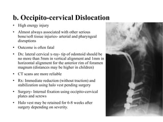
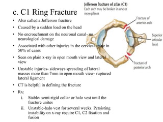
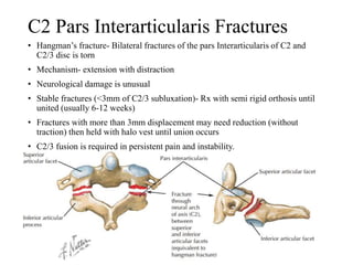
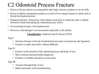
SlideShare a Scribd company logo
SPINAL DISORDERS
PRESENTER: Kosgei K. Victor
MBCh.B V
SUPERVISOR: Dr. Muteti E. MBCh.B, Mmed(Ortho), FCS, Spine
Specialist- Moi University-Eldoret, Kenya.
Introduction to the Spine
• Made of 33 vertebrae; 7 cervical, 12 thoracic, 5 lumbar, 5
sacral (fused) and 4 coccygeal (fused).
• Vertebrae form a functional column.
• Made of 3 columns:
a. Anterior- ALL & anterior 2/3 of vertebral body
b. Middle- PLL & posterior 1/3 of vertebral body
c. Posterior- pedicles, lamina, spinous processes and
ligaments
• Spinal curves: normal curves
a. Cervical lordosis
b. Thoracic kyphosis
c. Lumbar lordosis
d. Sacral kyphosis
• Functions:
a. Stability, support and movement
b. Haemopoietic function
c. Spinal cord and nerve root transmission
Spinal disorders- Introduction
• Spinal disorders can be:
A. Traumatic
B. Non traumatic
A. Traumatic Disorders
1. Cervical Spine Injuries
a. Occipito-condyle fracture
• High energy fracture
• Usually associated with skull fractures
• CT scan is essential in diagnosis (likely to be
missed in plain x-ray.
• Rx:
i. Impacted and undisplaced fractures- brace
immobilization for 8-12 weeks.
ii. Displaced fractures- halo vest or operative fixation.
b. Occipito-cervical Dislocation
• High energy injury
• Almost always associated with other serious
bone/soft tissue injuries- arterial and pharyngeal
disruptions
• Outcome is often fatal
• Dx: lateral cervical x-ray- tip of odontoid should be
no more than 5mm in vertical alignment and 1mm in
horizontal alignment for the anterior rim of foramen
magnum (distances may be higher in children)
• CT scans are more reliable
• Rx: Immediate reduction (without traction) and
stabilization using halo vest pending surgery
• Surgery- Internal fixation using occipito-cervical
plates and screws
• Halo vest may be retained for 6-8 weeks after
surgery depending on severity.
c. C1 Ring Fracture
• Also called a Jefferson fracture
• Caused by a sudden load on the head
• No encroachment on the neuronal canal- no
neurological damage
• Associated with other injuries in the cervical spine in
50% of cases
• Seen on plain x-ray in open mouth view and lateral
view
• Unstable injuries- sideways spreading of lateral
masses more than 7mm in open mouth view- ruptured
lateral ligament
• CT is helpful in defining the fracture
• Rx:
i. Stable- semi-rigid collar or halo vest until the
fracture unites
ii. Unstable-halo vest for several weeks. Persisting
instability on x-ray require C1, C2 fixation and
fusion
C2 Pars Interarticularis Fractures
• Hangman’s fracture- Bilateral fractures of the pars Interarticularis of C2 and
C2/3 disc is torn
• Mechanism- extension with distraction
• Neurological damage is unusual
• Stable fractures (<3mm of C2/3 subluxation)- Rx with semi rigid orthosis until
united (usually 6-12 weeks)
• Fractures with more than 3mm displacement may need reduction (without
traction) then held with halo vest until union occurs
• C2/3 fusion is required in persistent pain and instability.
C2 Odontoid Process Fracture
• Occur as flexion injuries in young adults after high velocity accidents or severe falls
• Occur in elderly osteoporotic people as a result of low energy trauma in which neck is
forced into hyperextension
• Displaced fracture- dislocation of the atlanto-axial joint in which the atlas is shifted
forward or backward taking the odontoid process with it
• No neurological injury with displacement
• However, cord damage is not uncommon especially in the elderly
Classification- Anderson and D’Alonzo (1974)
Type I
• Avulsion fracture at the tip of odontoid process due to traction by alar ligaments
• Fracture is stable and unites without difficulty
Type II
• Fracture at the junction of the odontoid process and body of axis
• Most common and potentially dangerous
• Fracture is unstable and prone to non union
Type III
• Fracture through body of axis
• Stable and unites with immobilization
Clinical Features
• Hx- severe neck strain followed by pain and stiffness due to
muscle spasm
• Neurological symptoms may occur
• Plain x-rays show the fracture
• MRI useful as it may reveal rupture of transverse ligaments
Treatment
Type I
• Immobilization in a rigid collar
Type II
• Undisplaced- held by halo vest, elderly- rigid collar
• Displaced- Reduced by traction then operative fusion of C1/2
Type III
• Undisplaced- halo vest for 8-12 weeks
• Displaced- reduction by halo traction the and immobilization by
halo vest for 8-12 weeks
Posterior Ligament Fracture
• Result from sudden flexion of mid cervical spine
• Upper vertebra tilts forward on the one below opening an
interspinous space posteriorly
• Patient complains of pain and there may be tenderness
• X-ray may reveal an increased gap space between adjacent spines
• Unstable injury- angulation more than 110, anterior translation of
vertebra more than 3.5mm or if facets are fractured and displaced
• Stable- semi rigid collar for 6 weeks is adequate
• Unstable- posterior fixation and fusion is advisable
Wedge Compression Fracture
• Result from flexion leading to compression fracture of vertebral
body
• Middle and posterior elements remain intact and injury is stable
• Rx- collar for 6-12 weeks
• Axial CT/MRI should be done to identify associated injuries.
Burst and Compression-flexion Fractures
Due to axial compression of cervical spine
Burst fracture- vertebral body is crushed in neutral position of head
Tear drop fracture- antero-inferior fragment of vertebral body is sheared off
due to combined compression and flexion
There’s risk of posterior displacement of vertebral fragment and spinal cord
injury
Plain x-ray show crushed vertebral body or a triangular fragment separated
from antero-inferior edge of fractured vertebra
Traction should be applied immediately
CT and MRI should be done to look for retropulsion of bone fragments into
spinal canal
Rx:
i. No neurological deficit- surgical or confinement to bed and traction for 2-4
weeks followed by halo vest immobilization for 6-8 weeks
- Halo vest unsuitable in initial Rx because it does not provide axial traction
ii. Neurological symptoms- urgent anterior decompression- anterior
corpectomy, bone grafting and plate fixations
- Sometimes posterior stabilization is needed.
Fracture Dislocation
• Caused by severe flexion or flexion rotation injuries
• Inferior articular facets of one vertebra ride forward
over superior facets of the vertebra below
• Posterior ligaments are ruptured and spine is unstable;
often there's cord damage
• Lateral x-ray shows forward displacement of a vertebra
on the one below of greater than half the vertebra’s
antero-posterior width
• Displacement must be reduced urgently using skull
traction for 6 weeks
• MRI should be performed to rule out presence of disc
disruption
• Collar and halo vest immobilization is advised for
further 12 weeks
• Posterior open reduction and fusion is advised if closed
reduction fails.
Hyperextension Injury
• Caused by acceleration forces
• Bone and joint disruptions are rare- strains of soft tissue
• Stable in neutral position
• Should be held by collar for 6-8 weeks
Avulsion Injury of the Spinous Process
• May occur in severe voluntary contraction of muscles at
the back of the neck
• Known as clay- shoveller’s fracture (C7 spinous process
fracture)
• Immobilized with a collar
Cervical Disc Herniation
• May cause severe pain radiating to one or both limbs
and neurological symptoms
• Diagnosis is confirmed by MRI or CT myelography
• Paresis may need surgical decompression
Sprained Neck (Whiplash injury)
• Common following RTAs
• Caused by sudden acceleration forces in vehicles without seat
headrest followed by recoil in flexion
• Women are more affected than male- neck muscles are more
gracile
• Pathology involves anterior spinal ligament, capsular fibers
straining
Clinical features
• Pain and neck stiffness appear 12-24 hours
• Pain radiates to the shoulders associated with headache, dizziness,
blurring of vision and paresthesia
• Neck muscles are tender and movements are restricted
• Neurological deficits are uncommon
• X- rays may show straightening of normal cervical lordosis due to
muscle spasms
• MRI may show disc degeneration
Grade Clinical Pattern
0 No neck symptoms or signs
1 Neck pain, stiffness and tenderness
No physical signs
2 Neck symptoms and musculoskeletal signs
3 Neck symptoms and neurological signs
4 Neck symptoms and fracture or dislocation
Grading
• DDx
- Vertebral fracture
- Mid cervical subluxation
• Rx
- Analgesics
- Physiotherapy
- Osteopathy an d chiropractic treatments- helpful
Thoracolumbar Fractures
Flexion compression Injuries
• Most common vertebral fracture
• Caused by severe spinal flexion
• Posterior ligaments remain intact
• Pain may be severe but fracture is usually stable
• Neurological injury is rare
• Rx:
i. Minimal wedging- bed rest for two weeks until
pain subsides
ii. Moderate wedging- thoracolumbar brace or cast
applied with the spine in extension for 1-2 weeks
• If instability persists, surgical fixation is indicated
Axial Compression or Burst Injury
• Severe axial compression may ‘explode’ the vertebral body
causing failure of both anterior and middle columns
• Posterior column is usually but not always undamaged
• Posterior part of vertebral body is shattered and fragments of
bone and disc may be displaced into spinal canal
• Injury is usually unstable
• AP x-rays show spreading of vertebral body with an increase
in interpedicular distance
• CT scan is essential to see retropulsion of bone fragments
into spinal canal
• Rx:
- No neurological damage- bed immobilization and
thoracolumbar brace or cast for 12 weeks
- Neurological symptoms- anterior decompression and
stabilization
Jack-Knife Injury
• Combined flexion and posterior distraction may cause mid
lumbar spine to jack-knife at an axis anterior to the vertebral
column
• Seen in lap seat belt injuries
• Unstable in flexion, though neurological damage is
uncommon
• X-rays show transverse fractures in the pedicles or
transverse process
• Lateral view shows opening of disc space posteriorly
• Heals rapidly and requires 3 months in a body cast and well
fitting brace
Fracture Dislocation
• Segmental displacement may occur with various
combinations of flexion, compression, rotation and shear
• All three columns are affected and spine is grossly unstable
• Often associated with neurological damage
• X-rays show fractures through the vertebral body, pedicles,
articular processes and laminae
• Varying degrees of subluxation or bilateral dislocation
• Associated fractures of transverse processes of ribs
• CT is helpful in demonstrating degree of spinal occlusion
• Fracture dislocation without neurological deficit- surgical
stabilization
• Fracture dislocation with partial neurological deficit-
surgical decompression and stabilization
• Fracture dislocation with paraplegia- shorten hospital stay,
rehabilitate and reduce painful deformity
Non-Traumatic Disorders
Scoliosis
• Lateral curvature of the spine
• Postural scoliosis- deformity is compensatory or secondary to
some condition outside the spine such as short leg or pelvic tilt.
Curvature disappears when patient sits
• Structural scoliosis- non correctable deformity of the affected
segment
• Spinous processes swing towards concavity of the fracture and
transverse processes rotate posteriorly
• Ribs on the convex side swing out producing a rib hump
• Deformity is liable to increase throughout the the growth period
• Types:
• Idiopathic- infantile or juvenile
• Osteopathic/congenital
• Neuropathic
• myopathic
Clinical Features
• Deformity is the presenting feature
• Pain is a rare complain- alert for neural tumors
• Family history is common
• Associated with abnormalities during childbirth or
pregnancy
• Spine is deviated from midline
Treatment
• Non operative
• Exercises
• Bracing- Milwaukee and Boston braces
• Operative
• Indications- rapidly deteriorating and cosmetic (deformities
>300 )
• Methods
- Harrington system
- Rod and Sub laminar wiring
- Cotrel dubousset system
Kyphosis
• Excessive thoracic curvature- hyper kyphosis, kyphos, gibbous
• Caused by sharp posterior angulation due to localized posterior collapse or
wedging of one or more vertebrae
• May result from congenital, fracture or spinal tuberculosis
Postural Kyphosis
• Associated with other postural defects like flat feet
• Voluntarily corrected by posture training and exercises
• Compensatory kyphosis due to other deformities- usually increased lumbosacral
lordosis
Structural Kyphosis
• Fixed and associated with changes in shape of vertebrae
• Children- congenital vertebral defects, skeletal dysplasia such as achondroplasia
and in osteogenesis imperfecta
• Older children- tuberculous spondylitis
• Adolescence- Scheurmann’s disease
• Adults- ankylosing spondylitis, TB spondylitis, spinal trauma or childhood
disorder
• Elderly- osteoporosis
• Treatment- dependent on the cause
Tuberculosis
• Most common site of skeletal tuberculosis
• Account for 50% of all musculoskeletal TB
Pathology
• Blood borne infection settles in vertebral body adjacent to
intervertebral disc
• Bone destruction and cessation with infection spreading to
adjacent disc space
• Paravertebral abscess may form and then track along muscle plane
and involve sacroiliac or hip joint or along the psoas muscle to the
thigh
• Gibbous (kyphos) develop as vertebral body collapse into each
other
• Risk of cord damage due to pressure by the abscess, granulation,
sequestra or displaced bone or ischemia from spinal artery
thrombosis
• With healing vertebrae re-calcify and bony fusion may occur
• Resultant kyphosis risk to cord compression
Clinical Features
• Long history of ill health and backache
• Gibbous formation is dominant
• Concurrent pulmonary TB common in children under 10 years
• Spinal movements are restricted
• Children under 10 years usually develop pigeon chest (pectus carinatum)
• Sensory/motor changes may occur in lower limb
• MRI and CT scan are suitable in investigation- can point out abscesses cord
compression, vertebral fractures
• Other tests- ESR- raised
- Montoux- positive
- Needle biopsy- limited to no neurological symptoms
• DDx- malignancies- metastases
- Pyogenic infections
- Hydatid disease
Treatment
• Objectives- eradicate or arrest disease
- prevent/correct deformity
- Prevent or treat complication
• Pharmacology- anti-TBs for six months
• Surgical- drainage and stabilization of vertebra
Pyogenic Osteomyelitis
• Uncommon- diagnosis usually delayed
• Risk groups- elderly, chronically ill and
immunosuppressed
Pathology
• Staph. Aureus is responsible for 50-60%
• Immunosuppressed patients- E. coli, Pseudomonas
• Sources of infection- hematogenous, inoculation
• Infection begins at vertebral end plates with spread to
disc and adjacent vertebra
• Can also spread along anterior longitudinal ligament
and into paravertebral tissues
• Spinal canal involvement (epidural abscess)- surgical
emmergency
Clinical Features
• Localized pain associated with muscle spasm and restricted
movements
• Point tenderness over affected area
• History of invasive procedure or distant infection
• Pyrexia and tachycardia may be present
• X-ray- loss of disc height, irregular disc space, erosion of
vertebral end plate
• Radionuclide scans- increased activity at site
• MRI- characteristic changes in vertebral end plates disc and
paravertebral tissues
• Needle biopsy- culture and sensitivity
• Other test- ESR, CRP and antistaphyloccocal antibodies
Treatment
• Bed rest, pain relief
• Iv antibiotics
• Operative- indications- poor response to conservative Rx,
presence of neurological signs and need to drain abscess
Discitis
• Infection limited to intervertebral disc
• Due to direct inoculation following discography,
chemonucleolysis or discectomy
• Vertebral end plates rapidly attacked and infection
spreads into vertebral body
• Tenderness over affected disc
• ESR is elevated
• Children- infection assumed to be blood borne
• X-ray features same as pyogenic spondylitis
• Rx- broad spectrum antibiotics
Intervertebral Disc Degeneration
• Major cause of chronic backache
• Age related- 50% occur in 50+ years
• Disc gradually dries out with ageing
• Nucleolus pulposus changes for turgid gelatinous bulb to brownish
dissociated structure
• Annulus fibrosus develop fissures parallel to the vertebral end
plate and herniations of nuclear material squeeze through the
fissures
• Disc cells then die at an increased rate
• Glycosaminoglycans production is diminished leading to poor
water retention and drying off
• Usually asymptomatic
• Pain is secondary to effects of disc prolapse
• X-rays- flattening out of disc space and osteophyte formation
• MRI- bulging of annulus fibrosus
• Rx-asymptomatic- no treatment
• Symptomatic- management of secondary effects
Intervertebral Disc Prolapse
• Physical stress is the probable cause
• May also result from hydrophilic disturbance of nuclear material
• Protrusion- posterior bulging of disc with intact annulus
• Extrusion- fibrocartlaginous material released posteriorly due to rupture of
annulus
• Sequestration- part of nucleus may separate and lie freely or move into the
intervertebral foramen
• Large central rupture may cause compression of Cauda equine
• Postero-lateral rupture compresses nerve root proximal to point of exit
• Major cause of acute back pain
• Sciatica- pain in buttocks and posterior thigh and calf due to nerve root
compression
• Paresthesia, numbness and weakness are common signs
• Back pain and sciatica made worse by coughing
• Cauda equine compression may cause urinary retention and perineal numbness
• X-rays- narrow disc, osteophytes
• CT and MRI are more reliable
Treatment
• Heat and analgesics soothe
• Exercise strengthen muscles
• Principles- rest, reduction, removal and rehabilitation
• Rest- patient kept in bed with hips and knees slightly flexed
• Reduction- continuous bed res for 2 weeks may reduce prolapse
• Epidural injection of corticosteroid and local anesthesia may help
• Chemonucleolysis- dissolution of nucleus pulposus using a
proteolytic enzyme (chymopapain)
• Removal- indications
1. Cauda equine compression syndrome
2. Neurological deterioration
3. Persistent pain and sciatic tension
• Operative methods- laminotomy and micro discectomy
• Rehabilitate-isometric exercises
Features of Cauda Equina Syndrome
1. Bladder and bowel incontinence
2. Perineal numbness
3. Bilateral sciatica
4. Lower limb weakness
5. Crossed straight leg sign
Chronic Back Pain Syndrome
• Self perpetuated
• Accompanied by non- organic physical signs
1. Pain and tenderness of bizarre degree of distribution
2. Pain on performing impressive but non-stressful
movements
3. Variations in raising the leg while distracting patient
attention
4. Sensory and motor function do not fit the
physiological patterns
5. Over determined behavior during physical
examination
• However, pathologies should be excluded before
making this diagnosis
Thank you…
Questions/comments
References
1. Apley’s system of orthopedics and fractures 9th
edition
2. Netters concise atlas of orthopedic anatomy

More Related Content

What's hot

Recurrent Dislocation of patella -PAWAN
Recurrent Dislocation of patella -PAWANRecurrent Dislocation of patella -PAWAN
Recurrent Dislocation of patella -PAWAN
Pawan Yadav
 
Shoulder dislocation: Types and Management Methods of Reduction
Shoulder dislocation: Types and Management Methods of ReductionShoulder dislocation: Types and Management Methods of Reduction
Shoulder dislocation: Types and Management Methods of Reduction
Uzair Siddiqui
 
Angular deformities around the knee seminar
Angular deformities around the knee seminarAngular deformities around the knee seminar
Angular deformities around the knee seminar
Prashanth Kumar
 
Radial club hand (Radial Dysplasia)
Radial club hand (Radial Dysplasia)Radial club hand (Radial Dysplasia)
Radial club hand (Radial Dysplasia)
Aiman Ali
 
Galleazi fracture dislocation
Galleazi fracture dislocationGalleazi fracture dislocation
Galleazi fracture dislocation
Fawas Muhammad
 
Clavicle fractures
Clavicle fractures Clavicle fractures
Clavicle fractures
Hardik Pawar
 
Genu Varum
Genu Varum Genu Varum
Genu Varum
Ayobami Ayodele
 
Proximal humerus fractures
Proximal humerus fracturesProximal humerus fractures
Proximal humerus fractures
mithilesh216
 
chondromalacia patellae
chondromalacia patellae chondromalacia patellae
chondromalacia patellae orthoprince
 
Median nerve injuries
Median nerve injuriesMedian nerve injuries
Median nerve injuriesNabil Khalil
 
Charcot joint arthropathy and rehabilitation management
Charcot joint arthropathy and rehabilitation managementCharcot joint arthropathy and rehabilitation management
Charcot joint arthropathy and rehabilitation management
Dr Harshanand Popalwar
 
Meniscal injury
Meniscal injuryMeniscal injury
Meniscal injury
manoj das
 
Iliotibial Band Syndrome (Itbs)
Iliotibial Band Syndrome (Itbs)Iliotibial Band Syndrome (Itbs)
Iliotibial Band Syndrome (Itbs)
colinmasterson
 
Colles' fracture & physiotherapy management
Colles' fracture & physiotherapy management Colles' fracture & physiotherapy management
Colles' fracture & physiotherapy management
ANNIE BLESSIE
 
Peripheral nerve injury
Peripheral nerve injuryPeripheral nerve injury
Peripheral nerve injury
Ahmed Shawky
 
Peripheral nerve injuries
Peripheral nerve injuriesPeripheral nerve injuries
Peripheral nerve injuries
Binod Chaudhary
 
Acromio clavicular joint injury
Acromio clavicular joint injuryAcromio clavicular joint injury
Acromio clavicular joint injury
Shri Guru Ram Rai Institute of Medical Science
 
Brachial plexus injuries by Dr. Rashi Goel PT
Brachial plexus injuries by Dr. Rashi Goel PTBrachial plexus injuries by Dr. Rashi Goel PT
Brachial plexus injuries by Dr. Rashi Goel PT
Orthocure Chain of Clinics
 
Shoulder dislocation
Shoulder dislocationShoulder dislocation
Shoulder dislocation
SCGH ED CME
 
Median nerve injury
Median nerve injuryMedian nerve injury
Median nerve injury
Biswajit Deka
 

What's hot (20)

Recurrent Dislocation of patella -PAWAN
Recurrent Dislocation of patella -PAWANRecurrent Dislocation of patella -PAWAN
Recurrent Dislocation of patella -PAWAN
 
Shoulder dislocation: Types and Management Methods of Reduction
Shoulder dislocation: Types and Management Methods of ReductionShoulder dislocation: Types and Management Methods of Reduction
Shoulder dislocation: Types and Management Methods of Reduction
 
Angular deformities around the knee seminar
Angular deformities around the knee seminarAngular deformities around the knee seminar
Angular deformities around the knee seminar
 
Radial club hand (Radial Dysplasia)
Radial club hand (Radial Dysplasia)Radial club hand (Radial Dysplasia)
Radial club hand (Radial Dysplasia)
 
Galleazi fracture dislocation
Galleazi fracture dislocationGalleazi fracture dislocation
Galleazi fracture dislocation
 
Clavicle fractures
Clavicle fractures Clavicle fractures
Clavicle fractures
 
Genu Varum
Genu Varum Genu Varum
Genu Varum
 
Proximal humerus fractures
Proximal humerus fracturesProximal humerus fractures
Proximal humerus fractures
 
chondromalacia patellae
chondromalacia patellae chondromalacia patellae
chondromalacia patellae
 
Median nerve injuries
Median nerve injuriesMedian nerve injuries
Median nerve injuries
 
Charcot joint arthropathy and rehabilitation management
Charcot joint arthropathy and rehabilitation managementCharcot joint arthropathy and rehabilitation management
Charcot joint arthropathy and rehabilitation management
 
Meniscal injury
Meniscal injuryMeniscal injury
Meniscal injury
 
Iliotibial Band Syndrome (Itbs)
Iliotibial Band Syndrome (Itbs)Iliotibial Band Syndrome (Itbs)
Iliotibial Band Syndrome (Itbs)
 
Colles' fracture & physiotherapy management
Colles' fracture & physiotherapy management Colles' fracture & physiotherapy management
Colles' fracture & physiotherapy management
 
Peripheral nerve injury
Peripheral nerve injuryPeripheral nerve injury
Peripheral nerve injury
 
Peripheral nerve injuries
Peripheral nerve injuriesPeripheral nerve injuries
Peripheral nerve injuries
 
Acromio clavicular joint injury
Acromio clavicular joint injuryAcromio clavicular joint injury
Acromio clavicular joint injury
 
Brachial plexus injuries by Dr. Rashi Goel PT
Brachial plexus injuries by Dr. Rashi Goel PTBrachial plexus injuries by Dr. Rashi Goel PT
Brachial plexus injuries by Dr. Rashi Goel PT
 
Shoulder dislocation
Shoulder dislocationShoulder dislocation
Shoulder dislocation
 
Median nerve injury
Median nerve injuryMedian nerve injury
Median nerve injury
 

Similar to Spinal Disorders 2017

Cervical spine
Cervical spineCervical spine
Cervical spine
HadeyaQ
 
CME SPINAL INJURY.pptx
CME SPINAL INJURY.pptxCME SPINAL INJURY.pptx
CME SPINAL INJURY.pptx
mieyoi
 
Spinal injuries
Spinal injuriesSpinal injuries
Spinal injuries
Rashmi Jain
 
The elbow fracture
The elbow fractureThe elbow fracture
The elbow fracture
MONTHER ALKHAWLANY
 
Subaxial spine
Subaxial spineSubaxial spine
Subaxial spine
SHAMEEJ MUHAMED KV
 
Cervicolumber Injury.pptx
Cervicolumber Injury.pptxCervicolumber Injury.pptx
Cervicolumber Injury.pptx
IrfanNashad1
 
spinal injuries presentation orthopaedics
spinal injuries presentation orthopaedicsspinal injuries presentation orthopaedics
spinal injuries presentation orthopaedics
seervidivyanshu18869
 
Rehab cervical through cocegeal power pt
Rehab cervical through cocegeal power ptRehab cervical through cocegeal power pt
Rehab cervical through cocegeal power ptMeklelle university
 
Clavicle fracture
Clavicle fractureClavicle fracture
Clavicle fracture
Yeswanth Mohan
 
CME Orthopedic.pptx
CME Orthopedic.pptxCME Orthopedic.pptx
CME Orthopedic.pptx
Parveen739769
 
Elbow Injuries.pptx
Elbow Injuries.pptxElbow Injuries.pptx
Elbow Injuries.pptx
esicOrtho1
 
Cervical spine injury antomy and management.pptx
Cervical spine injury antomy and management.pptxCervical spine injury antomy and management.pptx
Cervical spine injury antomy and management.pptx
MisStrom
 
Traumatic spinal injury
Traumatic spinal injuryTraumatic spinal injury
Traumatic spinal injury
Dr Abdul Qayyum Khan
 
Thoraco lumbar fractures of spine
Thoraco lumbar fractures of spine Thoraco lumbar fractures of spine
Thoraco lumbar fractures of spine
Sunil Santhosh
 
Upperlimb fractures bpt
Upperlimb fractures bptUpperlimb fractures bpt
Upperlimb fractures bptvaruntandra
 
Vertebral Fracture and Spinal Cord Injury.pptx
Vertebral Fracture and Spinal Cord Injury.pptxVertebral Fracture and Spinal Cord Injury.pptx
Vertebral Fracture and Spinal Cord Injury.pptx
VenoshaGunasekaran
 
Emergency management of common dislocations
Emergency management of common dislocationsEmergency management of common dislocations
Emergency management of common dislocations
Makafui Yigah
 
Cervical spine injuries and its management
Cervical spine injuries and its managementCervical spine injuries and its management
Cervical spine injuries and its management
Prashanth Kumar
 
cervicalspineinjuriesanditsmanagement-161119063840.pdf
cervicalspineinjuriesanditsmanagement-161119063840.pdfcervicalspineinjuriesanditsmanagement-161119063840.pdf
cervicalspineinjuriesanditsmanagement-161119063840.pdf
deepanraj369475
 
Presentation1.pptx, radiological imaging of spinal trauma and spinal cord inj...
Presentation1.pptx, radiological imaging of spinal trauma and spinal cord inj...Presentation1.pptx, radiological imaging of spinal trauma and spinal cord inj...
Presentation1.pptx, radiological imaging of spinal trauma and spinal cord inj...Abdellah Nazeer
 

Similar to Spinal Disorders 2017 (20)

Cervical spine
Cervical spineCervical spine
Cervical spine
 
CME SPINAL INJURY.pptx
CME SPINAL INJURY.pptxCME SPINAL INJURY.pptx
CME SPINAL INJURY.pptx
 
Spinal injuries
Spinal injuriesSpinal injuries
Spinal injuries
 
The elbow fracture
The elbow fractureThe elbow fracture
The elbow fracture
 
Subaxial spine
Subaxial spineSubaxial spine
Subaxial spine
 
Cervicolumber Injury.pptx
Cervicolumber Injury.pptxCervicolumber Injury.pptx
Cervicolumber Injury.pptx
 
spinal injuries presentation orthopaedics
spinal injuries presentation orthopaedicsspinal injuries presentation orthopaedics
spinal injuries presentation orthopaedics
 
Rehab cervical through cocegeal power pt
Rehab cervical through cocegeal power ptRehab cervical through cocegeal power pt
Rehab cervical through cocegeal power pt
 
Clavicle fracture
Clavicle fractureClavicle fracture
Clavicle fracture
 
CME Orthopedic.pptx
CME Orthopedic.pptxCME Orthopedic.pptx
CME Orthopedic.pptx
 
Elbow Injuries.pptx
Elbow Injuries.pptxElbow Injuries.pptx
Elbow Injuries.pptx
 
Cervical spine injury antomy and management.pptx
Cervical spine injury antomy and management.pptxCervical spine injury antomy and management.pptx
Cervical spine injury antomy and management.pptx
 
Traumatic spinal injury
Traumatic spinal injuryTraumatic spinal injury
Traumatic spinal injury
 
Thoraco lumbar fractures of spine
Thoraco lumbar fractures of spine Thoraco lumbar fractures of spine
Thoraco lumbar fractures of spine
 
Upperlimb fractures bpt
Upperlimb fractures bptUpperlimb fractures bpt
Upperlimb fractures bpt
 
Vertebral Fracture and Spinal Cord Injury.pptx
Vertebral Fracture and Spinal Cord Injury.pptxVertebral Fracture and Spinal Cord Injury.pptx
Vertebral Fracture and Spinal Cord Injury.pptx
 
Emergency management of common dislocations
Emergency management of common dislocationsEmergency management of common dislocations
Emergency management of common dislocations
 
Cervical spine injuries and its management
Cervical spine injuries and its managementCervical spine injuries and its management
Cervical spine injuries and its management
 
cervicalspineinjuriesanditsmanagement-161119063840.pdf
cervicalspineinjuriesanditsmanagement-161119063840.pdfcervicalspineinjuriesanditsmanagement-161119063840.pdf
cervicalspineinjuriesanditsmanagement-161119063840.pdf
 
Presentation1.pptx, radiological imaging of spinal trauma and spinal cord inj...
Presentation1.pptx, radiological imaging of spinal trauma and spinal cord inj...Presentation1.pptx, radiological imaging of spinal trauma and spinal cord inj...
Presentation1.pptx, radiological imaging of spinal trauma and spinal cord inj...
 

Recently uploaded

How STIs Influence the Development of Pelvic Inflammatory Disease.pptx
How STIs Influence the Development of Pelvic Inflammatory Disease.pptxHow STIs Influence the Development of Pelvic Inflammatory Disease.pptx
How STIs Influence the Development of Pelvic Inflammatory Disease.pptx
FFragrant
 
Ozempic: Preoperative Management of Patients on GLP-1 Receptor Agonists
Ozempic: Preoperative Management of Patients on GLP-1 Receptor Agonists  Ozempic: Preoperative Management of Patients on GLP-1 Receptor Agonists
Ozempic: Preoperative Management of Patients on GLP-1 Receptor Agonists
Saeid Safari
 
How to Give Better Lectures: Some Tips for Doctors
How to Give Better Lectures: Some Tips for DoctorsHow to Give Better Lectures: Some Tips for Doctors
How to Give Better Lectures: Some Tips for Doctors
LanceCatedral
 
Are There Any Natural Remedies To Treat Syphilis.pdf
Are There Any Natural Remedies To Treat Syphilis.pdfAre There Any Natural Remedies To Treat Syphilis.pdf
Are There Any Natural Remedies To Treat Syphilis.pdf
Little Cross Family Clinic
 
ARTHROLOGY PPT NCISM SYLLABUS AYURVEDA STUDENTS
ARTHROLOGY PPT NCISM SYLLABUS AYURVEDA STUDENTSARTHROLOGY PPT NCISM SYLLABUS AYURVEDA STUDENTS
ARTHROLOGY PPT NCISM SYLLABUS AYURVEDA STUDENTS
Dr. Vinay Pareek
 
Ophthalmology Clinical Tests for OSCE exam
Ophthalmology Clinical Tests for OSCE examOphthalmology Clinical Tests for OSCE exam
Ophthalmology Clinical Tests for OSCE exam
KafrELShiekh University
 
KDIGO 2024 guidelines for diabetologists
KDIGO 2024 guidelines for diabetologistsKDIGO 2024 guidelines for diabetologists
KDIGO 2024 guidelines for diabetologists
د.محمود نجيب
 
BRACHYTHERAPY OVERVIEW AND APPLICATORS
BRACHYTHERAPY OVERVIEW  AND  APPLICATORSBRACHYTHERAPY OVERVIEW  AND  APPLICATORS
BRACHYTHERAPY OVERVIEW AND APPLICATORS
Krishan Murari
 
Physiology of Chemical Sensation of smell.pdf
Physiology of Chemical Sensation of smell.pdfPhysiology of Chemical Sensation of smell.pdf
Physiology of Chemical Sensation of smell.pdf
MedicoseAcademics
 
Tom Selleck Health: A Comprehensive Look at the Iconic Actor’s Wellness Journey
Tom Selleck Health: A Comprehensive Look at the Iconic Actor’s Wellness JourneyTom Selleck Health: A Comprehensive Look at the Iconic Actor’s Wellness Journey
Tom Selleck Health: A Comprehensive Look at the Iconic Actor’s Wellness Journey
greendigital
 
Evaluation of antidepressant activity of clitoris ternatea in animals
Evaluation of antidepressant activity of clitoris ternatea in animalsEvaluation of antidepressant activity of clitoris ternatea in animals
Evaluation of antidepressant activity of clitoris ternatea in animals
Shweta
 
Cervical & Brachial Plexus By Dr. RIG.pptx
Cervical & Brachial Plexus By Dr. RIG.pptxCervical & Brachial Plexus By Dr. RIG.pptx
Cervical & Brachial Plexus By Dr. RIG.pptx
Dr. Rabia Inam Gandapore
 
planning for change nursing Management ppt
planning for change nursing Management pptplanning for change nursing Management ppt
planning for change nursing Management ppt
Thangamjayarani
 
heat stroke and heat exhaustion in children
heat stroke and heat exhaustion in childrenheat stroke and heat exhaustion in children
heat stroke and heat exhaustion in children
SumeraAhmad5
 
Light House Retreats: Plant Medicine Retreat Europe
Light House Retreats: Plant Medicine Retreat EuropeLight House Retreats: Plant Medicine Retreat Europe
Light House Retreats: Plant Medicine Retreat Europe
Lighthouse Retreat
 
ARTIFICIAL INTELLIGENCE IN HEALTHCARE.pdf
ARTIFICIAL INTELLIGENCE IN  HEALTHCARE.pdfARTIFICIAL INTELLIGENCE IN  HEALTHCARE.pdf
ARTIFICIAL INTELLIGENCE IN HEALTHCARE.pdf
Anujkumaranit
 
Superficial & Deep Fascia of the NECK.pptx
Superficial & Deep Fascia of the NECK.pptxSuperficial & Deep Fascia of the NECK.pptx
Superficial & Deep Fascia of the NECK.pptx
Dr. Rabia Inam Gandapore
 
Knee anatomy and clinical tests 2024.pdf
Knee anatomy and clinical tests 2024.pdfKnee anatomy and clinical tests 2024.pdf
Knee anatomy and clinical tests 2024.pdf
vimalpl1234
 
TEST BANK for Operations Management, 14th Edition by William J. Stevenson, Ve...
TEST BANK for Operations Management, 14th Edition by William J. Stevenson, Ve...TEST BANK for Operations Management, 14th Edition by William J. Stevenson, Ve...
TEST BANK for Operations Management, 14th Edition by William J. Stevenson, Ve...
kevinkariuki227
 
Vision-1.pptx, Eye structure, basics of optics
Vision-1.pptx, Eye structure, basics of opticsVision-1.pptx, Eye structure, basics of optics
Vision-1.pptx, Eye structure, basics of optics
Sai Sailesh Kumar Goothy
 

Recently uploaded (20)

How STIs Influence the Development of Pelvic Inflammatory Disease.pptx
How STIs Influence the Development of Pelvic Inflammatory Disease.pptxHow STIs Influence the Development of Pelvic Inflammatory Disease.pptx
How STIs Influence the Development of Pelvic Inflammatory Disease.pptx
 
Ozempic: Preoperative Management of Patients on GLP-1 Receptor Agonists
Ozempic: Preoperative Management of Patients on GLP-1 Receptor Agonists  Ozempic: Preoperative Management of Patients on GLP-1 Receptor Agonists
Ozempic: Preoperative Management of Patients on GLP-1 Receptor Agonists
 
How to Give Better Lectures: Some Tips for Doctors
How to Give Better Lectures: Some Tips for DoctorsHow to Give Better Lectures: Some Tips for Doctors
How to Give Better Lectures: Some Tips for Doctors
 
Are There Any Natural Remedies To Treat Syphilis.pdf
Are There Any Natural Remedies To Treat Syphilis.pdfAre There Any Natural Remedies To Treat Syphilis.pdf
Are There Any Natural Remedies To Treat Syphilis.pdf
 
ARTHROLOGY PPT NCISM SYLLABUS AYURVEDA STUDENTS
ARTHROLOGY PPT NCISM SYLLABUS AYURVEDA STUDENTSARTHROLOGY PPT NCISM SYLLABUS AYURVEDA STUDENTS
ARTHROLOGY PPT NCISM SYLLABUS AYURVEDA STUDENTS
 
Ophthalmology Clinical Tests for OSCE exam
Ophthalmology Clinical Tests for OSCE examOphthalmology Clinical Tests for OSCE exam
Ophthalmology Clinical Tests for OSCE exam
 
KDIGO 2024 guidelines for diabetologists
KDIGO 2024 guidelines for diabetologistsKDIGO 2024 guidelines for diabetologists
KDIGO 2024 guidelines for diabetologists
 
BRACHYTHERAPY OVERVIEW AND APPLICATORS
BRACHYTHERAPY OVERVIEW  AND  APPLICATORSBRACHYTHERAPY OVERVIEW  AND  APPLICATORS
BRACHYTHERAPY OVERVIEW AND APPLICATORS
 
Physiology of Chemical Sensation of smell.pdf
Physiology of Chemical Sensation of smell.pdfPhysiology of Chemical Sensation of smell.pdf
Physiology of Chemical Sensation of smell.pdf
 
Tom Selleck Health: A Comprehensive Look at the Iconic Actor’s Wellness Journey
Tom Selleck Health: A Comprehensive Look at the Iconic Actor’s Wellness JourneyTom Selleck Health: A Comprehensive Look at the Iconic Actor’s Wellness Journey
Tom Selleck Health: A Comprehensive Look at the Iconic Actor’s Wellness Journey
 
Evaluation of antidepressant activity of clitoris ternatea in animals
Evaluation of antidepressant activity of clitoris ternatea in animalsEvaluation of antidepressant activity of clitoris ternatea in animals
Evaluation of antidepressant activity of clitoris ternatea in animals
 
Cervical & Brachial Plexus By Dr. RIG.pptx
Cervical & Brachial Plexus By Dr. RIG.pptxCervical & Brachial Plexus By Dr. RIG.pptx
Cervical & Brachial Plexus By Dr. RIG.pptx
 
planning for change nursing Management ppt
planning for change nursing Management pptplanning for change nursing Management ppt
planning for change nursing Management ppt
 
heat stroke and heat exhaustion in children
heat stroke and heat exhaustion in childrenheat stroke and heat exhaustion in children
heat stroke and heat exhaustion in children
 
Light House Retreats: Plant Medicine Retreat Europe
Light House Retreats: Plant Medicine Retreat EuropeLight House Retreats: Plant Medicine Retreat Europe
Light House Retreats: Plant Medicine Retreat Europe
 
ARTIFICIAL INTELLIGENCE IN HEALTHCARE.pdf
ARTIFICIAL INTELLIGENCE IN  HEALTHCARE.pdfARTIFICIAL INTELLIGENCE IN  HEALTHCARE.pdf
ARTIFICIAL INTELLIGENCE IN HEALTHCARE.pdf
 
Superficial & Deep Fascia of the NECK.pptx
Superficial & Deep Fascia of the NECK.pptxSuperficial & Deep Fascia of the NECK.pptx
Superficial & Deep Fascia of the NECK.pptx
 
Knee anatomy and clinical tests 2024.pdf
Knee anatomy and clinical tests 2024.pdfKnee anatomy and clinical tests 2024.pdf
Knee anatomy and clinical tests 2024.pdf
 
TEST BANK for Operations Management, 14th Edition by William J. Stevenson, Ve...
TEST BANK for Operations Management, 14th Edition by William J. Stevenson, Ve...TEST BANK for Operations Management, 14th Edition by William J. Stevenson, Ve...
TEST BANK for Operations Management, 14th Edition by William J. Stevenson, Ve...
 
Vision-1.pptx, Eye structure, basics of optics
Vision-1.pptx, Eye structure, basics of opticsVision-1.pptx, Eye structure, basics of optics
Vision-1.pptx, Eye structure, basics of optics
 

Spinal Disorders 2017

  • 1. SPINAL DISORDERS PRESENTER: Kosgei K. Victor MBCh.B V SUPERVISOR: Dr. Muteti E. MBCh.B, Mmed(Ortho), FCS, Spine Specialist- Moi University-Eldoret, Kenya.
  • 2. Introduction to the Spine • Made of 33 vertebrae; 7 cervical, 12 thoracic, 5 lumbar, 5 sacral (fused) and 4 coccygeal (fused). • Vertebrae form a functional column. • Made of 3 columns: a. Anterior- ALL & anterior 2/3 of vertebral body b. Middle- PLL & posterior 1/3 of vertebral body c. Posterior- pedicles, lamina, spinous processes and ligaments • Spinal curves: normal curves a. Cervical lordosis b. Thoracic kyphosis c. Lumbar lordosis d. Sacral kyphosis • Functions: a. Stability, support and movement b. Haemopoietic function c. Spinal cord and nerve root transmission
  • 3. Spinal disorders- Introduction • Spinal disorders can be: A. Traumatic B. Non traumatic
  • 4. A. Traumatic Disorders 1. Cervical Spine Injuries a. Occipito-condyle fracture • High energy fracture • Usually associated with skull fractures • CT scan is essential in diagnosis (likely to be missed in plain x-ray. • Rx: i. Impacted and undisplaced fractures- brace immobilization for 8-12 weeks. ii. Displaced fractures- halo vest or operative fixation.
  • 5. b. Occipito-cervical Dislocation • High energy injury • Almost always associated with other serious bone/soft tissue injuries- arterial and pharyngeal disruptions • Outcome is often fatal • Dx: lateral cervical x-ray- tip of odontoid should be no more than 5mm in vertical alignment and 1mm in horizontal alignment for the anterior rim of foramen magnum (distances may be higher in children) • CT scans are more reliable • Rx: Immediate reduction (without traction) and stabilization using halo vest pending surgery • Surgery- Internal fixation using occipito-cervical plates and screws • Halo vest may be retained for 6-8 weeks after surgery depending on severity.
  • 6. c. C1 Ring Fracture • Also called a Jefferson fracture • Caused by a sudden load on the head • No encroachment on the neuronal canal- no neurological damage • Associated with other injuries in the cervical spine in 50% of cases • Seen on plain x-ray in open mouth view and lateral view • Unstable injuries- sideways spreading of lateral masses more than 7mm in open mouth view- ruptured lateral ligament • CT is helpful in defining the fracture • Rx: i. Stable- semi-rigid collar or halo vest until the fracture unites ii. Unstable-halo vest for several weeks. Persisting instability on x-ray require C1, C2 fixation and fusion
  • 7. C2 Pars Interarticularis Fractures • Hangman’s fracture- Bilateral fractures of the pars Interarticularis of C2 and C2/3 disc is torn • Mechanism- extension with distraction • Neurological damage is unusual • Stable fractures (<3mm of C2/3 subluxation)- Rx with semi rigid orthosis until united (usually 6-12 weeks) • Fractures with more than 3mm displacement may need reduction (without traction) then held with halo vest until union occurs • C2/3 fusion is required in persistent pain and instability.
  • 8. C2 Odontoid Process Fracture • Occur as flexion injuries in young adults after high velocity accidents or severe falls • Occur in elderly osteoporotic people as a result of low energy trauma in which neck is forced into hyperextension • Displaced fracture- dislocation of the atlanto-axial joint in which the atlas is shifted forward or backward taking the odontoid process with it • No neurological injury with displacement • However, cord damage is not uncommon especially in the elderly Classification- Anderson and D’Alonzo (1974) Type I • Avulsion fracture at the tip of odontoid process due to traction by alar ligaments • Fracture is stable and unites without difficulty Type II • Fracture at the junction of the odontoid process and body of axis • Most common and potentially dangerous • Fracture is unstable and prone to non union Type III • Fracture through body of axis • Stable and unites with immobilization
  • 9. Clinical Features • Hx- severe neck strain followed by pain and stiffness due to muscle spasm • Neurological symptoms may occur • Plain x-rays show the fracture • MRI useful as it may reveal rupture of transverse ligaments Treatment Type I • Immobilization in a rigid collar Type II • Undisplaced- held by halo vest, elderly- rigid collar • Displaced- Reduced by traction then operative fusion of C1/2 Type III • Undisplaced- halo vest for 8-12 weeks • Displaced- reduction by halo traction the and immobilization by halo vest for 8-12 weeks
  • 10. Posterior Ligament Fracture • Result from sudden flexion of mid cervical spine • Upper vertebra tilts forward on the one below opening an interspinous space posteriorly • Patient complains of pain and there may be tenderness • X-ray may reveal an increased gap space between adjacent spines • Unstable injury- angulation more than 110, anterior translation of vertebra more than 3.5mm or if facets are fractured and displaced • Stable- semi rigid collar for 6 weeks is adequate • Unstable- posterior fixation and fusion is advisable Wedge Compression Fracture • Result from flexion leading to compression fracture of vertebral body • Middle and posterior elements remain intact and injury is stable • Rx- collar for 6-12 weeks • Axial CT/MRI should be done to identify associated injuries.
  • 11. Burst and Compression-flexion Fractures Due to axial compression of cervical spine Burst fracture- vertebral body is crushed in neutral position of head Tear drop fracture- antero-inferior fragment of vertebral body is sheared off due to combined compression and flexion There’s risk of posterior displacement of vertebral fragment and spinal cord injury Plain x-ray show crushed vertebral body or a triangular fragment separated from antero-inferior edge of fractured vertebra Traction should be applied immediately CT and MRI should be done to look for retropulsion of bone fragments into spinal canal Rx: i. No neurological deficit- surgical or confinement to bed and traction for 2-4 weeks followed by halo vest immobilization for 6-8 weeks - Halo vest unsuitable in initial Rx because it does not provide axial traction ii. Neurological symptoms- urgent anterior decompression- anterior corpectomy, bone grafting and plate fixations - Sometimes posterior stabilization is needed.
  • 12. Fracture Dislocation • Caused by severe flexion or flexion rotation injuries • Inferior articular facets of one vertebra ride forward over superior facets of the vertebra below • Posterior ligaments are ruptured and spine is unstable; often there's cord damage • Lateral x-ray shows forward displacement of a vertebra on the one below of greater than half the vertebra’s antero-posterior width • Displacement must be reduced urgently using skull traction for 6 weeks • MRI should be performed to rule out presence of disc disruption • Collar and halo vest immobilization is advised for further 12 weeks • Posterior open reduction and fusion is advised if closed reduction fails.
  • 13. Hyperextension Injury • Caused by acceleration forces • Bone and joint disruptions are rare- strains of soft tissue • Stable in neutral position • Should be held by collar for 6-8 weeks Avulsion Injury of the Spinous Process • May occur in severe voluntary contraction of muscles at the back of the neck • Known as clay- shoveller’s fracture (C7 spinous process fracture) • Immobilized with a collar Cervical Disc Herniation • May cause severe pain radiating to one or both limbs and neurological symptoms • Diagnosis is confirmed by MRI or CT myelography • Paresis may need surgical decompression
  • 14. Sprained Neck (Whiplash injury) • Common following RTAs • Caused by sudden acceleration forces in vehicles without seat headrest followed by recoil in flexion • Women are more affected than male- neck muscles are more gracile • Pathology involves anterior spinal ligament, capsular fibers straining Clinical features • Pain and neck stiffness appear 12-24 hours • Pain radiates to the shoulders associated with headache, dizziness, blurring of vision and paresthesia • Neck muscles are tender and movements are restricted • Neurological deficits are uncommon • X- rays may show straightening of normal cervical lordosis due to muscle spasms • MRI may show disc degeneration
  • 15. Grade Clinical Pattern 0 No neck symptoms or signs 1 Neck pain, stiffness and tenderness No physical signs 2 Neck symptoms and musculoskeletal signs 3 Neck symptoms and neurological signs 4 Neck symptoms and fracture or dislocation Grading • DDx - Vertebral fracture - Mid cervical subluxation • Rx - Analgesics - Physiotherapy - Osteopathy an d chiropractic treatments- helpful
  • 16. Thoracolumbar Fractures Flexion compression Injuries • Most common vertebral fracture • Caused by severe spinal flexion • Posterior ligaments remain intact • Pain may be severe but fracture is usually stable • Neurological injury is rare • Rx: i. Minimal wedging- bed rest for two weeks until pain subsides ii. Moderate wedging- thoracolumbar brace or cast applied with the spine in extension for 1-2 weeks • If instability persists, surgical fixation is indicated
  • 17. Axial Compression or Burst Injury • Severe axial compression may ‘explode’ the vertebral body causing failure of both anterior and middle columns • Posterior column is usually but not always undamaged • Posterior part of vertebral body is shattered and fragments of bone and disc may be displaced into spinal canal • Injury is usually unstable • AP x-rays show spreading of vertebral body with an increase in interpedicular distance • CT scan is essential to see retropulsion of bone fragments into spinal canal • Rx: - No neurological damage- bed immobilization and thoracolumbar brace or cast for 12 weeks - Neurological symptoms- anterior decompression and stabilization
  • 18. Jack-Knife Injury • Combined flexion and posterior distraction may cause mid lumbar spine to jack-knife at an axis anterior to the vertebral column • Seen in lap seat belt injuries • Unstable in flexion, though neurological damage is uncommon • X-rays show transverse fractures in the pedicles or transverse process • Lateral view shows opening of disc space posteriorly • Heals rapidly and requires 3 months in a body cast and well fitting brace
  • 19. Fracture Dislocation • Segmental displacement may occur with various combinations of flexion, compression, rotation and shear • All three columns are affected and spine is grossly unstable • Often associated with neurological damage • X-rays show fractures through the vertebral body, pedicles, articular processes and laminae • Varying degrees of subluxation or bilateral dislocation • Associated fractures of transverse processes of ribs • CT is helpful in demonstrating degree of spinal occlusion • Fracture dislocation without neurological deficit- surgical stabilization • Fracture dislocation with partial neurological deficit- surgical decompression and stabilization • Fracture dislocation with paraplegia- shorten hospital stay, rehabilitate and reduce painful deformity
  • 20. Non-Traumatic Disorders Scoliosis • Lateral curvature of the spine • Postural scoliosis- deformity is compensatory or secondary to some condition outside the spine such as short leg or pelvic tilt. Curvature disappears when patient sits • Structural scoliosis- non correctable deformity of the affected segment • Spinous processes swing towards concavity of the fracture and transverse processes rotate posteriorly • Ribs on the convex side swing out producing a rib hump • Deformity is liable to increase throughout the the growth period • Types: • Idiopathic- infantile or juvenile • Osteopathic/congenital • Neuropathic • myopathic
  • 21. Clinical Features • Deformity is the presenting feature • Pain is a rare complain- alert for neural tumors • Family history is common • Associated with abnormalities during childbirth or pregnancy • Spine is deviated from midline Treatment • Non operative • Exercises • Bracing- Milwaukee and Boston braces • Operative • Indications- rapidly deteriorating and cosmetic (deformities >300 ) • Methods - Harrington system - Rod and Sub laminar wiring - Cotrel dubousset system
  • 22. Kyphosis • Excessive thoracic curvature- hyper kyphosis, kyphos, gibbous • Caused by sharp posterior angulation due to localized posterior collapse or wedging of one or more vertebrae • May result from congenital, fracture or spinal tuberculosis Postural Kyphosis • Associated with other postural defects like flat feet • Voluntarily corrected by posture training and exercises • Compensatory kyphosis due to other deformities- usually increased lumbosacral lordosis Structural Kyphosis • Fixed and associated with changes in shape of vertebrae • Children- congenital vertebral defects, skeletal dysplasia such as achondroplasia and in osteogenesis imperfecta • Older children- tuberculous spondylitis • Adolescence- Scheurmann’s disease • Adults- ankylosing spondylitis, TB spondylitis, spinal trauma or childhood disorder • Elderly- osteoporosis • Treatment- dependent on the cause
  • 23. Tuberculosis • Most common site of skeletal tuberculosis • Account for 50% of all musculoskeletal TB Pathology • Blood borne infection settles in vertebral body adjacent to intervertebral disc • Bone destruction and cessation with infection spreading to adjacent disc space • Paravertebral abscess may form and then track along muscle plane and involve sacroiliac or hip joint or along the psoas muscle to the thigh • Gibbous (kyphos) develop as vertebral body collapse into each other • Risk of cord damage due to pressure by the abscess, granulation, sequestra or displaced bone or ischemia from spinal artery thrombosis • With healing vertebrae re-calcify and bony fusion may occur • Resultant kyphosis risk to cord compression
  • 24. Clinical Features • Long history of ill health and backache • Gibbous formation is dominant • Concurrent pulmonary TB common in children under 10 years • Spinal movements are restricted • Children under 10 years usually develop pigeon chest (pectus carinatum) • Sensory/motor changes may occur in lower limb • MRI and CT scan are suitable in investigation- can point out abscesses cord compression, vertebral fractures • Other tests- ESR- raised - Montoux- positive - Needle biopsy- limited to no neurological symptoms • DDx- malignancies- metastases - Pyogenic infections - Hydatid disease Treatment • Objectives- eradicate or arrest disease - prevent/correct deformity - Prevent or treat complication • Pharmacology- anti-TBs for six months • Surgical- drainage and stabilization of vertebra
  • 25. Pyogenic Osteomyelitis • Uncommon- diagnosis usually delayed • Risk groups- elderly, chronically ill and immunosuppressed Pathology • Staph. Aureus is responsible for 50-60% • Immunosuppressed patients- E. coli, Pseudomonas • Sources of infection- hematogenous, inoculation • Infection begins at vertebral end plates with spread to disc and adjacent vertebra • Can also spread along anterior longitudinal ligament and into paravertebral tissues • Spinal canal involvement (epidural abscess)- surgical emmergency
  • 26. Clinical Features • Localized pain associated with muscle spasm and restricted movements • Point tenderness over affected area • History of invasive procedure or distant infection • Pyrexia and tachycardia may be present • X-ray- loss of disc height, irregular disc space, erosion of vertebral end plate • Radionuclide scans- increased activity at site • MRI- characteristic changes in vertebral end plates disc and paravertebral tissues • Needle biopsy- culture and sensitivity • Other test- ESR, CRP and antistaphyloccocal antibodies Treatment • Bed rest, pain relief • Iv antibiotics • Operative- indications- poor response to conservative Rx, presence of neurological signs and need to drain abscess
  • 27. Discitis • Infection limited to intervertebral disc • Due to direct inoculation following discography, chemonucleolysis or discectomy • Vertebral end plates rapidly attacked and infection spreads into vertebral body • Tenderness over affected disc • ESR is elevated • Children- infection assumed to be blood borne • X-ray features same as pyogenic spondylitis • Rx- broad spectrum antibiotics
  • 28. Intervertebral Disc Degeneration • Major cause of chronic backache • Age related- 50% occur in 50+ years • Disc gradually dries out with ageing • Nucleolus pulposus changes for turgid gelatinous bulb to brownish dissociated structure • Annulus fibrosus develop fissures parallel to the vertebral end plate and herniations of nuclear material squeeze through the fissures • Disc cells then die at an increased rate • Glycosaminoglycans production is diminished leading to poor water retention and drying off • Usually asymptomatic • Pain is secondary to effects of disc prolapse • X-rays- flattening out of disc space and osteophyte formation • MRI- bulging of annulus fibrosus • Rx-asymptomatic- no treatment • Symptomatic- management of secondary effects
  • 29. Intervertebral Disc Prolapse • Physical stress is the probable cause • May also result from hydrophilic disturbance of nuclear material • Protrusion- posterior bulging of disc with intact annulus • Extrusion- fibrocartlaginous material released posteriorly due to rupture of annulus • Sequestration- part of nucleus may separate and lie freely or move into the intervertebral foramen • Large central rupture may cause compression of Cauda equine • Postero-lateral rupture compresses nerve root proximal to point of exit • Major cause of acute back pain • Sciatica- pain in buttocks and posterior thigh and calf due to nerve root compression • Paresthesia, numbness and weakness are common signs • Back pain and sciatica made worse by coughing • Cauda equine compression may cause urinary retention and perineal numbness • X-rays- narrow disc, osteophytes • CT and MRI are more reliable
  • 30. Treatment • Heat and analgesics soothe • Exercise strengthen muscles • Principles- rest, reduction, removal and rehabilitation • Rest- patient kept in bed with hips and knees slightly flexed • Reduction- continuous bed res for 2 weeks may reduce prolapse • Epidural injection of corticosteroid and local anesthesia may help • Chemonucleolysis- dissolution of nucleus pulposus using a proteolytic enzyme (chymopapain) • Removal- indications 1. Cauda equine compression syndrome 2. Neurological deterioration 3. Persistent pain and sciatic tension • Operative methods- laminotomy and micro discectomy • Rehabilitate-isometric exercises
  • 31. Features of Cauda Equina Syndrome 1. Bladder and bowel incontinence 2. Perineal numbness 3. Bilateral sciatica 4. Lower limb weakness 5. Crossed straight leg sign
  • 32. Chronic Back Pain Syndrome • Self perpetuated • Accompanied by non- organic physical signs 1. Pain and tenderness of bizarre degree of distribution 2. Pain on performing impressive but non-stressful movements 3. Variations in raising the leg while distracting patient attention 4. Sensory and motor function do not fit the physiological patterns 5. Over determined behavior during physical examination • However, pathologies should be excluded before making this diagnosis
  • 33. Thank you… Questions/comments References 1. Apley’s system of orthopedics and fractures 9th edition 2. Netters concise atlas of orthopedic anatomy