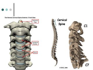
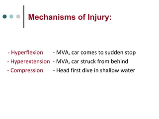
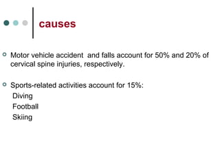
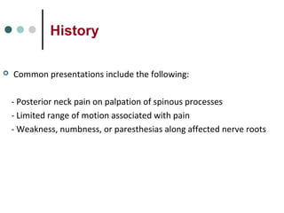
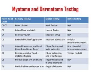
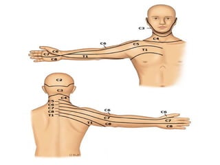
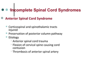
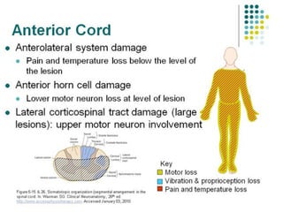
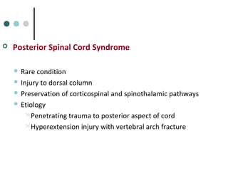
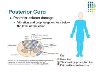
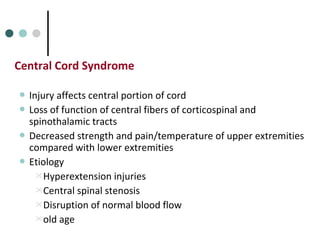
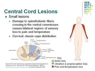
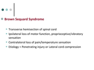
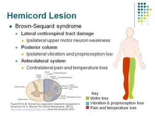
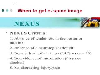
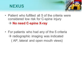
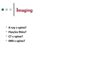
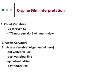
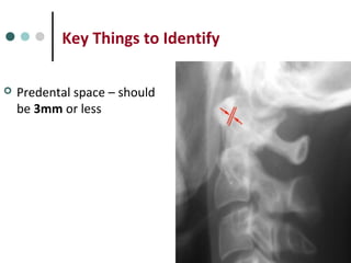
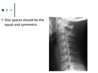
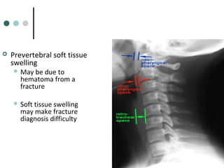
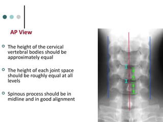
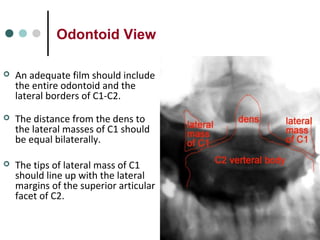
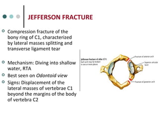
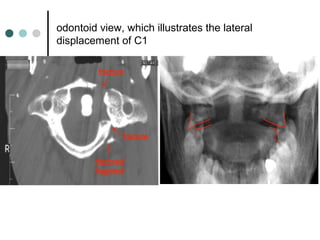
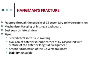
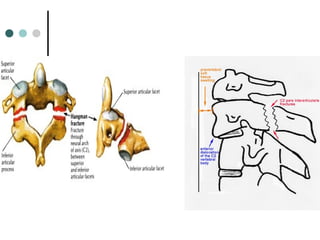
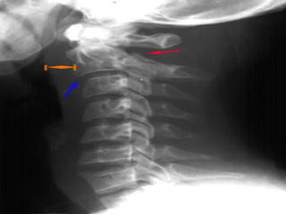
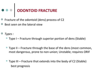
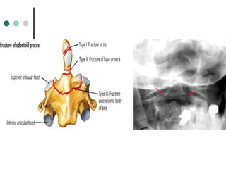
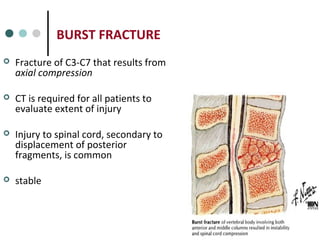
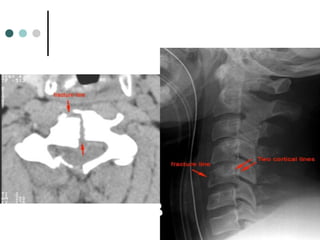
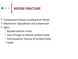
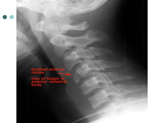
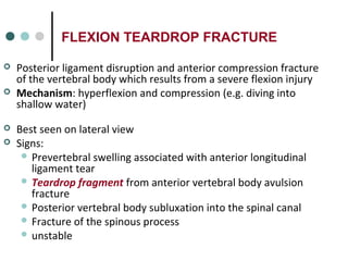
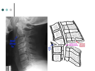
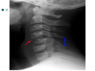
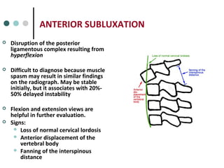
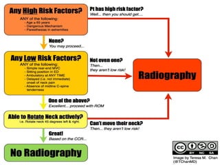
Cervical traumaCervical trauma
By
Dr. Hamdy Abdalla
 Spine injury must always be considered in any patients with multiple
injuries.
 Any patient with trauma who is not fully conscious should be assumed
to have cervical spine injury until proved otherwise.
 Collars alone are inadequate and they need to be supplemented by
manual stabilization or lateral support with blocks and forehead tape.
 Collars and neck support should not be removed unless cervical spine
injury has been excluded for sure.
Anatomy
• 7 Cervical Vertebrae
• C1 (Atlas) is a ring which articulates with the occiput
– C1 has no body
– C1 has no spinous process
• C2 (Axis) so named because it is the pivot on which the Atlas turns to
rotate the head
– The Axis has a vertical extension, the Dens,
which articulates with C1
Mechanisms of Injury:
- Hyperflexion - MVA, car comes to sudden stop
- Hyperextension - MVA, car struck from behind
- Compression - Head first dive in shallow water
causes
 Motor vehicle accident and falls account for 50% and 20% of
cervical spine injuries, respectively.
 Sports-related activities account for 15%:
Diving
Football
Skiing
History
 Common presentations include the following:
- Posterior neck pain on palpation of spinous processes
- Limited range of motion associated with pain
- Weakness, numbness, or paresthesias along affected nerve roots
Examination
 The examination should include :
Sensory
Motor
Proprioception
Perianal sensation
Rectal sphincter tone
Physical
- Common findings on physical examination in cervical spine injury include the
following:
- Spinal shock
Flaccidity Areflexia
Loss of anal sphincter tone Priapism
- Neurogenic shock
Hypotension Paradoxical bradycardia
Flushed, dry, and warm peripheral skin
- Associated with Paralysis of muscles of respiration
• Diaphragm invervated by C3-5
Management
 Diagnosis and management go hand in hand
 Inappropriate movement and examination can change the outcome
for the worse
 Early management
 Airway, Breathing and Circulation
 Exclude other life-threatening injury
 Properly immobilize patient
 Avoid hypothermia
 assess the patient’s neurologic status
Neurological level
- Most caudal level of motor / sensory function
- Motor and sensory may not be the same
- Sensory can vary on each side
Bony level
Site of vertebral column damage
 Complete injury
No motor or sensory function below injury level
 Incomplete injury
Any motor or sensory preservation below injury level
Incomplete Spinal Cord Syndromes
 Anterior Spinal Cord Syndrome
 Corticospinal and spinothalamic tracts
injured
 Preservation of posterior column pathway
 Etiology
Anterior spinal cord trauma
Flexion of cervical spine causing cord
contusion
Thrombosis of anterior spinal artery
 Posterior Spinal Cord Syndrome
 Rare condition
 Injury to dorsal column
 Preservation of corticospinal and spinothalamic pathways
 Etiology
Penetrating trauma to posterior aspect of cord
Hyperextension injury with vertebral arch fracture
Central Cord Syndrome
 Injury affects central portion of cord
 Loss of function of central fibers of corticospinal and
spinothalamic tracts
 Decreased strength and pain/temperature of upper extremities
compared with lower extremities
 Etiology
Hyperextension injuries
Central spinal stenosis
Disruption of normal blood flow
old age
 Brown Sequard Syndrome
 Transverse hemisection of spinal cord
 Ipsilateral loss of motor function, proprioceptive/vibratory
sensation
 Contralateral loss of pain/temperature sensation
 Etiology = Penetrating injury or Lateral cord compression
When to get c- spine image
Imaging
 X-ray c-spine?
 Flex/ex films?
 CT c-spine?
 MRI c-spine?
C-spine Film Interpretation
1. Count Vertebrae
-C1 through C7
-If T1 not seen do Swimmer’s view
2. Assess Curvature
3. Assess Vertebral Alignment (4 lines)
-ant vertebral line
-post vertebral line
-spinolaminal line
-post spinal line
C-spine Film Interpretation
4. Assess Bony Integrity
5. Assess Intervertebral Disk Spaces
6. Soft Tissues
 Lateral view
Top of T1 visible
Vertebral bodies of uniform height
Odontoid intact and closely applied to C1
 AP view
Spinous processes straight and spaced equally
Intervertebral spaces roughly equal
 Odontoid view
Odontoid intact
Equal spaces on either side of odontoid
Lateral margins of C1 and C2 align
Key Things to Identify
 Predental space – should
be 3mm or less
 Disc spaces should be the
equal and symmetric
 Prevertebral soft tissue
swelling
 May be due to
hematoma from a
fracture
 Soft tissue swelling
may make fracture
diagnosis difficulty
AP View
 The height of the cervical
vertebral bodies should be
approximately equal
 The height of each joint space
should be roughly equal at all
levels
 Spinous process should be in
midline and in good alignment
Odontoid View
 An adequate film should include
the entire odontoid and the
lateral borders of C1-C2.
 The distance from the dens to
the lateral masses of C1 should
be equal bilaterally.
 The tips of lateral mass of C1
should line up with the lateral
margins of the superior articular
facet of C2.
JEFFERSON FRACTURE
 Compression fracture of the
bony ring of C1, characterized
by lateral masses splitting and
transverse ligament tear
 Mechanism: Diving into shallow
water, RTA
 Best seen on Odontoid view
 Signs: Displacement of the
lateral masses of vertebrae C1
beyond the margins of the body
of vertebra C2
 Radiographic features:
the key radiographic view is the AP open mouth, which shows
displacement of the lateral masses of vertebrae C1 beyond the
margins of the body of vertebra C2. A lateral displacement of >2
mm or unilateral displacement may be indicative of a C1
fracture. CT is required to define the extent of fracture and to
detect fragments in the spinal canal.
Stability: unstable
 the lateral displacement of C1
indicates a Jefferson fracture.
odontoid view, which illustrates the lateral
displacement of C1
HANGMAN’S FRACTURE
 Fracture through the pedicle of C2 secondary to hyperextension
 Mechanism: Hanging or hitting a dashboard
 Best seen on lateral view
 Signs:
 Prevertebral soft tissue swelling
 Avulsion of anterior inferior corner of C2 associated with
rupture of the anterior longitudinal ligament
 Anterior dislocation of the C2 vertebral body
 Stability: unstable
ODONTOID FRACTURE
 Fracture of the odontoid (dens) process of C2
 Best seen on the lateral view
 Types :
 Type I – Fracture through superior portion of dens (Stable)
 Type II – Fracture through the base of the dens (most common,
most dangerous, prone to non-union; Unstable; requires ORIF
 Type III – Fracture that extends into the body of C2 (Stable)
best prognosis
BURST FRACTURE
 Fracture of C3-C7 that results from
axial compression
 CT is required for all patients to
evaluate extent of injury
 Injury to spinal cord, secondary to
displacement of posterior
fragments, is common
 stable
WEDGE FRACTURE
 Compression fracture resulting from flexion
 Mechanism: Hyperflexion and compression
 Signs:
 Buckled anterior cortex
 Loss of height of anterior vertebral body
 Anterosuperior fracture of vertebral body
 stable
FLEXION TEARDROP FRACTURE
 Posterior ligament disruption and anterior compression fracture
of the vertebral body which results from a severe flexion injury
 Mechanism: hyperflexion and compression (e.g. diving into
shallow water)
 Best seen on lateral view
 Signs:
 Prevertebral swelling associated with anterior longitudinal
ligament tear
 Teardrop fragment from anterior vertebral body avulsion
fracture
 Posterior vertebral body subluxation into the spinal canal
 Fracture of the spinous process
 unstable
BILATERAL FACET DISLOCATION
 Complete anterior dislocation of the vertebral body resulting
from extreme hyperflexion injury. It is associated with a very
high risk of cord damage
 Best seen on lateral view
 Signs:
 Complete anterior dislocation of affected vertebral body by
half or more of the vertebral body AP diameter
 Disruption of the posterior ligament complex and the
anterior longitudinal ligament
 unstable
UNILATERAL FACET DISLOCATION
 Facet joint dislocation and rupture of the joint ligaments
resulting from rotatory injury of the cervical vertebrae
 Best seen on lateral or oblique views
 Signs:
- Anterior dislocation of affected vertebral body by less than half
of the vertebral body AP diameter
- Discordant rotation above and below involved level
- Widening of the disk space
ANTERIOR SUBLUXATION
 Disruption of the posterior
ligamentous complex resulting from
hyperflexion
 Difficult to diagnose because muscle
spasm may result in similar findings
on the radiograph. May be stable
initially, but it associates with 20%-
50% delayed instability
 Flexion and extension views are
helpful in further evaluation.
 Signs:
 Loss of normal cervical lordosis
 Anterior displacement of the
vertebral body
 Fanning of the interspinous
distance
SCIWORA
 Significant Cord Injury without obvious radiological abnormality
- Higher incidence in pediatric population (34.8%)
 The relatively large size of the head
 inherent skeletal mobility
 cord vulnerable to damage
- Higher incidence above 60 yo
Posterior vertebral spurs due to spondylosis
Ligamentum flavum bulging due to loss of disc height
Risk of central cord syndrome after hyperextension injury
indications for surgical intervention
1. Neurologic deficit,
2. Spinal instability, and
3. Intractable pain.
Forget about all of this and
If you are not happy
do not discharge the patient.
THANK YOU FOR YOUR
ATTENTION !
Cervical trauma
Cervical trauma
Cervical trauma

Cervical trauma

  • 1.
  • 2.
     Spine injurymust always be considered in any patients with multiple injuries.  Any patient with trauma who is not fully conscious should be assumed to have cervical spine injury until proved otherwise.  Collars alone are inadequate and they need to be supplemented by manual stabilization or lateral support with blocks and forehead tape.  Collars and neck support should not be removed unless cervical spine injury has been excluded for sure.
  • 3.
    Anatomy • 7 CervicalVertebrae • C1 (Atlas) is a ring which articulates with the occiput – C1 has no body – C1 has no spinous process • C2 (Axis) so named because it is the pivot on which the Atlas turns to rotate the head – The Axis has a vertical extension, the Dens, which articulates with C1
  • 5.
    Mechanisms of Injury: -Hyperflexion - MVA, car comes to sudden stop - Hyperextension - MVA, car struck from behind - Compression - Head first dive in shallow water
  • 6.
    causes  Motor vehicleaccident and falls account for 50% and 20% of cervical spine injuries, respectively.  Sports-related activities account for 15%: Diving Football Skiing
  • 8.
    History  Common presentationsinclude the following: - Posterior neck pain on palpation of spinous processes - Limited range of motion associated with pain - Weakness, numbness, or paresthesias along affected nerve roots
  • 9.
    Examination  The examinationshould include : Sensory Motor Proprioception Perianal sensation Rectal sphincter tone
  • 10.
    Physical - Common findingson physical examination in cervical spine injury include the following: - Spinal shock Flaccidity Areflexia Loss of anal sphincter tone Priapism - Neurogenic shock Hypotension Paradoxical bradycardia Flushed, dry, and warm peripheral skin - Associated with Paralysis of muscles of respiration • Diaphragm invervated by C3-5
  • 11.
    Management  Diagnosis andmanagement go hand in hand  Inappropriate movement and examination can change the outcome for the worse  Early management  Airway, Breathing and Circulation  Exclude other life-threatening injury  Properly immobilize patient  Avoid hypothermia
  • 12.
     assess thepatient’s neurologic status Neurological level - Most caudal level of motor / sensory function - Motor and sensory may not be the same - Sensory can vary on each side Bony level Site of vertebral column damage
  • 15.
     Complete injury Nomotor or sensory function below injury level  Incomplete injury Any motor or sensory preservation below injury level
  • 17.
    Incomplete Spinal CordSyndromes  Anterior Spinal Cord Syndrome  Corticospinal and spinothalamic tracts injured  Preservation of posterior column pathway  Etiology Anterior spinal cord trauma Flexion of cervical spine causing cord contusion Thrombosis of anterior spinal artery
  • 19.
     Posterior SpinalCord Syndrome  Rare condition  Injury to dorsal column  Preservation of corticospinal and spinothalamic pathways  Etiology Penetrating trauma to posterior aspect of cord Hyperextension injury with vertebral arch fracture
  • 21.
    Central Cord Syndrome Injury affects central portion of cord  Loss of function of central fibers of corticospinal and spinothalamic tracts  Decreased strength and pain/temperature of upper extremities compared with lower extremities  Etiology Hyperextension injuries Central spinal stenosis Disruption of normal blood flow old age
  • 23.
     Brown SequardSyndrome  Transverse hemisection of spinal cord  Ipsilateral loss of motor function, proprioceptive/vibratory sensation  Contralateral loss of pain/temperature sensation  Etiology = Penetrating injury or Lateral cord compression
  • 25.
    When to getc- spine image
  • 27.
    Imaging  X-ray c-spine? Flex/ex films?  CT c-spine?  MRI c-spine?
  • 28.
    C-spine Film Interpretation 1.Count Vertebrae -C1 through C7 -If T1 not seen do Swimmer’s view 2. Assess Curvature 3. Assess Vertebral Alignment (4 lines) -ant vertebral line -post vertebral line -spinolaminal line -post spinal line
  • 29.
    C-spine Film Interpretation 4.Assess Bony Integrity 5. Assess Intervertebral Disk Spaces 6. Soft Tissues
  • 31.
     Lateral view Topof T1 visible Vertebral bodies of uniform height Odontoid intact and closely applied to C1  AP view Spinous processes straight and spaced equally Intervertebral spaces roughly equal  Odontoid view Odontoid intact Equal spaces on either side of odontoid Lateral margins of C1 and C2 align
  • 33.
    Key Things toIdentify  Predental space – should be 3mm or less
  • 34.
     Disc spacesshould be the equal and symmetric
  • 35.
     Prevertebral softtissue swelling  May be due to hematoma from a fracture  Soft tissue swelling may make fracture diagnosis difficulty
  • 36.
    AP View  Theheight of the cervical vertebral bodies should be approximately equal  The height of each joint space should be roughly equal at all levels  Spinous process should be in midline and in good alignment
  • 37.
    Odontoid View  Anadequate film should include the entire odontoid and the lateral borders of C1-C2.  The distance from the dens to the lateral masses of C1 should be equal bilaterally.  The tips of lateral mass of C1 should line up with the lateral margins of the superior articular facet of C2.
  • 38.
    JEFFERSON FRACTURE  Compressionfracture of the bony ring of C1, characterized by lateral masses splitting and transverse ligament tear  Mechanism: Diving into shallow water, RTA  Best seen on Odontoid view  Signs: Displacement of the lateral masses of vertebrae C1 beyond the margins of the body of vertebra C2
  • 39.
     Radiographic features: thekey radiographic view is the AP open mouth, which shows displacement of the lateral masses of vertebrae C1 beyond the margins of the body of vertebra C2. A lateral displacement of >2 mm or unilateral displacement may be indicative of a C1 fracture. CT is required to define the extent of fracture and to detect fragments in the spinal canal. Stability: unstable
  • 40.
     the lateraldisplacement of C1 indicates a Jefferson fracture.
  • 41.
    odontoid view, whichillustrates the lateral displacement of C1
  • 42.
    HANGMAN’S FRACTURE  Fracturethrough the pedicle of C2 secondary to hyperextension  Mechanism: Hanging or hitting a dashboard  Best seen on lateral view  Signs:  Prevertebral soft tissue swelling  Avulsion of anterior inferior corner of C2 associated with rupture of the anterior longitudinal ligament  Anterior dislocation of the C2 vertebral body  Stability: unstable
  • 45.
    ODONTOID FRACTURE  Fractureof the odontoid (dens) process of C2  Best seen on the lateral view  Types :  Type I – Fracture through superior portion of dens (Stable)  Type II – Fracture through the base of the dens (most common, most dangerous, prone to non-union; Unstable; requires ORIF  Type III – Fracture that extends into the body of C2 (Stable) best prognosis
  • 47.
    BURST FRACTURE  Fractureof C3-C7 that results from axial compression  CT is required for all patients to evaluate extent of injury  Injury to spinal cord, secondary to displacement of posterior fragments, is common  stable
  • 49.
    WEDGE FRACTURE  Compressionfracture resulting from flexion  Mechanism: Hyperflexion and compression  Signs:  Buckled anterior cortex  Loss of height of anterior vertebral body  Anterosuperior fracture of vertebral body  stable
  • 51.
    FLEXION TEARDROP FRACTURE Posterior ligament disruption and anterior compression fracture of the vertebral body which results from a severe flexion injury  Mechanism: hyperflexion and compression (e.g. diving into shallow water)  Best seen on lateral view  Signs:  Prevertebral swelling associated with anterior longitudinal ligament tear  Teardrop fragment from anterior vertebral body avulsion fracture  Posterior vertebral body subluxation into the spinal canal  Fracture of the spinous process  unstable
  • 54.
    BILATERAL FACET DISLOCATION Complete anterior dislocation of the vertebral body resulting from extreme hyperflexion injury. It is associated with a very high risk of cord damage  Best seen on lateral view  Signs:  Complete anterior dislocation of affected vertebral body by half or more of the vertebral body AP diameter  Disruption of the posterior ligament complex and the anterior longitudinal ligament  unstable
  • 56.
    UNILATERAL FACET DISLOCATION Facet joint dislocation and rupture of the joint ligaments resulting from rotatory injury of the cervical vertebrae  Best seen on lateral or oblique views  Signs: - Anterior dislocation of affected vertebral body by less than half of the vertebral body AP diameter - Discordant rotation above and below involved level - Widening of the disk space
  • 58.
    ANTERIOR SUBLUXATION  Disruptionof the posterior ligamentous complex resulting from hyperflexion  Difficult to diagnose because muscle spasm may result in similar findings on the radiograph. May be stable initially, but it associates with 20%- 50% delayed instability  Flexion and extension views are helpful in further evaluation.  Signs:  Loss of normal cervical lordosis  Anterior displacement of the vertebral body  Fanning of the interspinous distance
  • 59.
    SCIWORA  Significant CordInjury without obvious radiological abnormality - Higher incidence in pediatric population (34.8%)  The relatively large size of the head  inherent skeletal mobility  cord vulnerable to damage - Higher incidence above 60 yo Posterior vertebral spurs due to spondylosis Ligamentum flavum bulging due to loss of disc height Risk of central cord syndrome after hyperextension injury
  • 60.
    indications for surgicalintervention 1. Neurologic deficit, 2. Spinal instability, and 3. Intractable pain.
  • 61.
    Forget about allof this and If you are not happy do not discharge the patient.
  • 62.
    THANK YOU FORYOUR ATTENTION !