monteggia fracture
•Download as PPTX, PDF•
8 likes•6,805 views

MONTEGGIA FRACTURE DISLOCATION LECTURE FOR UNDERGRADUATE

Recommended
More Related Content
What's hot
What's hot (20)
Similar to monteggia fracture
Similar to monteggia fracture (20)
Recently uploaded
Recently uploaded (20)
monteggia fracture
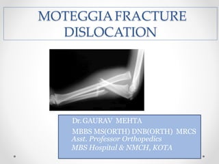
- 1. MOTEGGIAFRACTURE DISLOCATION Dr.GAURAV MEHTA MBBS MS(ORTH) DNB(ORTH) MRCS Asst. Professor Orthopedics MBS Hospital & NMCH, KOTA
- 2. MONTEGGIA FRACTURE DISLOCATION Historically described as : “fracture proximal third of shaft of ulna with dislocation of radial head”
- 3. HISTORY In 1814 Giovanni Batista Monteggia, a surgical pathologist and public health official in Milan, first described Monteggia fractures. He observed injuries in cadavers and provided the description: “Traumatic lesion distinguished by a fracture of the proximal third of the ulna and an anterior dislocation of the proximal epiphysis of the radius.”
- 4. EPIDEMIOLOGY Monteggia fractures constitute about 1 to 2% of forearm fractures.
- 5. CLASSIFICATION Bado's classification * Jose Louis Bado in 1958 divided Monteggia fractures into four types of true Monteggia lesions. Bado also classified certain injuries as equivalents to the classic or true Monteggia lesions because of their similar mechanism of injuries, radiographic pattern, or methods of treatment. * Bado JL. The Monteggia lesion. Clin Orthop 1967;50: 71 to 86.
- 7. BADO’s Classification Type I : Anterior dislocation: The radial head is dislocated anteriorly and the ulna has a fracture in the diaphyseal or proximal metaphyseal area. Most common type
- 8. BADO’s Classification Type II : Posterior dislocation: The radial head is posterior/posterolaterally dislocated, the ulna is usually fractured in the metaphysis. Associated with nerve palsy (PIN) and poor prognosis
- 9. BADO’s Classification Type III: Lateral dislocation : There is lateral dislocation of the radial head with a metaphyseal fracture of the ulna.
- 10. Type IV : Anterior dislocation with radius shaft fracture : the pattern of injury is the same as with a type I injury, with the inclusion of a radius shaft fracture below the level of the ulnar fracture. BADO’s Classification
- 11. Lett’s Classification Classified Monteggia fractures in children based on both the direction of the radial head dislocation and the type of ulnar fracture. The Bado type I class was subdivided into three sub types. Type A is anterior bowing of the ulna due toplastic deformation with anterior dislocation of the radial head. Type B includes a greenstick fracture of the ulna, type C has a complete ulnar Fracture.
- 13. MECHANISM OF INJURY Type I - forced pronation of forearm Type II - axial loading of forearm with flexed elbow Type III – forced abduction of elbow Type IV - Type I mechanism in which radial shaft additionally fails
- 14. Clinical Evaluation Fusiform swelling around the elbow. Painful restriction of movements i.e. elbow flexion , extension, pronation and supination. Careful neurovascular examination essential Radial nerve & posterior interosseous nerve injury common in Type II fracture pattern
- 15. RADIOGRAPHIC EVALUATION Anteroposterior (AP) and Lateral x-rays of the forearm. Radiographs of the joints at either end of the forearm wrist, particularly the position of the radial head Radiocapitellar Relation: Best defined by a true lateral view of the elbow. A line drawn down the long axis of the radius bisects the capitellum of the humerus regardless of the degree of flexion or extension of the elbow.
- 17. MANAGEMENT NON OPERATIVE • Reserved for pediatric population
- 18. MAINTENANCE OF REDUCTION Reduction is maintained by above elbow cast in flexion for 6-8 weeks duration. The degree of flexion varies depending on the direction of the radial head dislocation. When the radius is in a straight lateral or anterolateral position, flexion to 110 to 120 degrees improves stability. If there is a posterolateral component to the dislocation, a position of only 70 to 80 degrees of flexion has been recommended. Forearm rotation usually is in supination, which tightens the interosseous membrane and further stabilizes the reduction.
- 19. OPERATIVE TREATMENT MANAGEMENT Treatment of choice ORIF of ulna fracture with 3*5 mm contoured plate/ intramedullary nailing(less favored) in pediatric population Plate applied on tension side Closed reduction of radial head is rule once ulnar length achieved by ORIF Failure of closed reduction of radial head may require open reduction and radio-capitellar pinning • Post –op posterior elbow splint applied followed by physio
- 20. 1. Neglected Monteggia Fracture. 2. Non-union : most often seen in Bado type II 3. Nerve Injuries: radial, median, anterior-posterior interosseus nerve injury either traumatic or iatrogenic 4. Periarticular Ossification. 5. Compartment Syndrome. 6. Radial head instability COMPLICATIONS