Downloaded 794 times
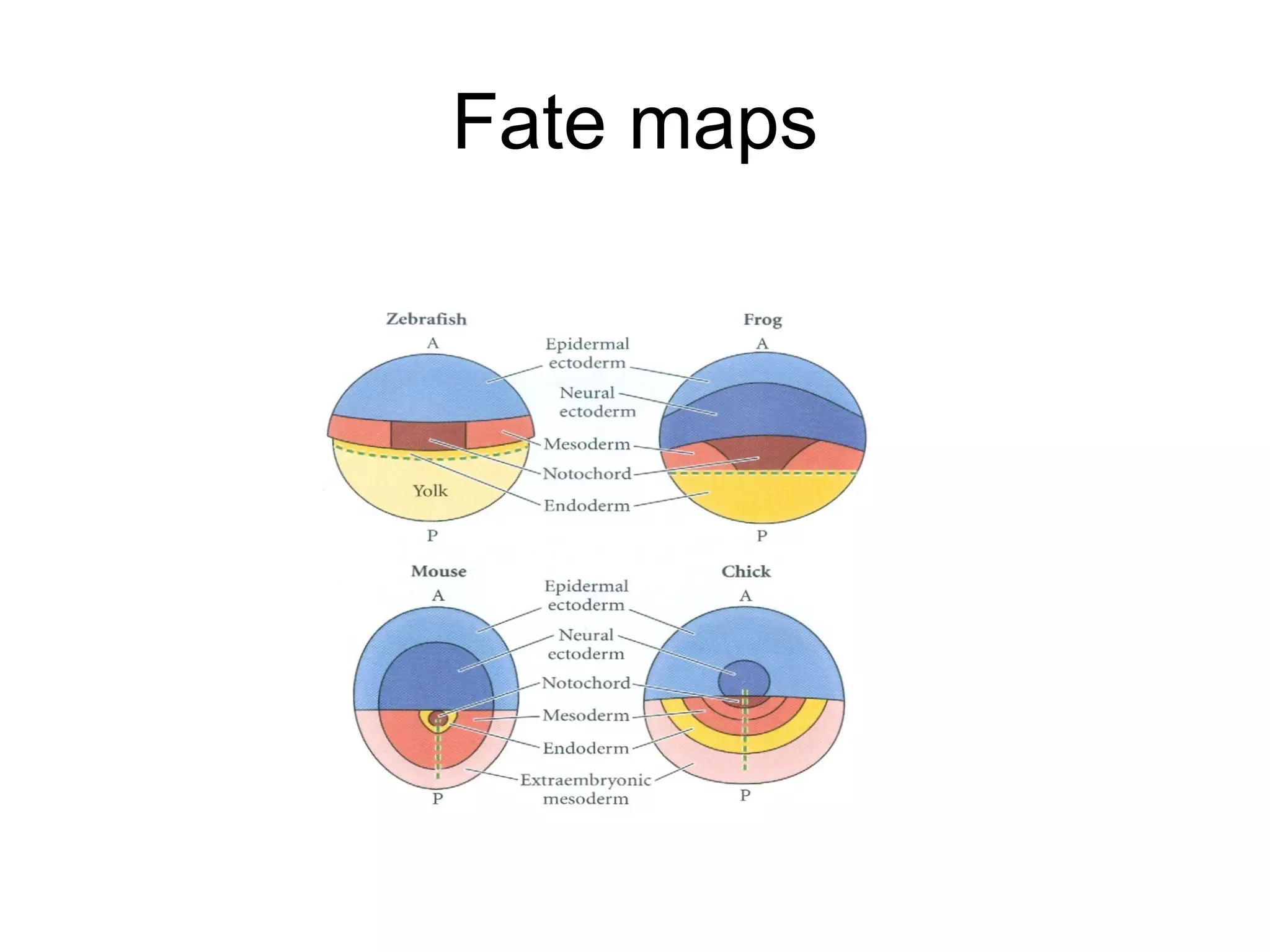
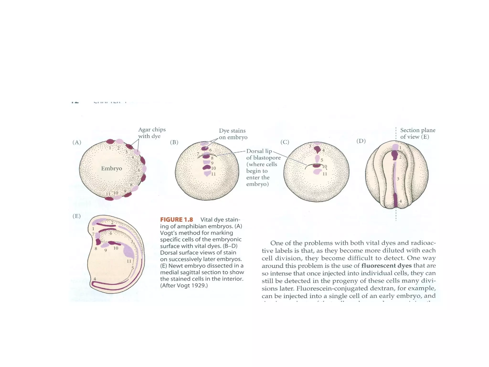
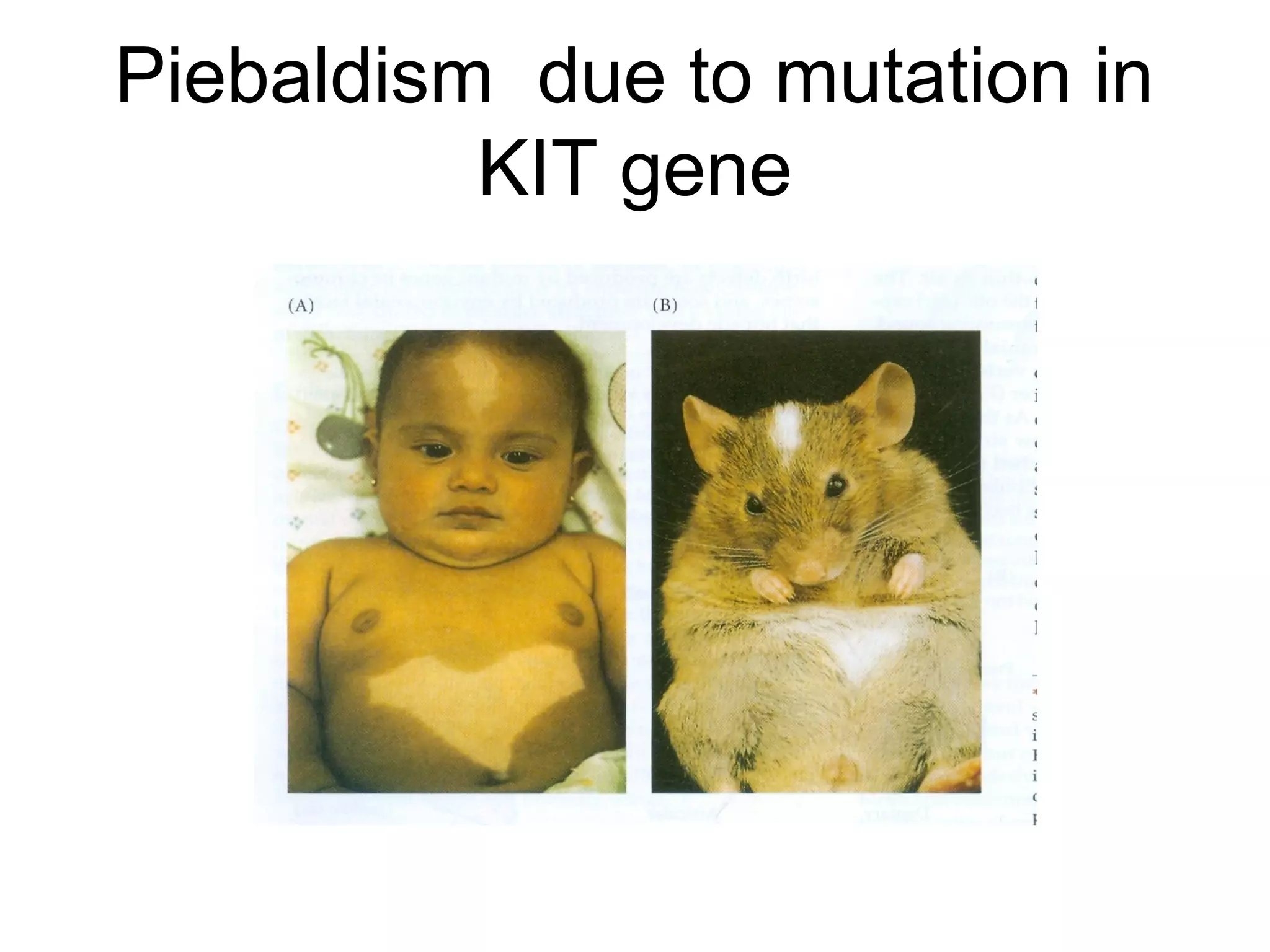


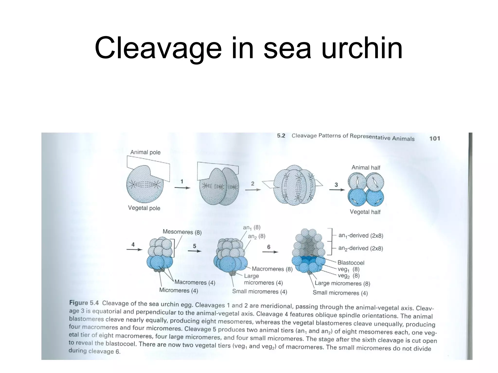
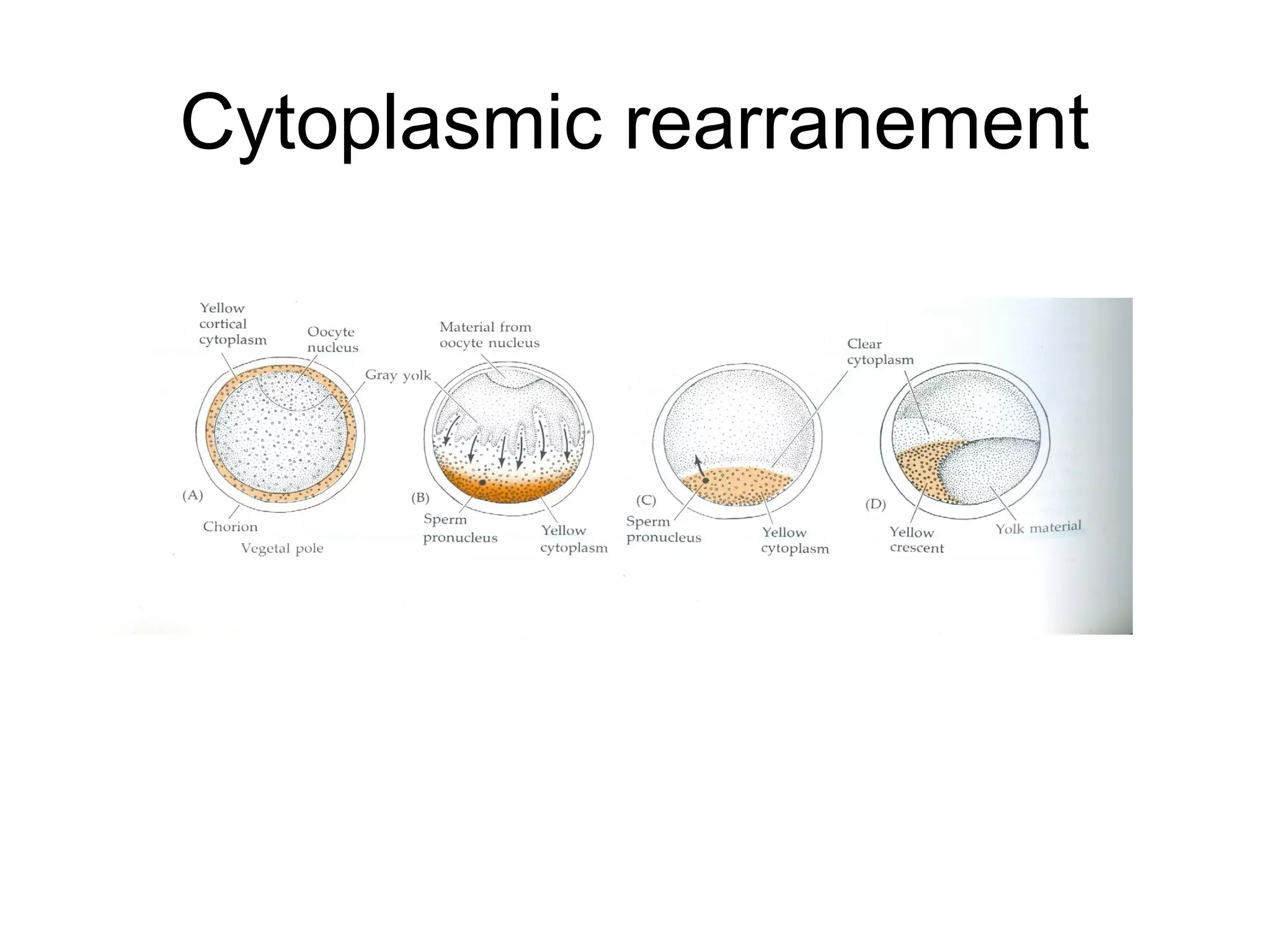
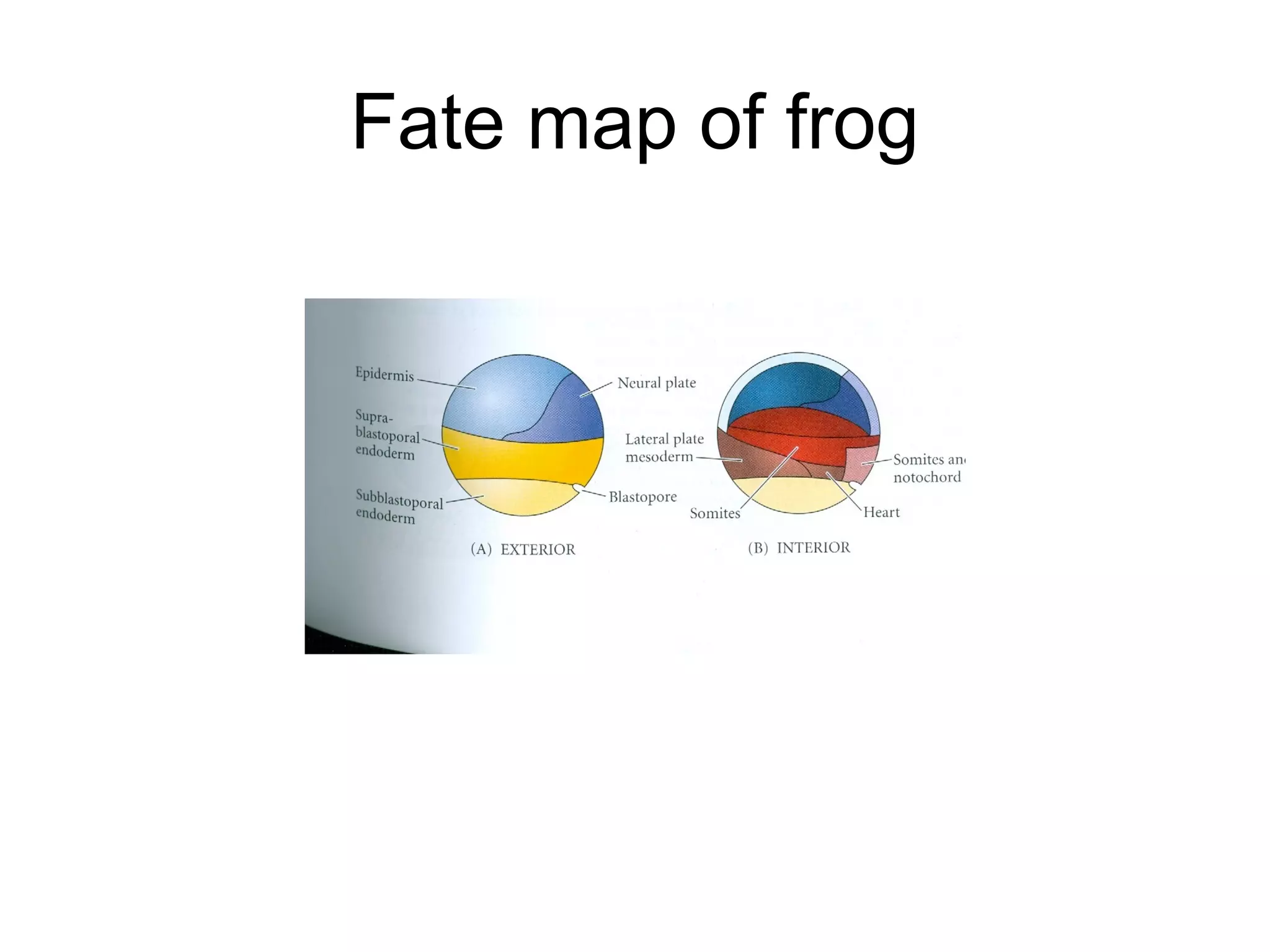
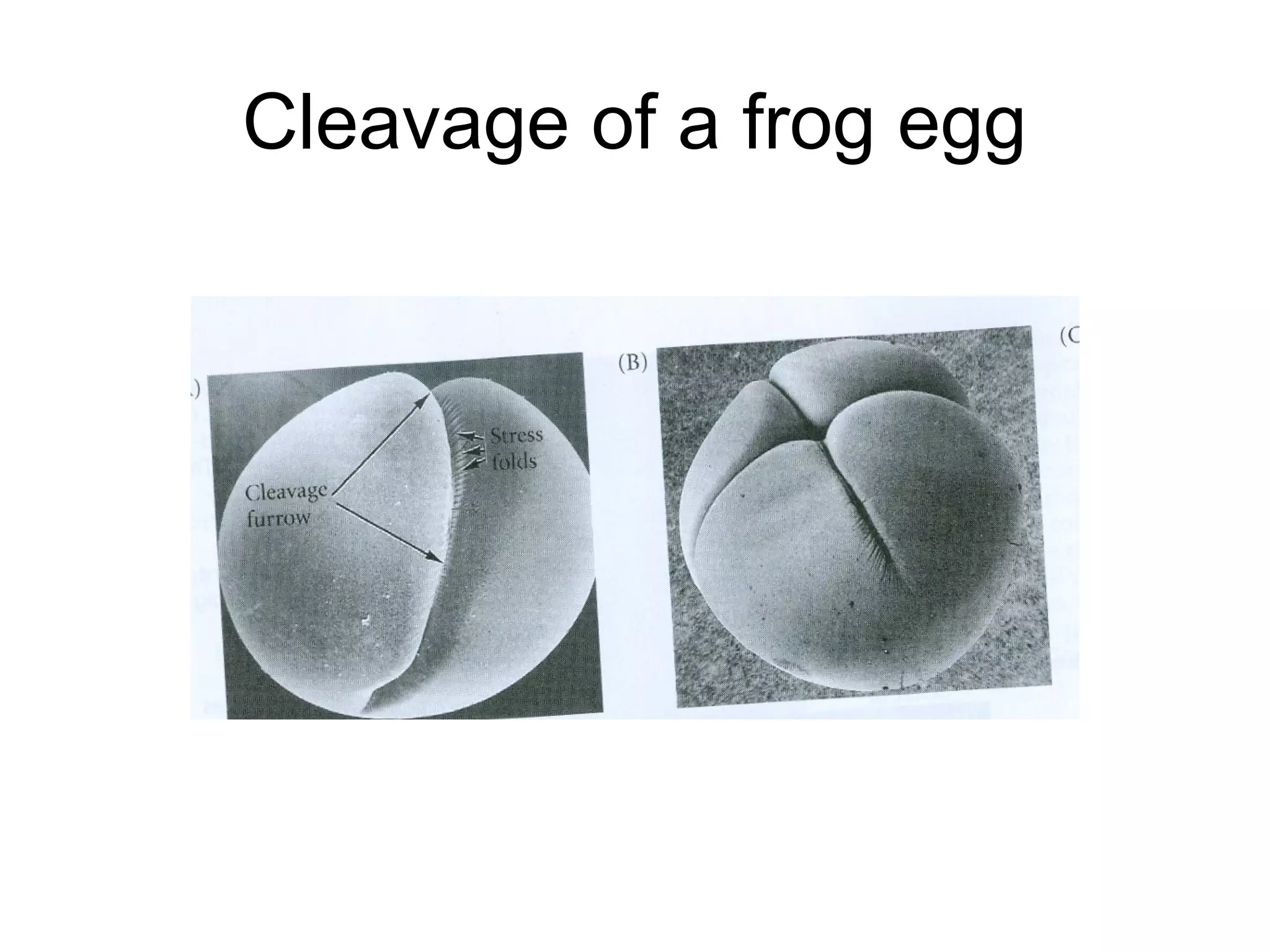
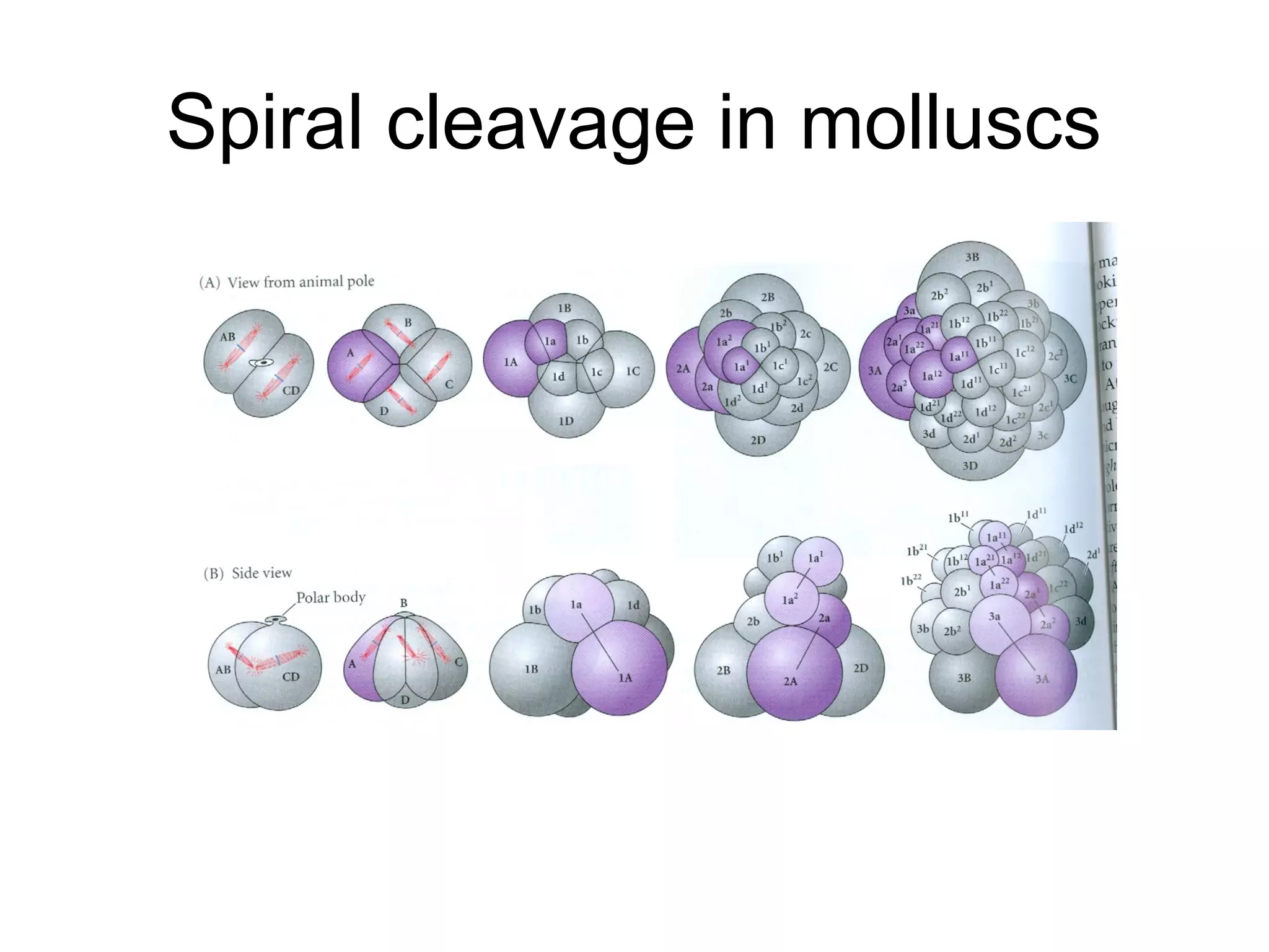
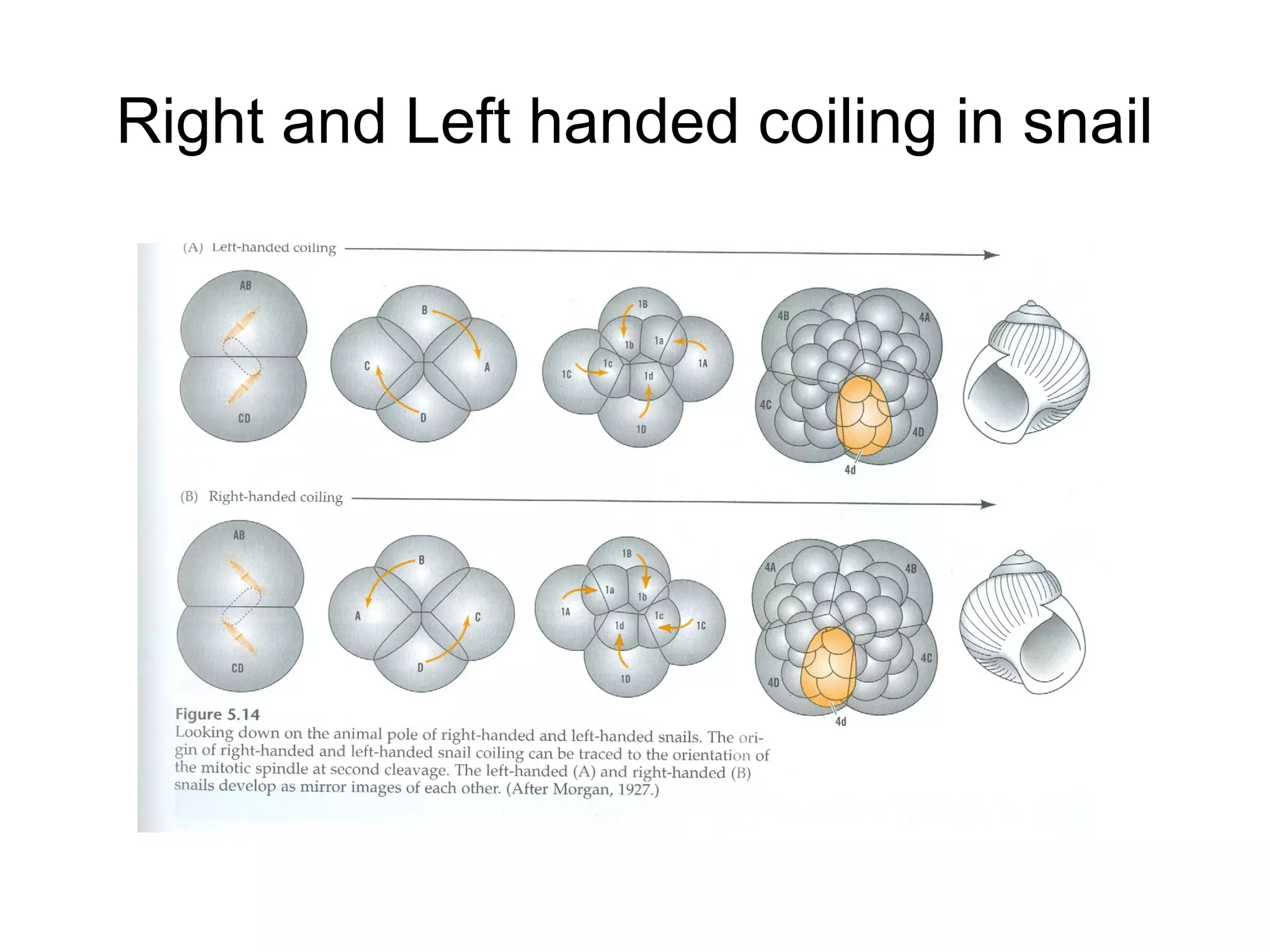
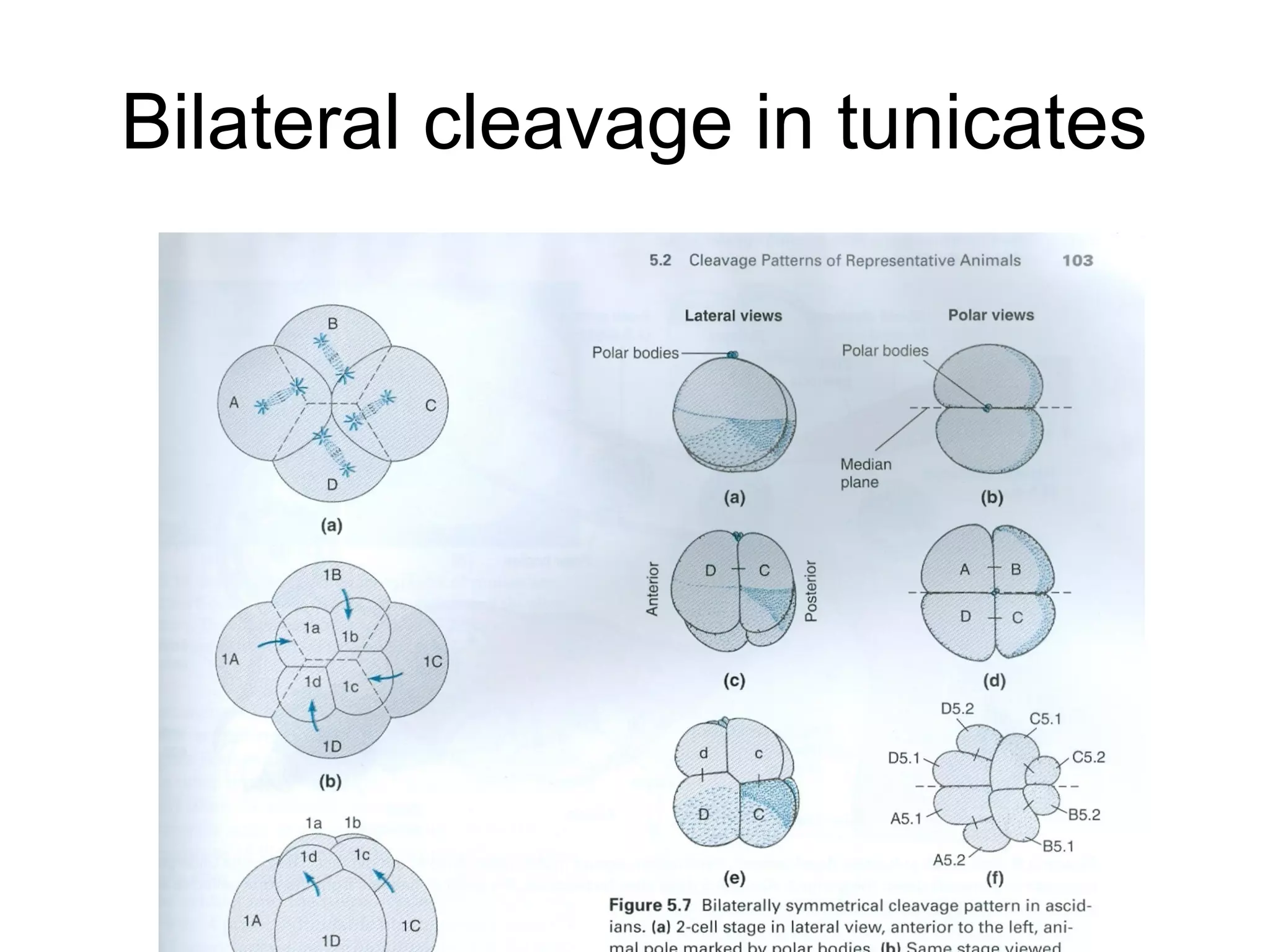
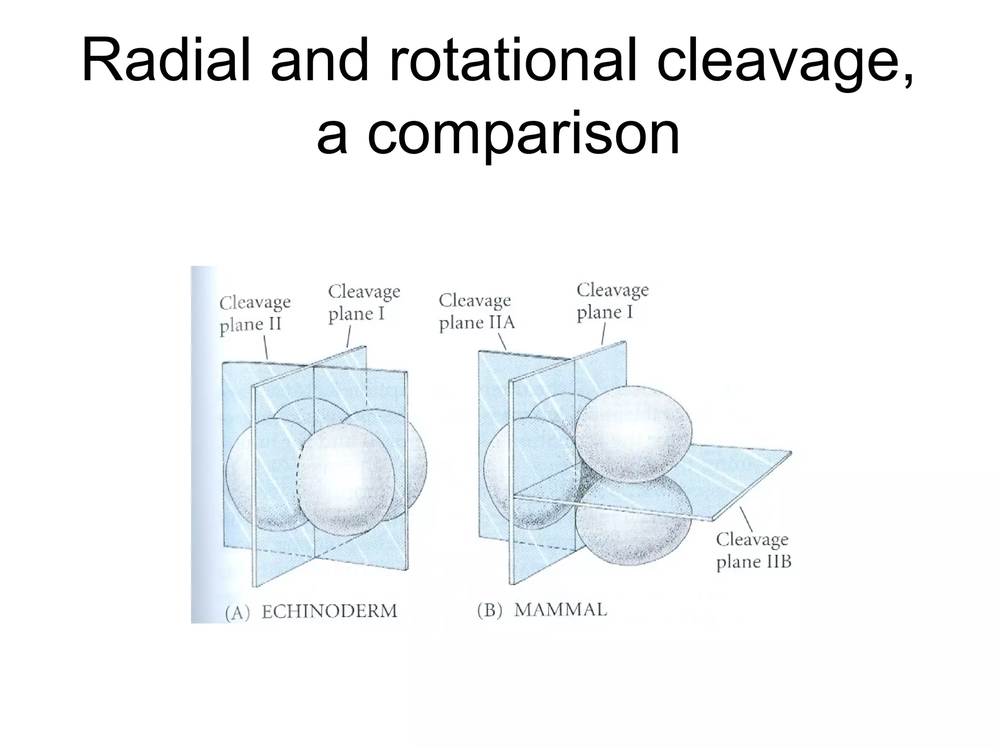
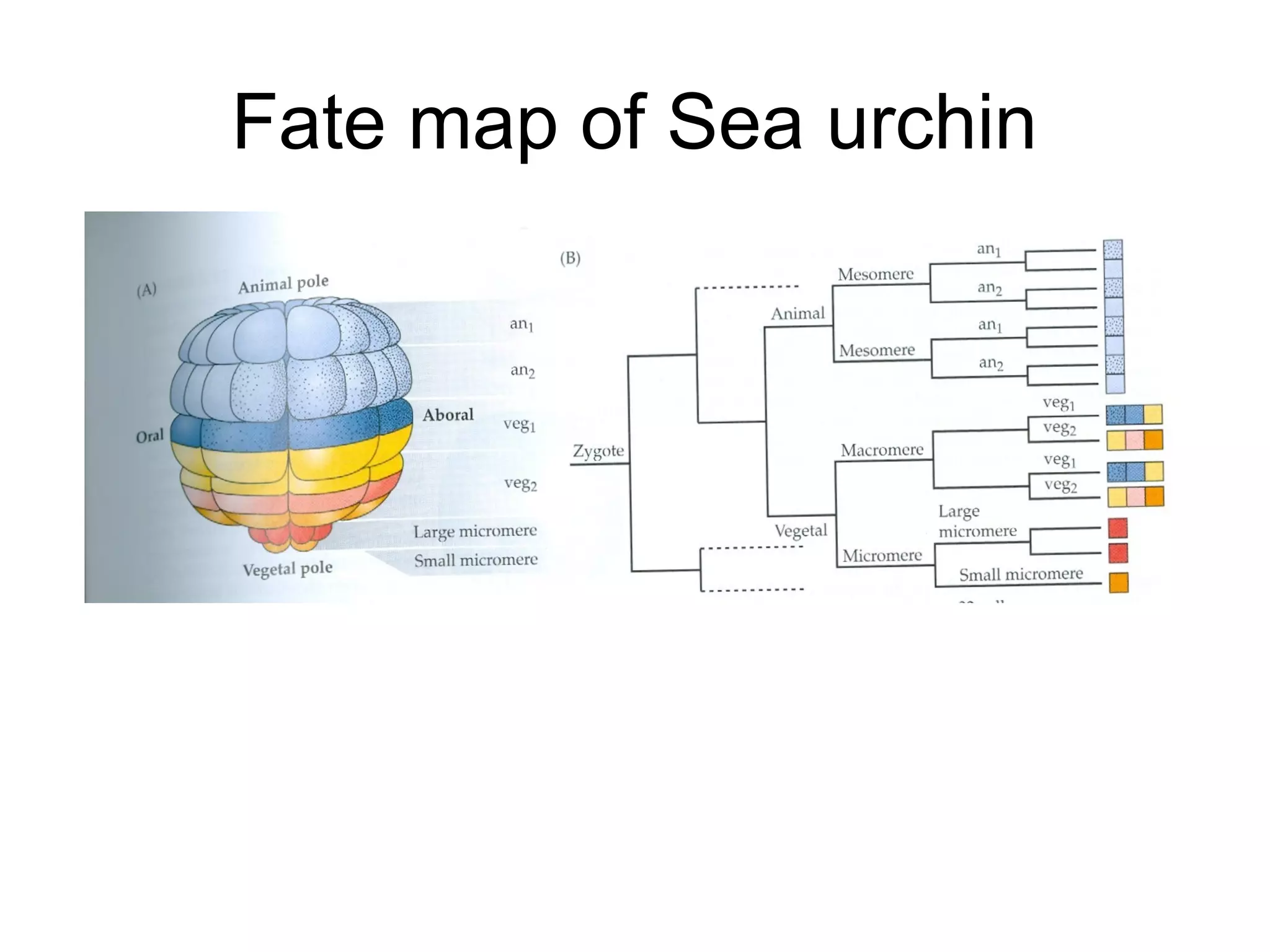
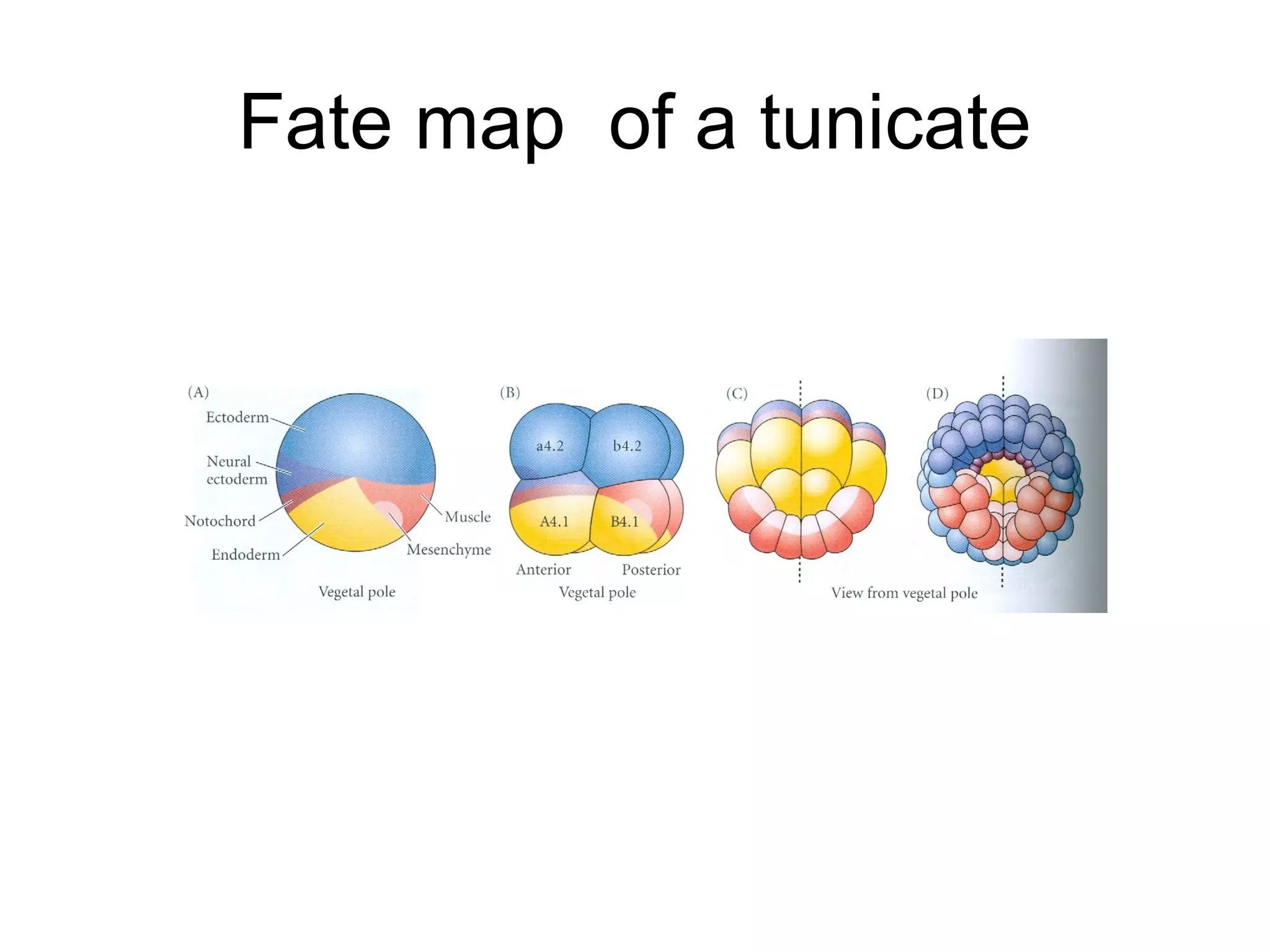
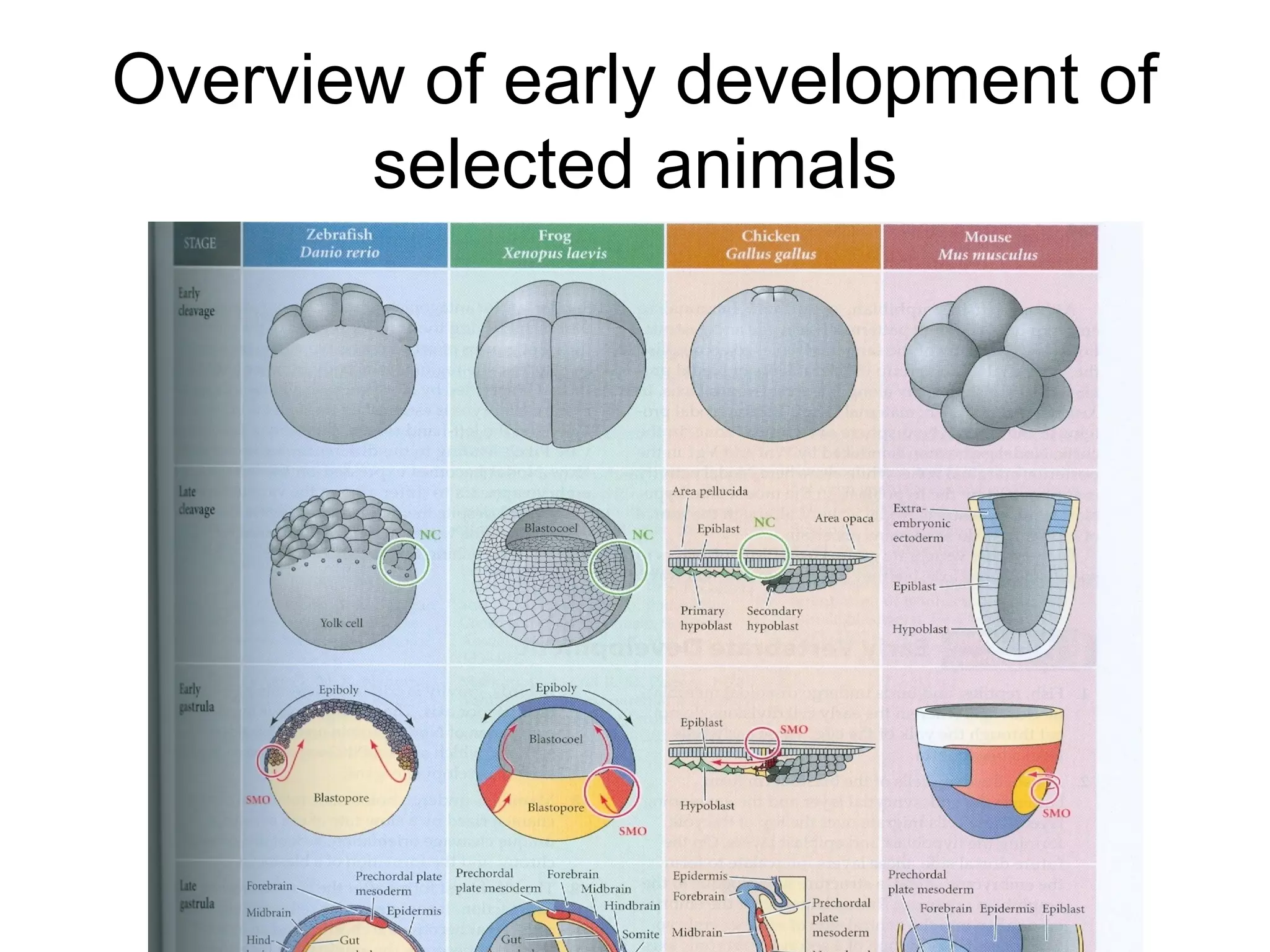
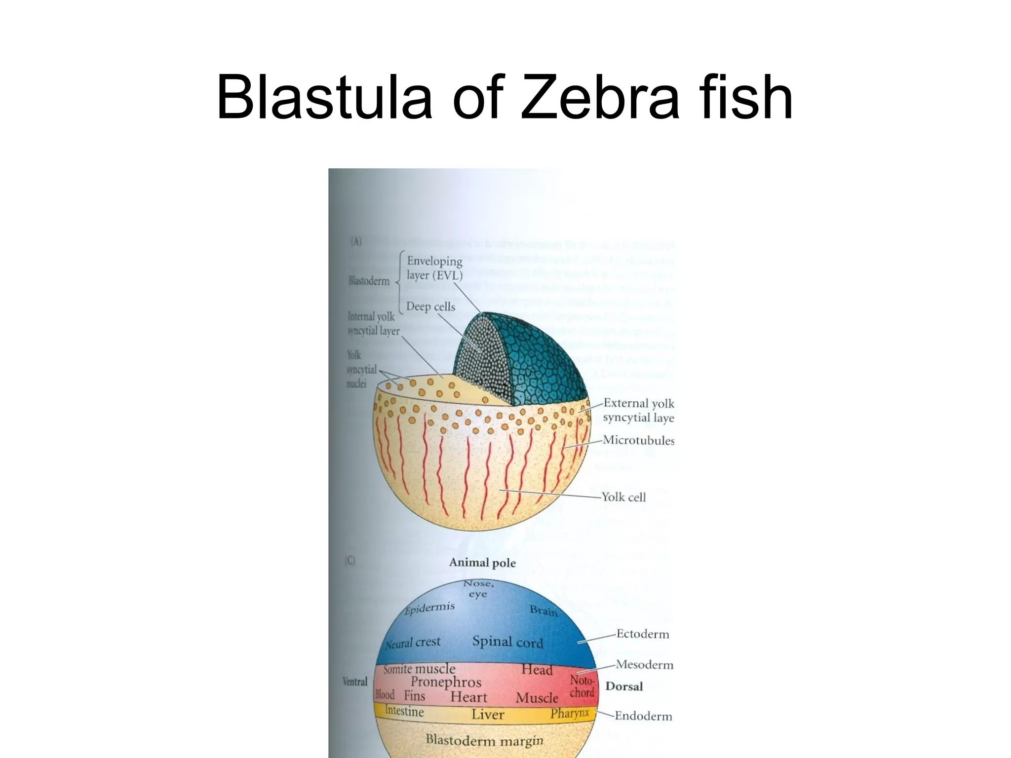
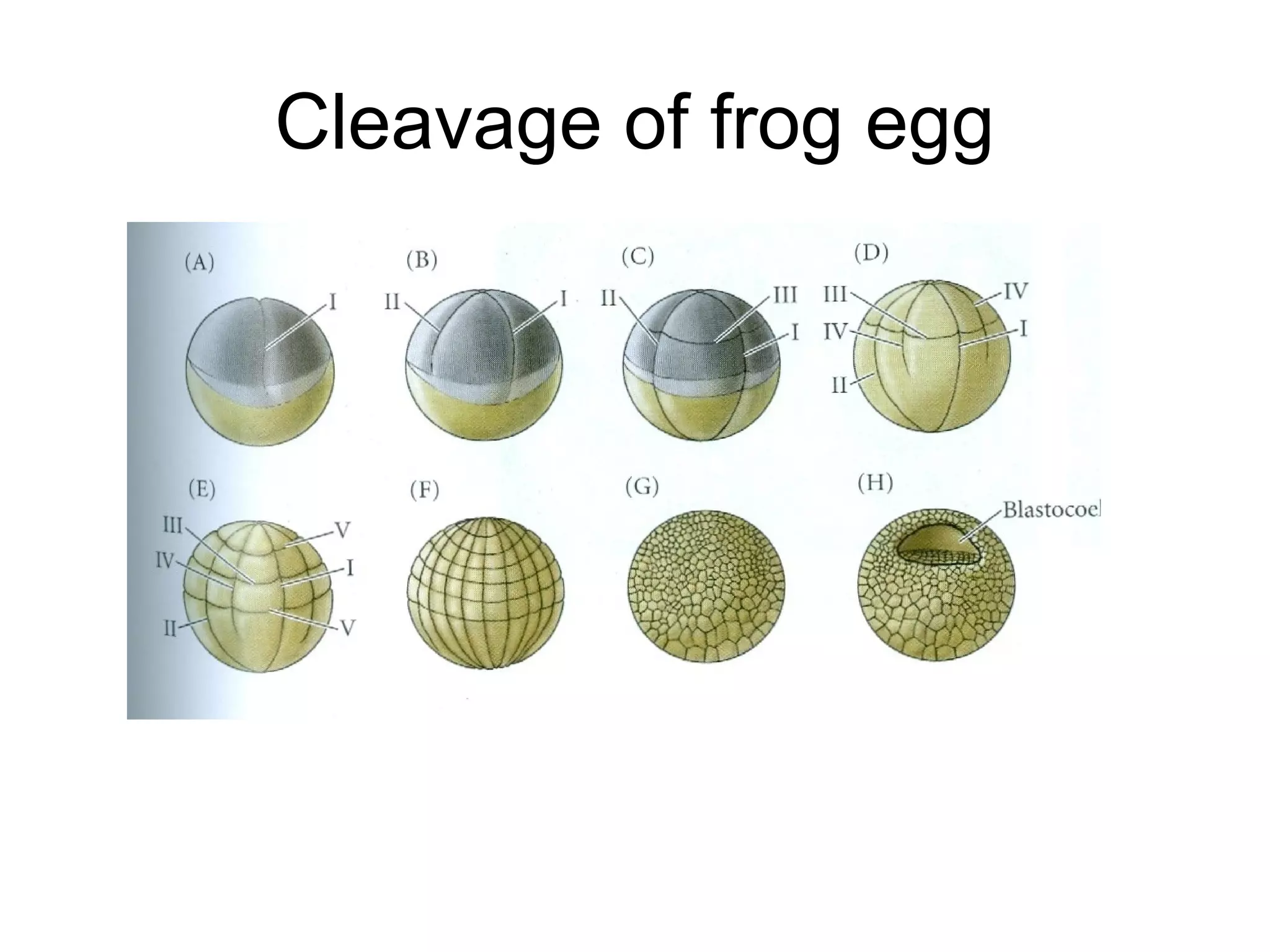
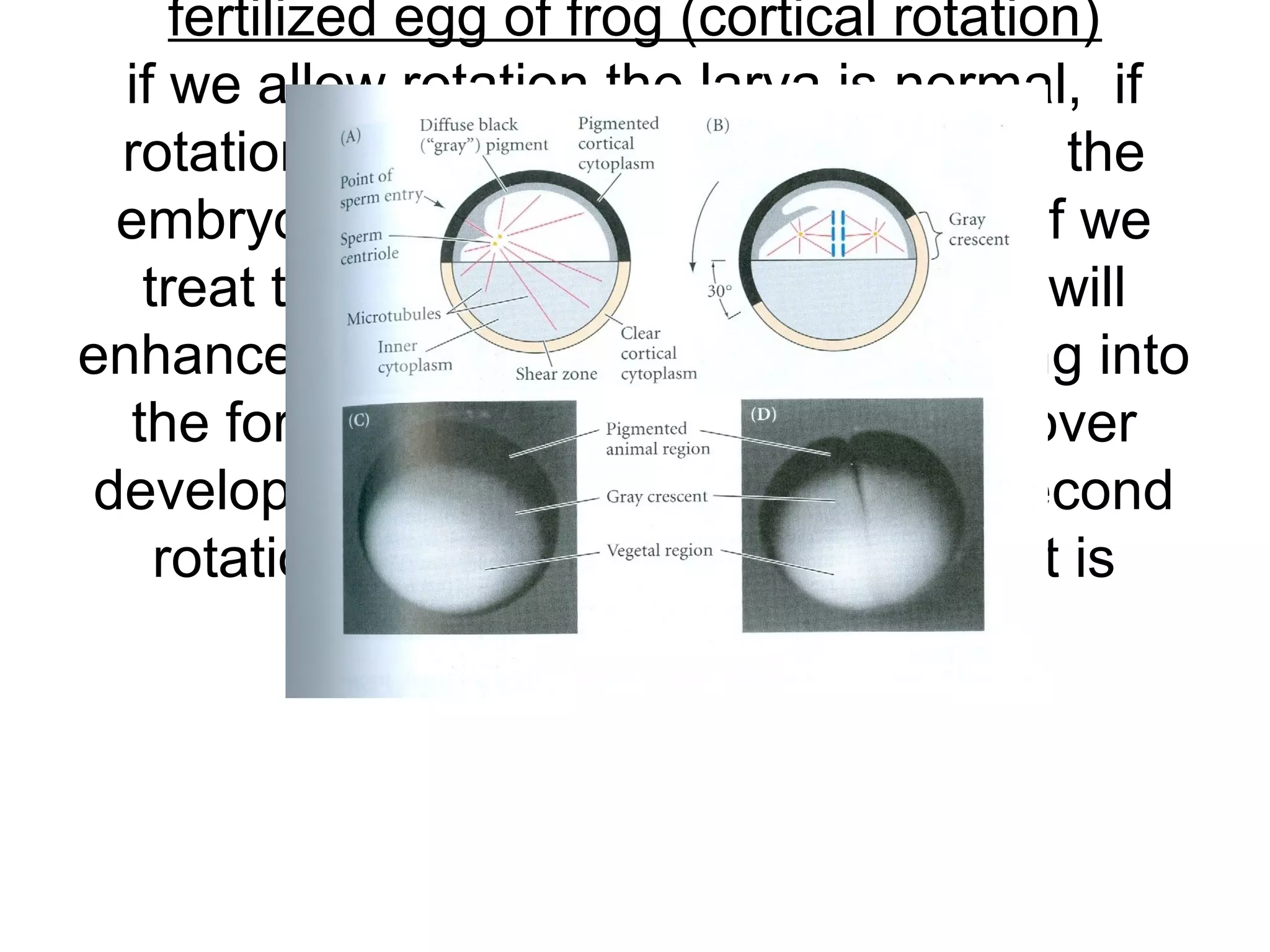
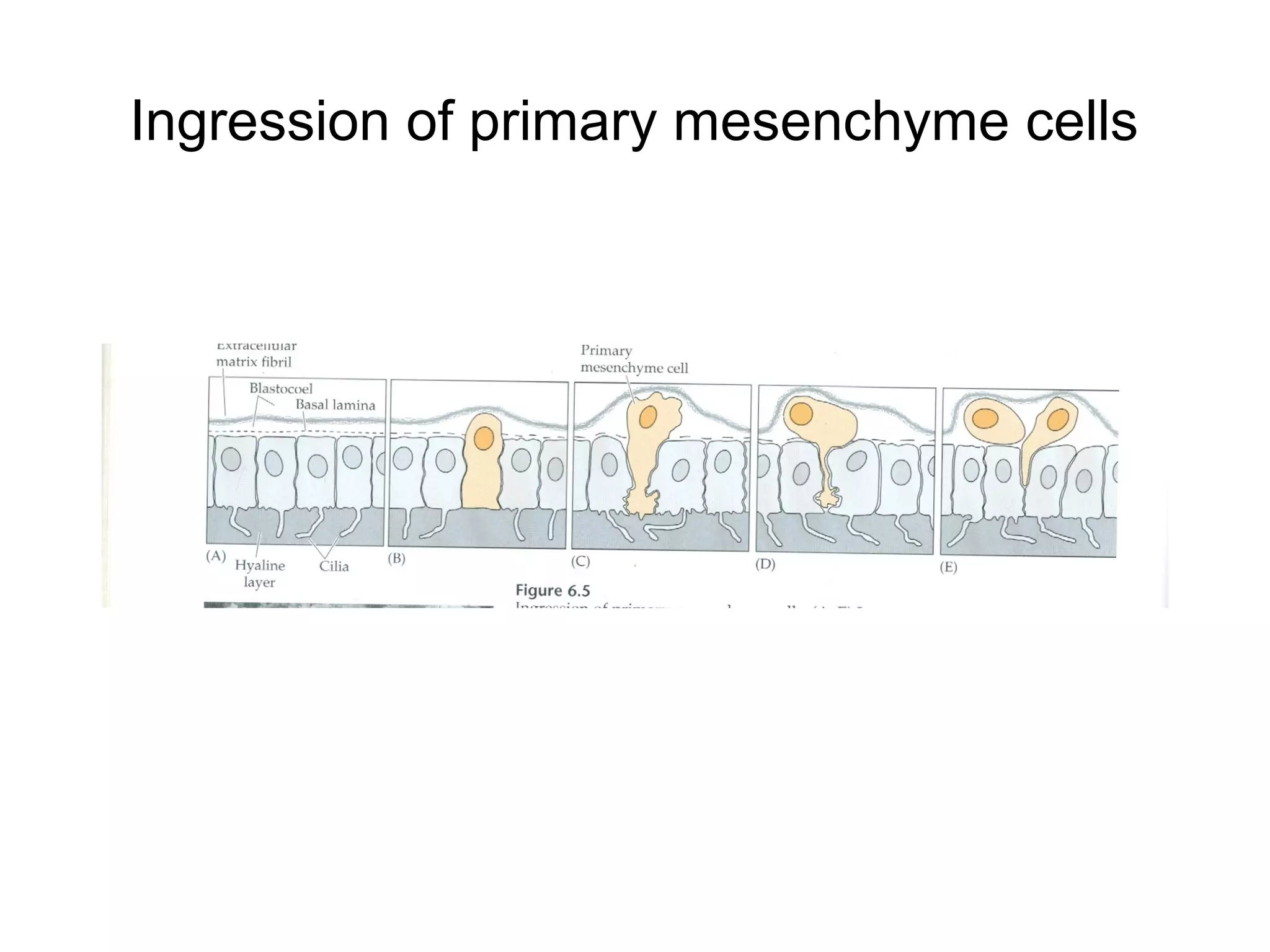
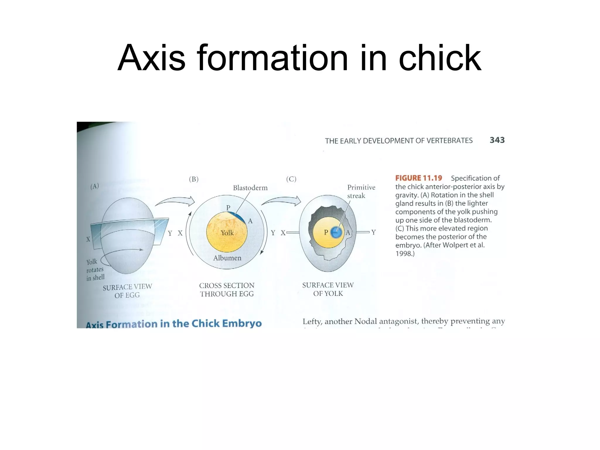
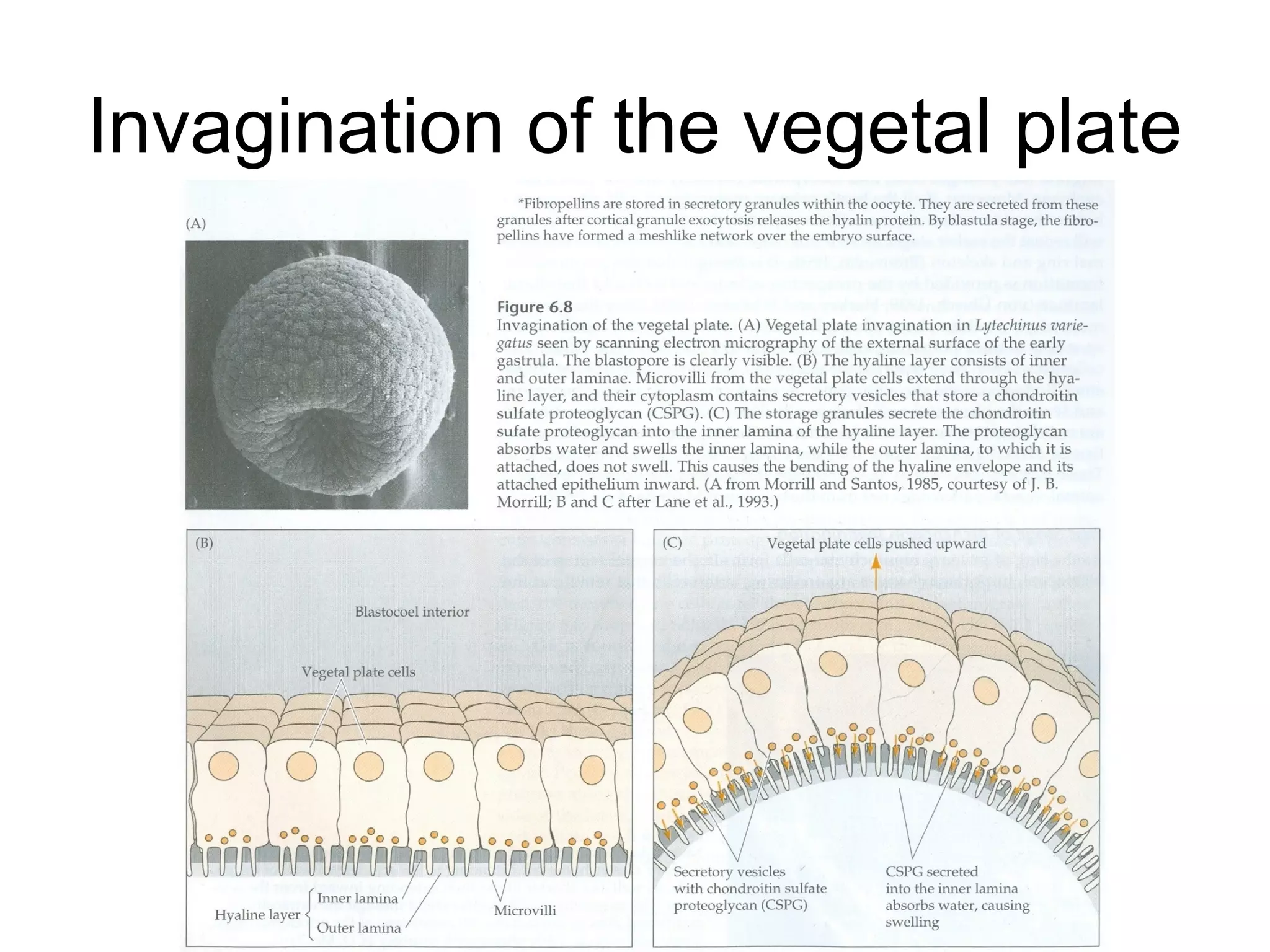
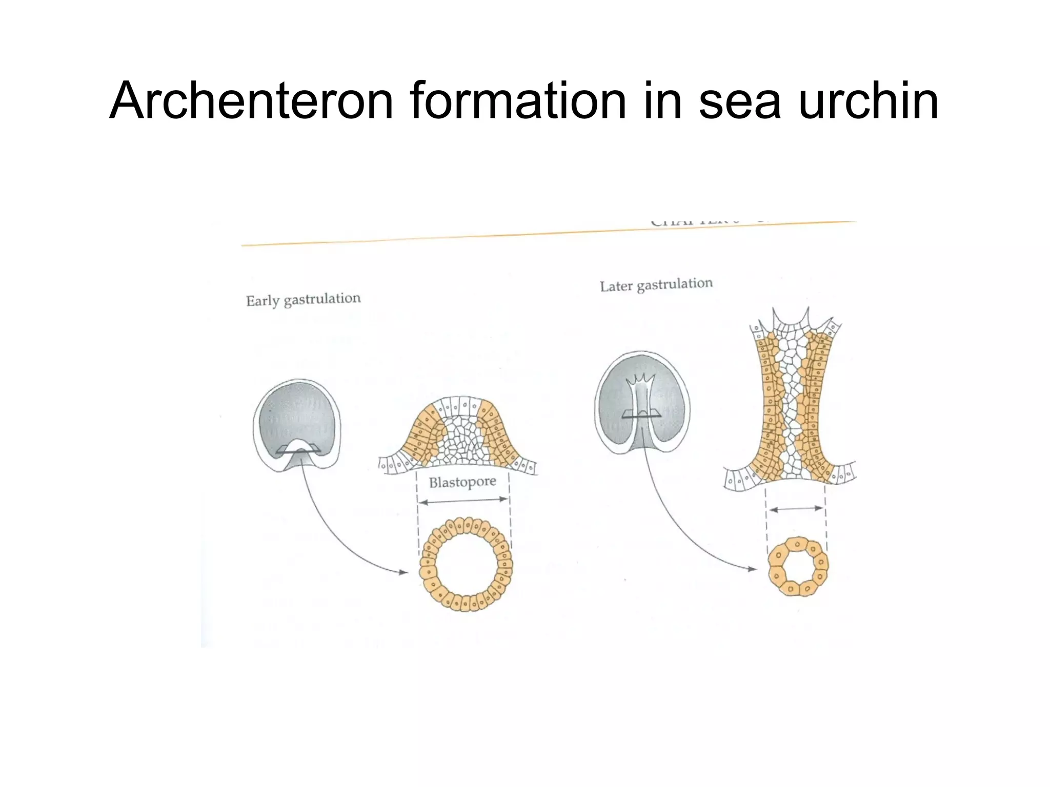
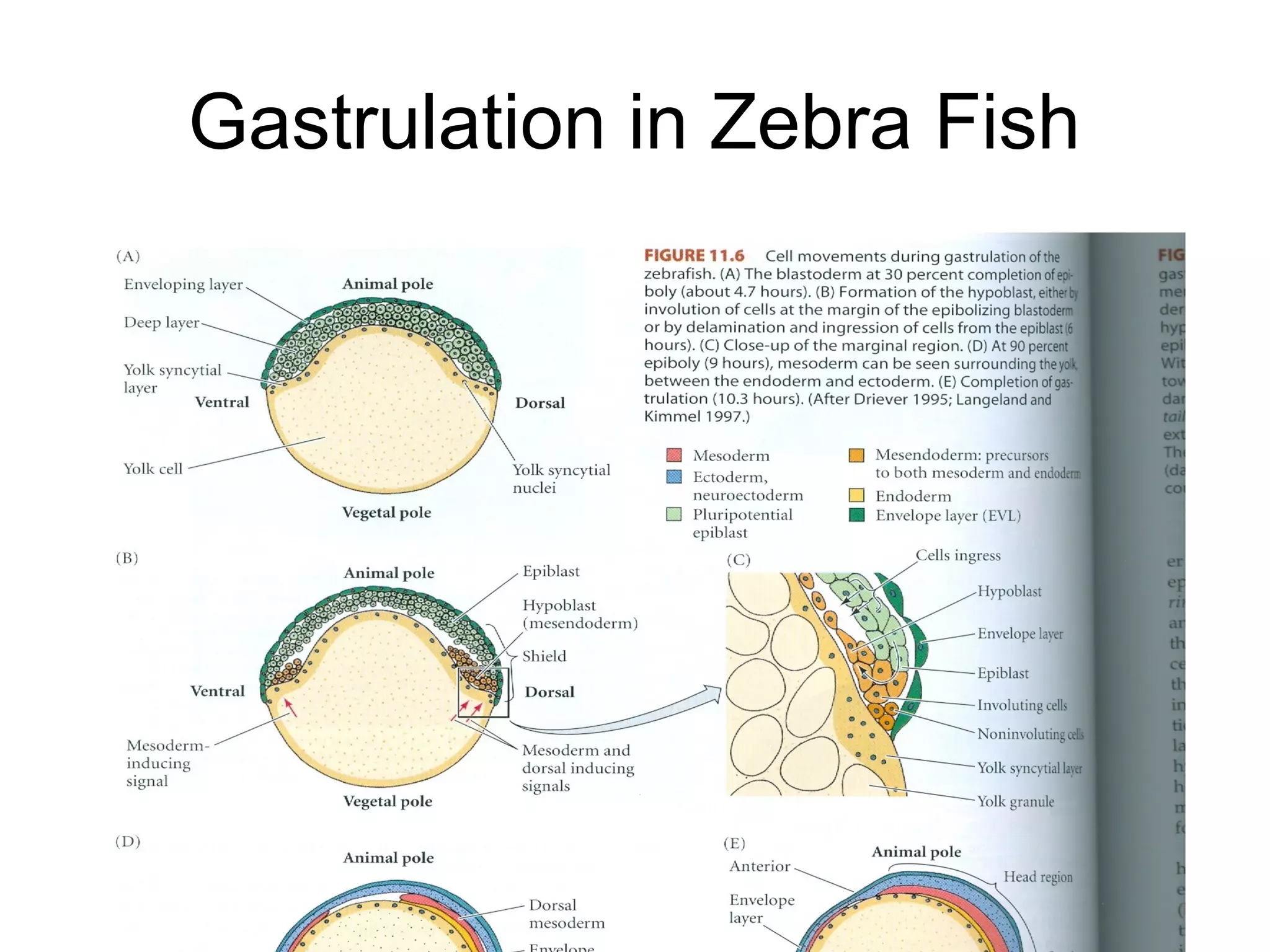
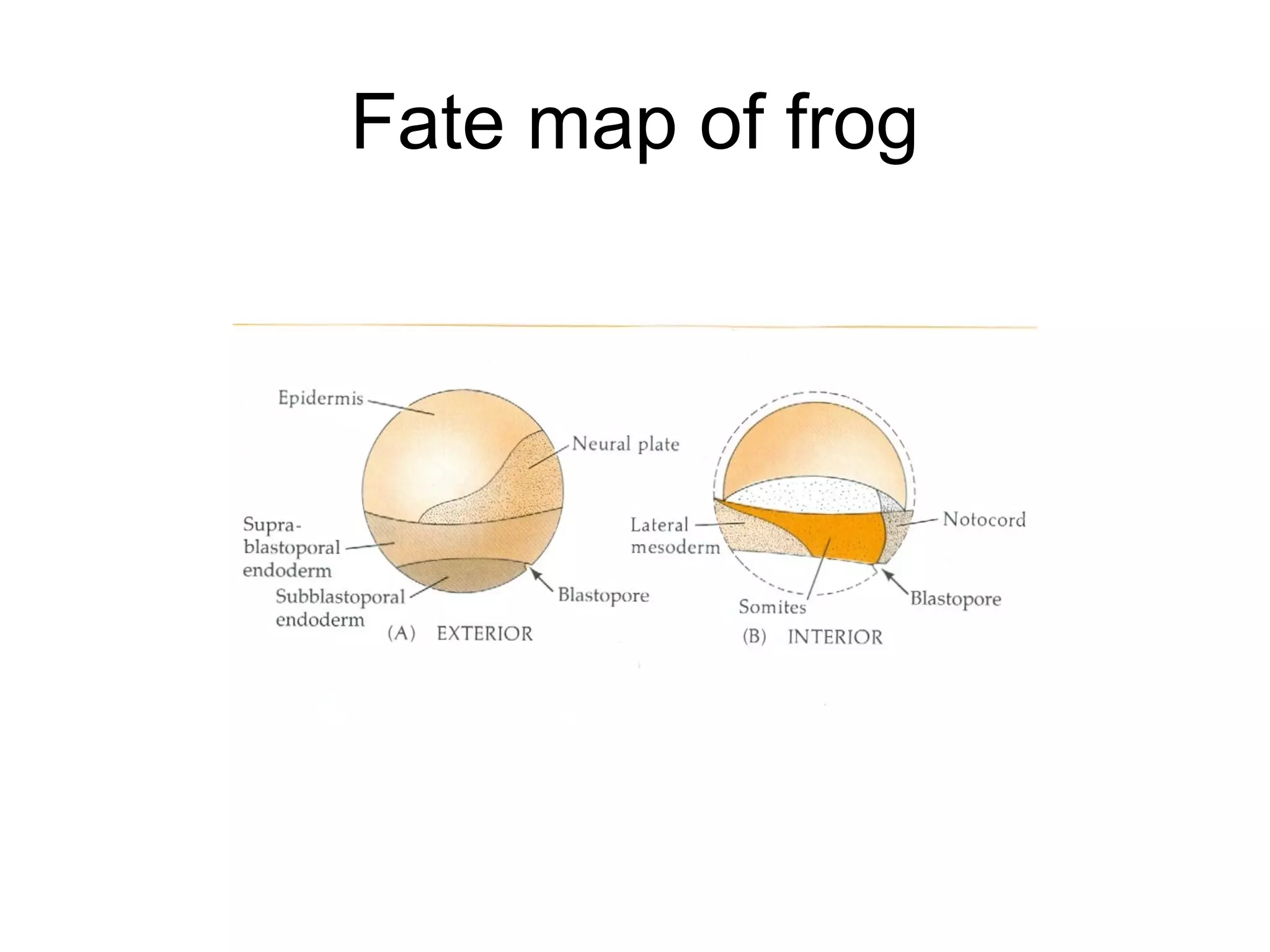
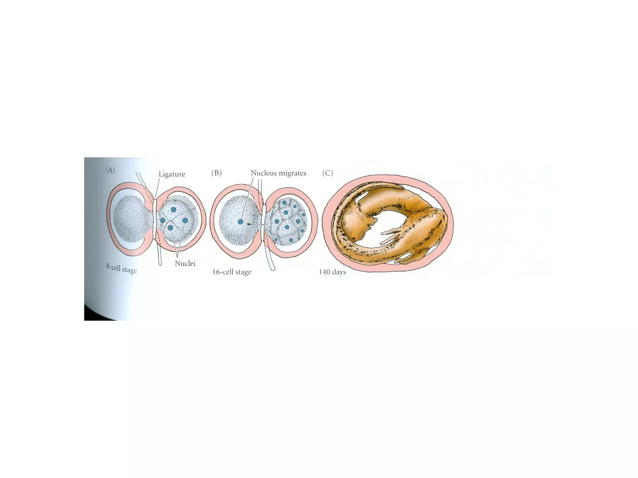

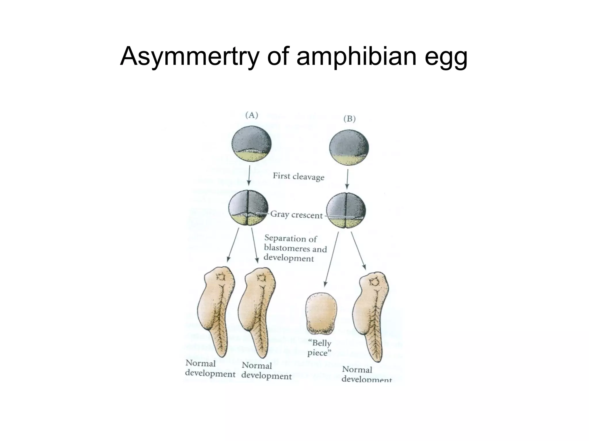
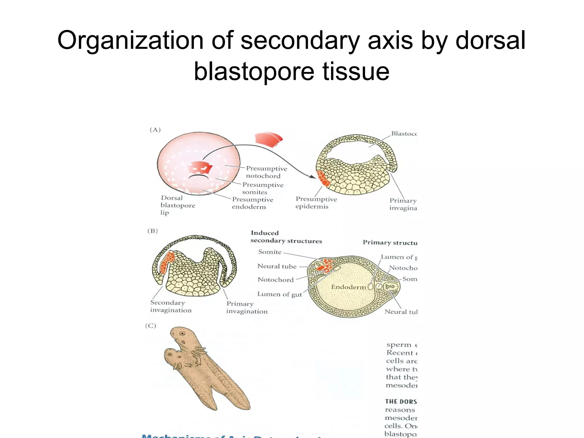
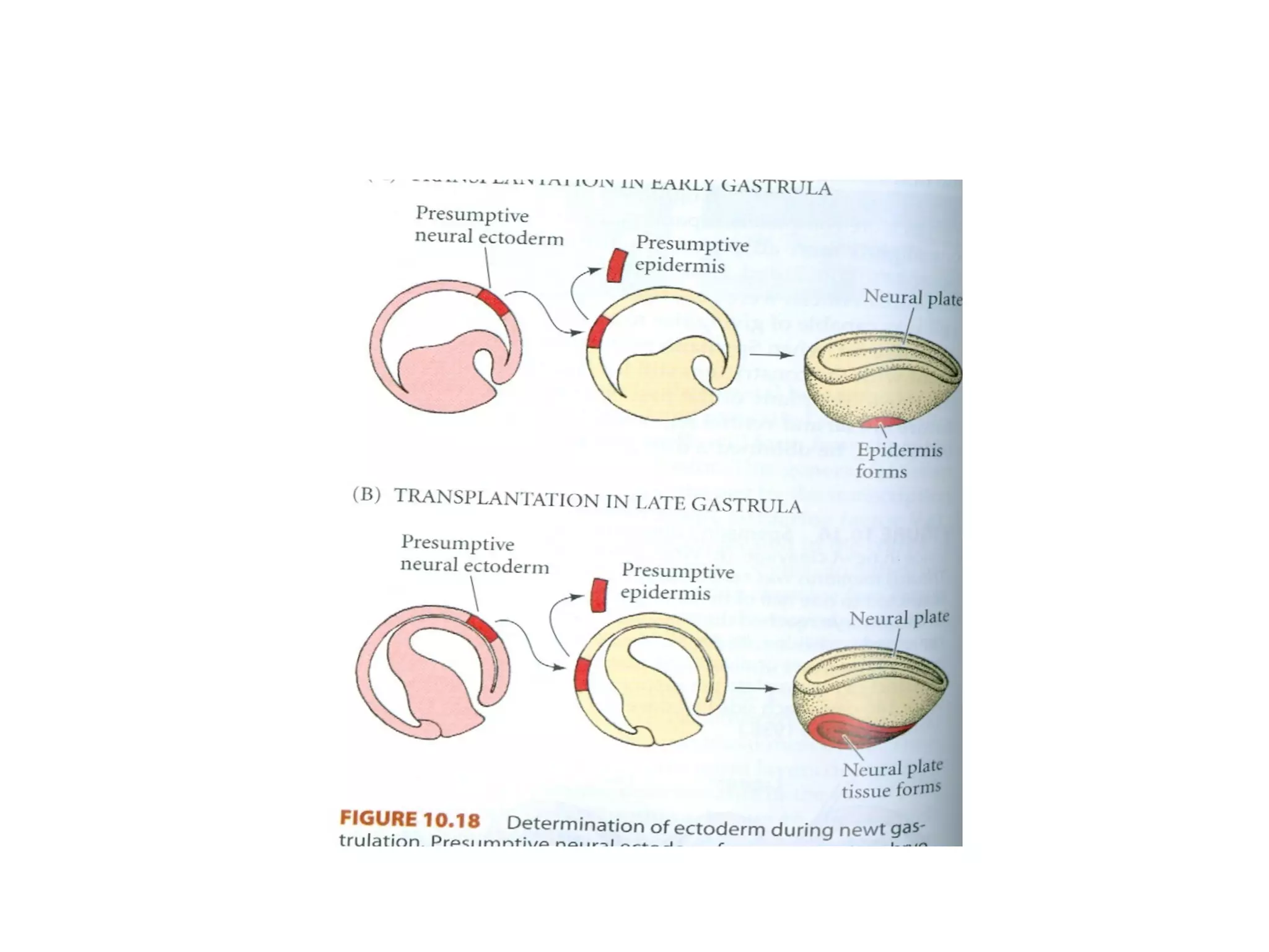
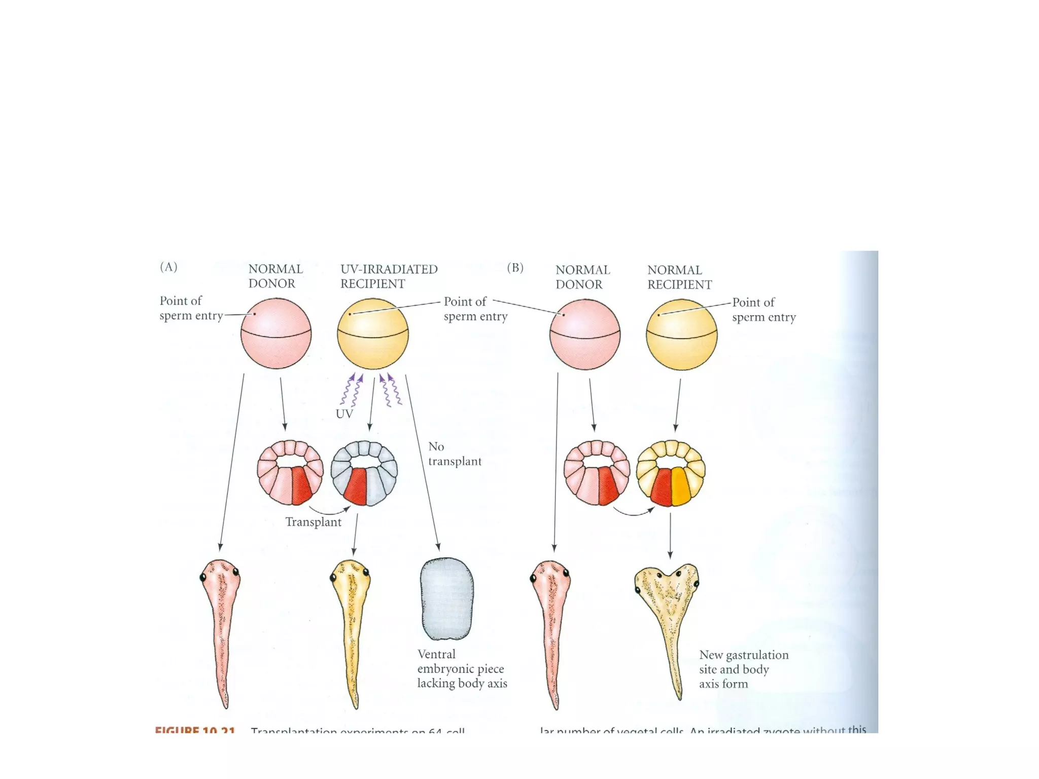
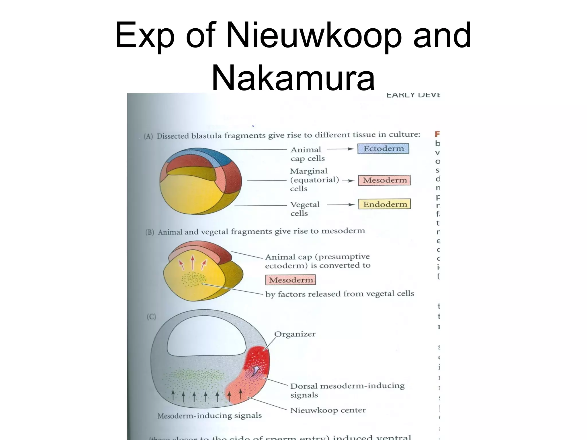
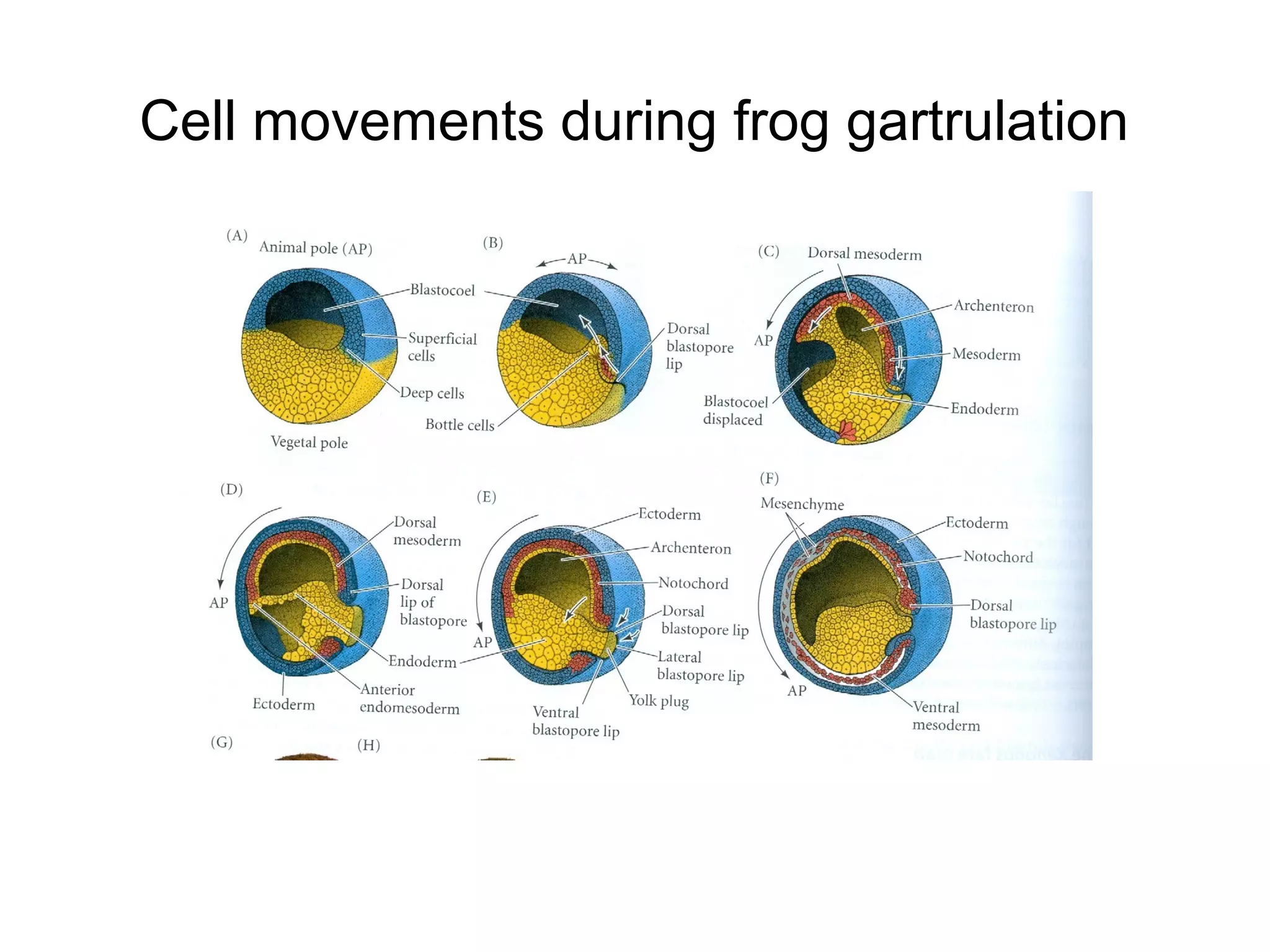
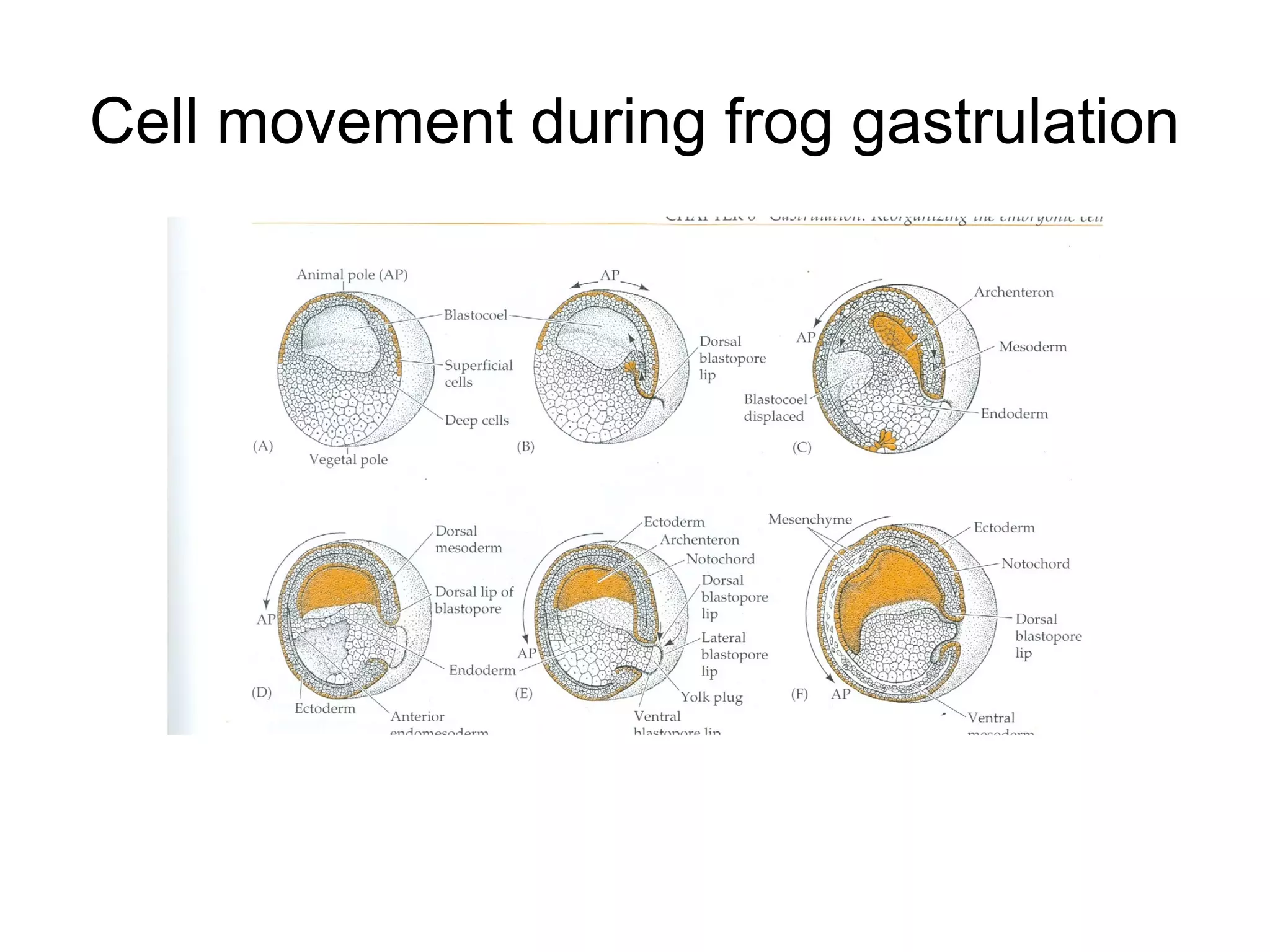
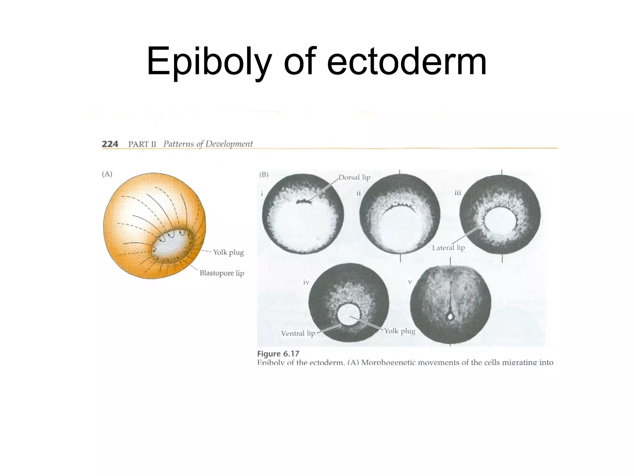
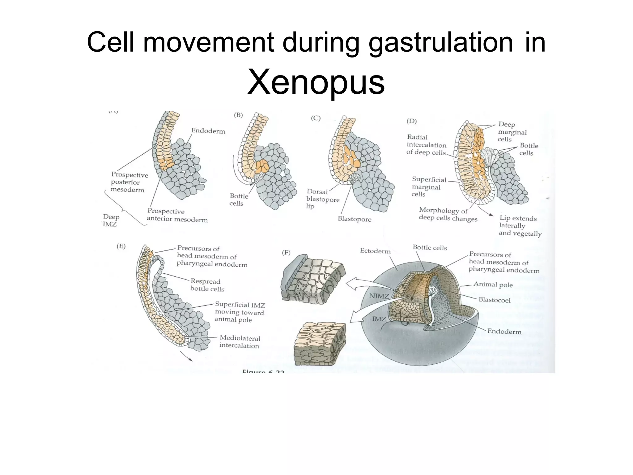
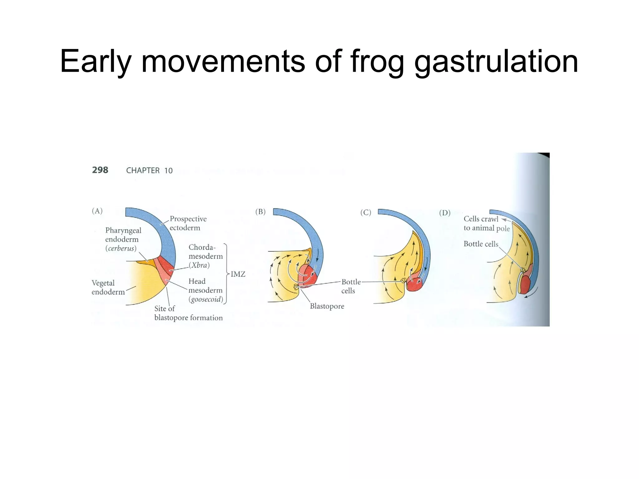

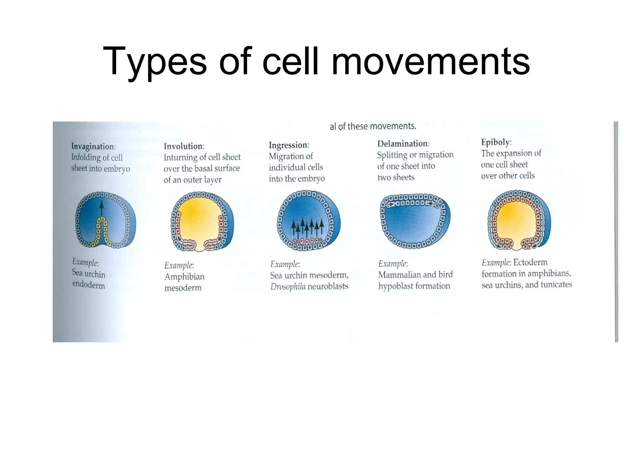
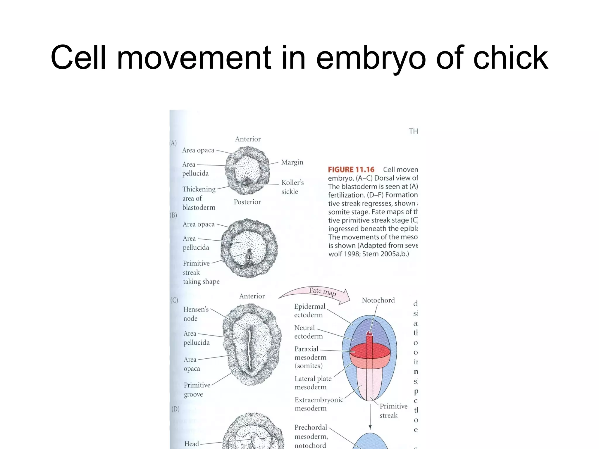
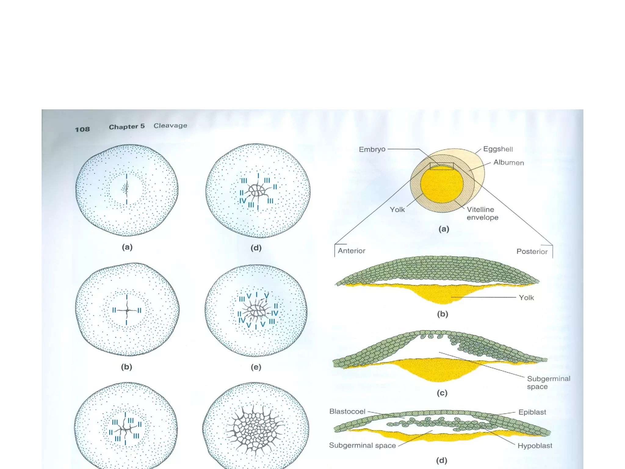
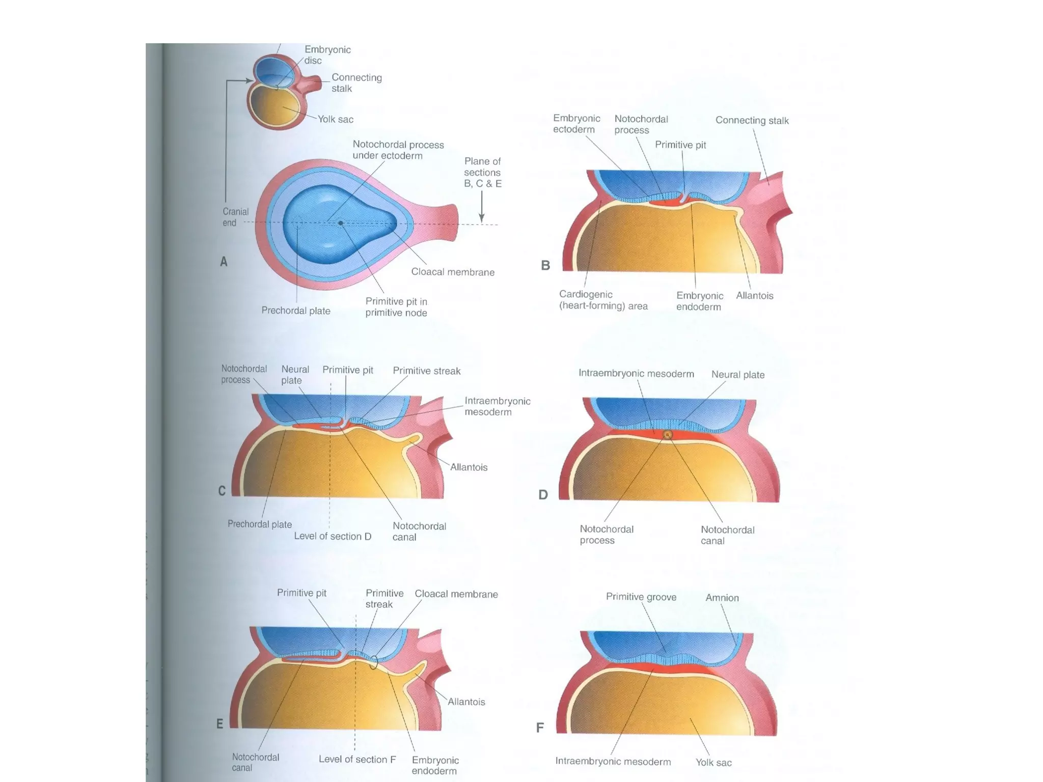
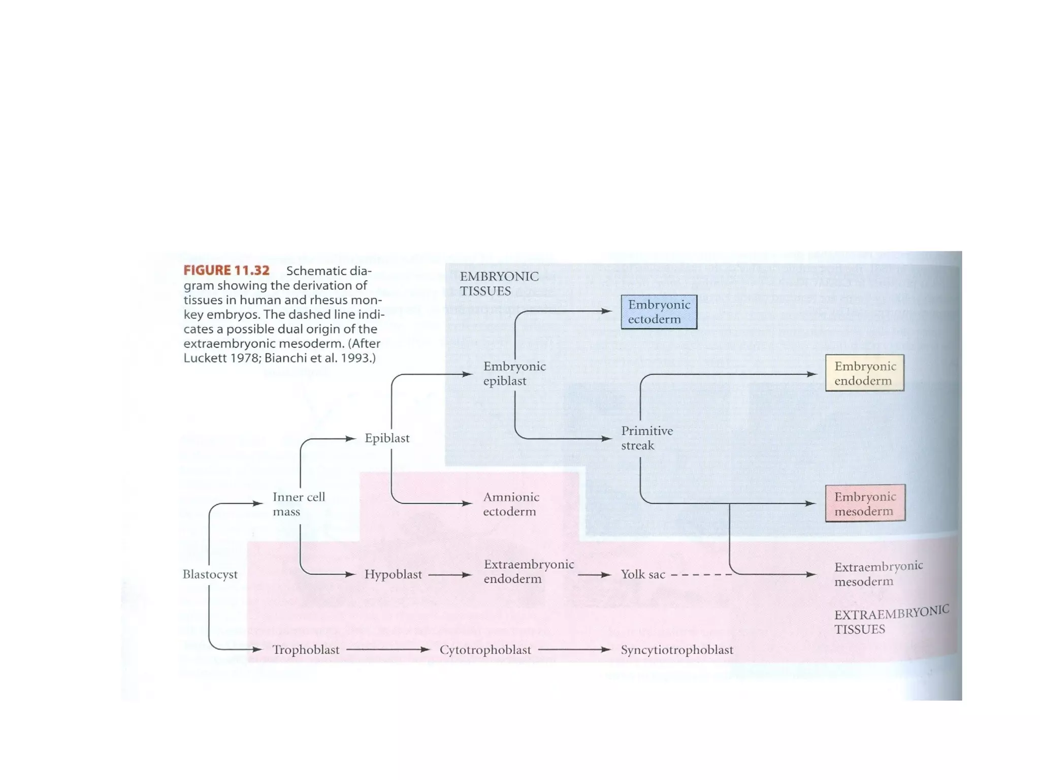
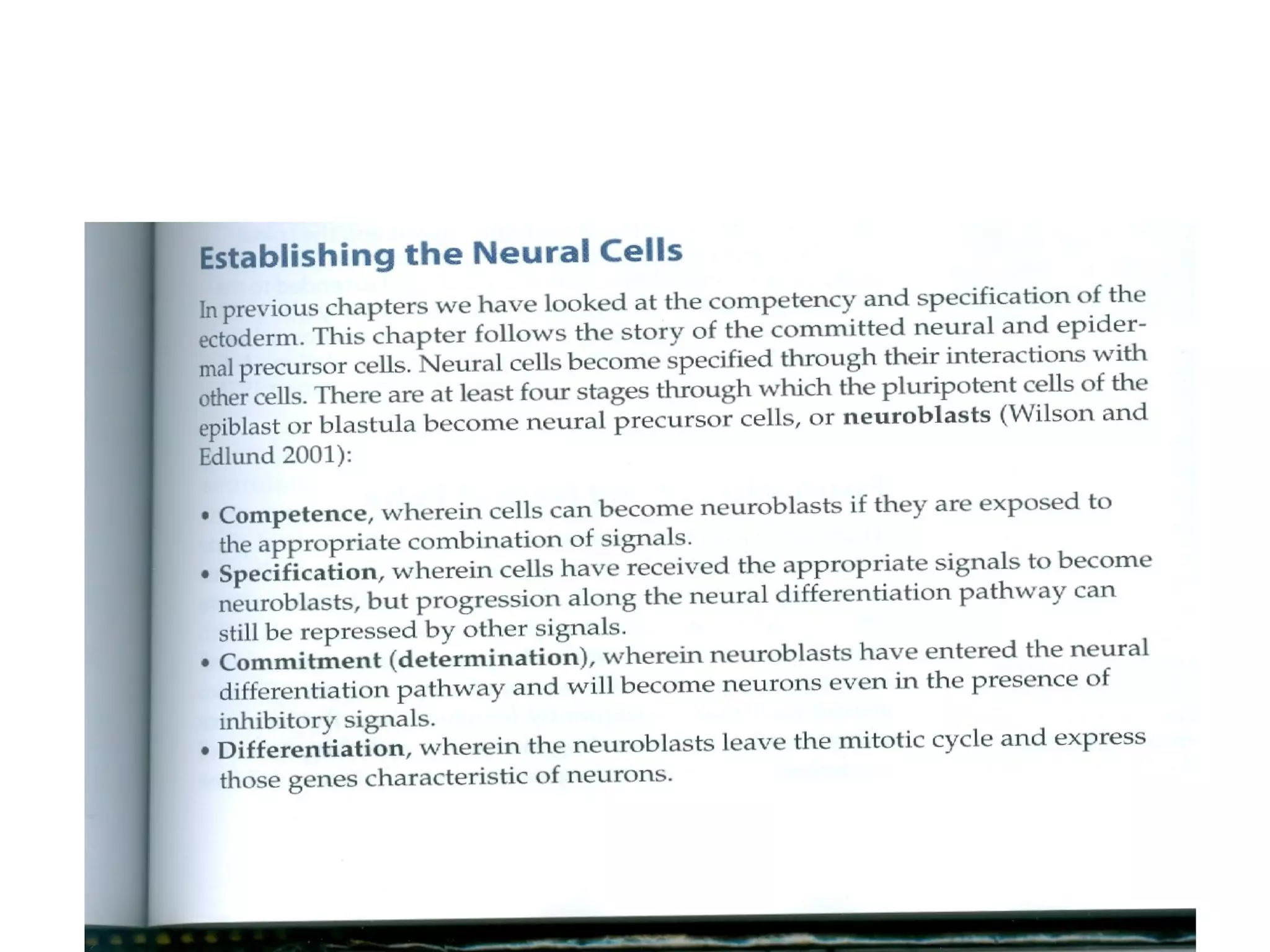
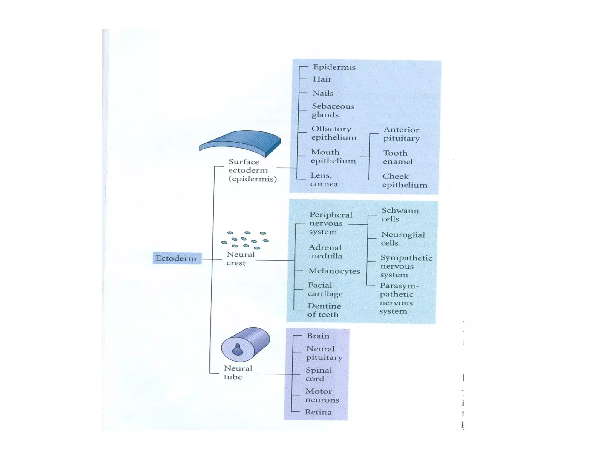

This document contains information on various topics related to early embryonic development, including: - Fate mapping, which determines the developmental fate of groups of cells. - Cleavage, the rapid cell divisions that occur after fertilization. Cleavage results in blastomeres and can be holoblastic, meroblastic, or spiral. - Gastrulation, the process of forming the three germ layers through cell movements and rearrangements. Types of cell movements during gastrulation are discussed for various animals. - Axis formation and patterning the early embryo. Mechanisms like dorsalization and secondary axis formation are covered. - Early stages of development for specific animals like frogs, sea urch