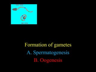
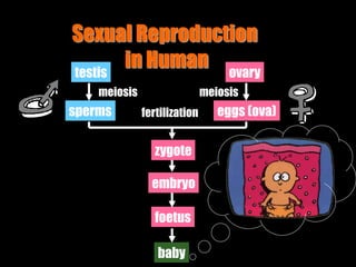
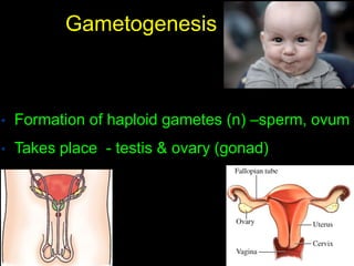
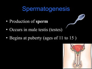
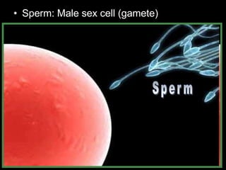
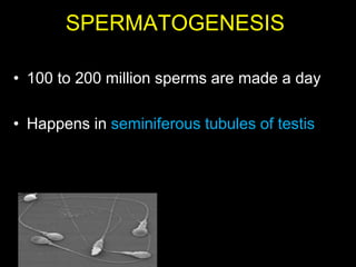
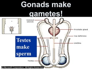
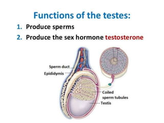
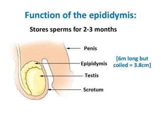
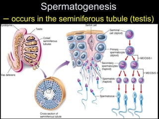
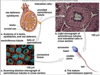
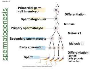
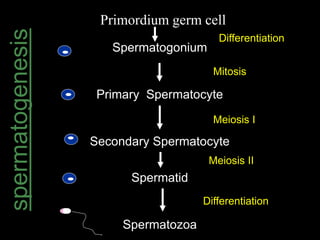
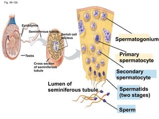
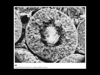
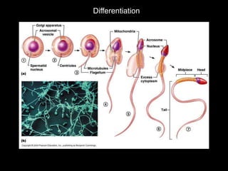
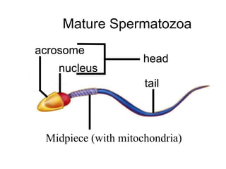
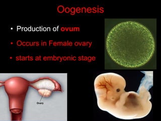
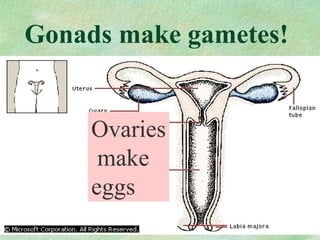
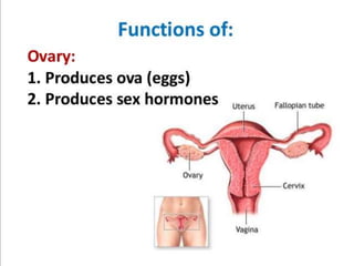
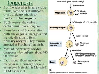
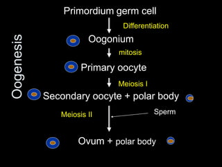
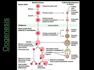
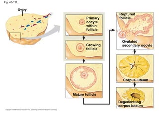
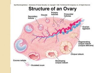
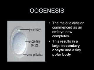
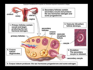
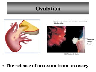
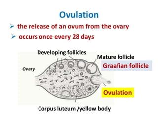
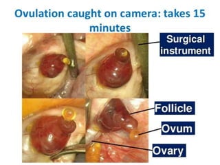
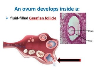
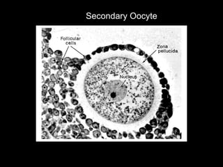
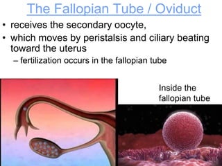
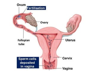
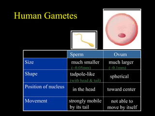
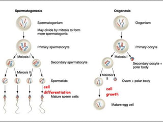
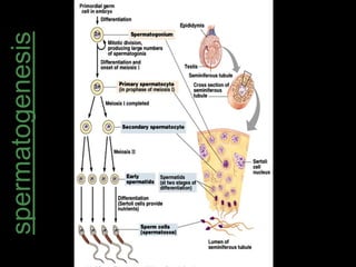
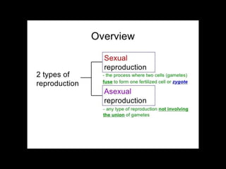
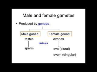
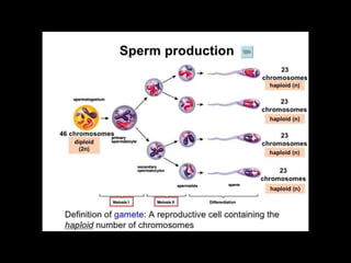
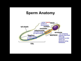
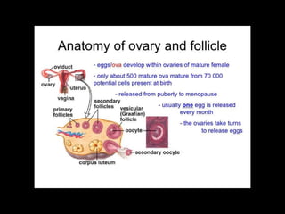
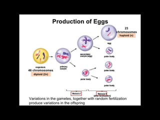
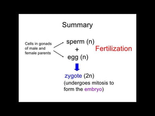
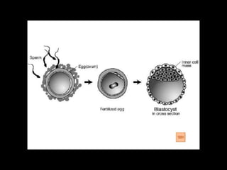
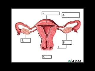
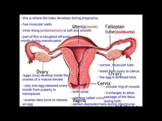
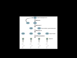
Reproduction 
GAMETOGENESIS
Formation of gametes 
A. Spermatogenesis 
B. Oogenesis
Sexual Reproduction 
in Human 
testis 
meiosis meiosis 
sperms 
ovary 
eggs (ova) 
fertilization 
zygote 
embryo 
foetus 
baby
Gametogenesis 
• Formation of haploid gametes (n) –sperm, ovum 
• Takes place - testis & ovary (gonad)
SPERMATOGENESIS
Spermatogenesis 
• Production of sperm 
• Occurs in male testis (testes) 
• Begins at puberty (ages of 11 to 15 )
• Sperm: Male sex cell (gamete)
SPERMATOGENESIS 
• 100 to 200 million sperms are made a day 
• Happens in seminiferous tubules of testis
Gonads make 
gametes! 
Testes 
make 
sperm
Spermatogenesis 
– occurs in the seminiferous tubule (testis)
Fig. 46-12c 
Primordial germ 
cell in embryo 
Differentiation 
Spermatogonium 
Mitosis 
Primary spermatocyte 
Meiosis I 
Secondary spermatocyte 
Meiosis II 
Early spermatid 
Differentiation 
(Sertoli 
cells provide 
nutrients) 
Sperm 
2n 
2n 
2n 
n n 
n n n n 
n n n n 
spermatogenesis
spermatogenesis 
Primordium germ cell 
Spermatogonium 
Differentiation 
Primary Spermatocyte 
Secondary Spermatocyte 
Spermatid 
Spermatozoa 
Mitosis 
Meiosis I 
Meiosis II 
Differentiation
Fig. 46-12b 
Epididymis 
Seminiferous tubule 
Sertoli cell 
nucleus 
Testis 
Cross section 
of seminiferous 
tubule 
Spermatogonium 
Primary 
spermatocyte 
Secondary 
spermatocyte 
Spermatids 
(two stages) 
Sperm 
Lumen of 
seminiferous tubule
Differentiation
VIDEO SPERMATOGENESIS
Mature Spermatozoa 
tail 
acrosome 
nucleus 
mitochondria 
head 
Midpiece (with mitochondria)
• Animation of flagella propelling sperm.
ANDROPAUSE ???
Abnormal Sperm
Male Infertility 
• Over the past 30 years, the sperm 
content of semen has dropped 
dramatically 
• In the 1970s sperm content of an 
average ejaculate was ~100 million / 
mL; now it is ~60 million / mL 
• It is thought that low sperm count is 
attributable to drug usage, alcohol 
abuse, cigarette smoking and possibly 
industrial and environmental toxins
Male Reproductive System 
• Hormones involved: 
–FSH stimulates the Sertoli to facilitate sperm 
development. 
–LH stimulates Leydig cells to secrete 
testosterone. 
• Negative feedback inhibition applies to 
FSH and LH secretion.
OOGENESIS
• Egg (Ovum): Female sex cell.
Oogenesis 
• Production of ovum 
• Occurs in Female ovary 
• starts at embryonic stage
Gonads make gametes! 
Ovaries 
make 
eggs
Ovary
Oogenesis 
Mitosis & Growth 
Meiosis1 
 5 or 6 weeks after female zygote 
forms, some cells in embryo’s 
ovary undergo mitosis to 
produce diploid oogonia 
 By 24 weeks, the embryo 
contains millions of oogonia 
 From then until 6 weeks after 
birth, the oogonia undergo a first 
meiotic division to form 
primary oocytes. They remain 
arrested at Prophase 1 at birth. 
 Most of the primary oocytes 
disappear – by puberty about 400 
000 remain. 
 Each month from puberty to 
menopause, 1 primary oocycte 
completes Meiosis1 & Meiosis II 
till Metaphase II.
Oogenesis 
Primordium germ cell 
Differentiation 
Oogonium 
mitosis 
Primary oocyte 
Meiosis I 
Secondary oocyte + polar body 
Meiosis II Sperm 
Ovum + polar body
Oogenesis
Fig. 46-12f 
Ovary 
Primary 
oocyte 
within 
follicle 
Ruptured 
follicle 
Growing 
follicle 
Mature follicle 
Ovulated 
secondary oocyte 
Corpus luteum 
Degenerating 
corpus luteum
Graafian follicle
OOGENESIS 
• The meiotic division 
commenced as an 
embryo now 
completes. 
• This results in a 
large secondary 
oocyte and a tiny 
polar body
Uterine tube 
Ovary 
Copyright © The McGraw-Hill Companies, Inc. Permission required for reproduction or display. 
Secondary 
oocyte 
© 2007 Landrum B. Shettles 
Ovulation 
• The release of an ovum from an ovary
VIDEO 
MATURATION 
OVULATION
Secondary Oocyte
Ovum 
• I ovum released per month. 
• Women release about 500 ova in a lifetime. 
• Larger 
• Stationary 
• contain food reserves (lipids, proteins, carbohydrates) 
to nourish the embryo after fertilisation
The Fallopian Tube / Oviduct 
• receives the secondary oocyte, 
• which moves by peristalsis and ciliary beating 
toward the uterus 
– fertilization occurs in the fallopian tube 
Inside the 
fallopian tube
Human Gametes 
much smaller 
(~0.05mm) 
much larger 
(~0.1mm) 
tadpole-like 
(with head & tail) 
spherical 
in the head toward center 
strongly mobile 
by its tail 
not able to 
move by itself 
Size 
Shape 
Position of nucleus 
Movement 
Sperm Ovum
Removing an ovarian cyst!
THE END
spermatogenesis
THE END
BIOLOGY FORM 5 CHAPTER 4 - 4.1C Spermatogenesis & 0ogenesis

BIOLOGY FORM 5 CHAPTER 4 - 4.1C Spermatogenesis & 0ogenesis

  • 1.
  • 2.
    Formation of gametes A. Spermatogenesis B. Oogenesis
  • 3.
    Sexual Reproduction inHuman testis meiosis meiosis sperms ovary eggs (ova) fertilization zygote embryo foetus baby
  • 4.
    Gametogenesis • Formationof haploid gametes (n) –sperm, ovum • Takes place - testis & ovary (gonad)
  • 5.
  • 6.
    Spermatogenesis • Productionof sperm • Occurs in male testis (testes) • Begins at puberty (ages of 11 to 15 )
  • 7.
    • Sperm: Malesex cell (gamete)
  • 8.
    SPERMATOGENESIS • 100to 200 million sperms are made a day • Happens in seminiferous tubules of testis
  • 9.
    Gonads make gametes! Testes make sperm
  • 17.
    Spermatogenesis – occursin the seminiferous tubule (testis)
  • 19.
    Fig. 46-12c Primordialgerm cell in embryo Differentiation Spermatogonium Mitosis Primary spermatocyte Meiosis I Secondary spermatocyte Meiosis II Early spermatid Differentiation (Sertoli cells provide nutrients) Sperm 2n 2n 2n n n n n n n n n n n spermatogenesis
  • 20.
    spermatogenesis Primordium germcell Spermatogonium Differentiation Primary Spermatocyte Secondary Spermatocyte Spermatid Spermatozoa Mitosis Meiosis I Meiosis II Differentiation
  • 21.
    Fig. 46-12b Epididymis Seminiferous tubule Sertoli cell nucleus Testis Cross section of seminiferous tubule Spermatogonium Primary spermatocyte Secondary spermatocyte Spermatids (two stages) Sperm Lumen of seminiferous tubule
  • 23.
  • 24.
  • 25.
    Mature Spermatozoa tail acrosome nucleus mitochondria head Midpiece (with mitochondria)
  • 31.
    • Animation offlagella propelling sperm.
  • 33.
  • 34.
  • 35.
    Male Infertility •Over the past 30 years, the sperm content of semen has dropped dramatically • In the 1970s sperm content of an average ejaculate was ~100 million / mL; now it is ~60 million / mL • It is thought that low sperm count is attributable to drug usage, alcohol abuse, cigarette smoking and possibly industrial and environmental toxins
  • 36.
    Male Reproductive System • Hormones involved: –FSH stimulates the Sertoli to facilitate sperm development. –LH stimulates Leydig cells to secrete testosterone. • Negative feedback inhibition applies to FSH and LH secretion.
  • 37.
  • 38.
    • Egg (Ovum):Female sex cell.
  • 39.
    Oogenesis • Productionof ovum • Occurs in Female ovary • starts at embryonic stage
  • 40.
    Gonads make gametes! Ovaries make eggs
  • 41.
  • 43.
    Oogenesis Mitosis &Growth Meiosis1  5 or 6 weeks after female zygote forms, some cells in embryo’s ovary undergo mitosis to produce diploid oogonia  By 24 weeks, the embryo contains millions of oogonia  From then until 6 weeks after birth, the oogonia undergo a first meiotic division to form primary oocytes. They remain arrested at Prophase 1 at birth.  Most of the primary oocytes disappear – by puberty about 400 000 remain.  Each month from puberty to menopause, 1 primary oocycte completes Meiosis1 & Meiosis II till Metaphase II.
  • 44.
    Oogenesis Primordium germcell Differentiation Oogonium mitosis Primary oocyte Meiosis I Secondary oocyte + polar body Meiosis II Sperm Ovum + polar body
  • 45.
  • 46.
    Fig. 46-12f Ovary Primary oocyte within follicle Ruptured follicle Growing follicle Mature follicle Ovulated secondary oocyte Corpus luteum Degenerating corpus luteum
  • 50.
  • 51.
    OOGENESIS • Themeiotic division commenced as an embryo now completes. • This results in a large secondary oocyte and a tiny polar body
  • 53.
    Uterine tube Ovary Copyright © The McGraw-Hill Companies, Inc. Permission required for reproduction or display. Secondary oocyte © 2007 Landrum B. Shettles Ovulation • The release of an ovum from an ovary
  • 57.
  • 58.
  • 60.
    Ovum • Iovum released per month. • Women release about 500 ova in a lifetime. • Larger • Stationary • contain food reserves (lipids, proteins, carbohydrates) to nourish the embryo after fertilisation
  • 62.
    The Fallopian Tube/ Oviduct • receives the secondary oocyte, • which moves by peristalsis and ciliary beating toward the uterus – fertilization occurs in the fallopian tube Inside the fallopian tube
  • 64.
    Human Gametes muchsmaller (~0.05mm) much larger (~0.1mm) tadpole-like (with head & tail) spherical in the head toward center strongly mobile by its tail not able to move by itself Size Shape Position of nucleus Movement Sperm Ovum
  • 66.
  • 67.
  • 68.
  • 85.