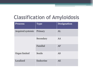
Amyloid


          Shahin Hameed
• The term amyloid, meaning starch-like, is a
  misnomer coined by Virchow when he observed
  that amyloid deposits would stain blue with the
  iodine reaction, suggesting the presence of
  starch or cellulose.
• Amyloid – is a protein that has an alternation in
  its secondary structure which imparts a
  particular insoluble form, called the beta-pleated
  sheet conformation.
• By definition, any protein deposits staining with
  Congo red and exhibiting green birefringence
  when viewed with polarized light are amyloid
Components of amyloid:
A. Fibril protein:

•   proteins make up 85-90% of the amyloid
    weight

•   responsible for tissue dysfunction due to
    pressure effects
B. P Component:

•   makes up about 10% of the amyloid’s weight

•   found on all amyloid fibrils, except those associated
    with Alzheimer’s senile plaques.

•   stabilizes fibril amyloid protein and decreases their
    clearance

•   plays a role in some of the other special stains that can
    demonstrate amyloid (TFT)
C. Non-amyloid components:

• Collagen, fibrin and reticulin

• Part of the amyloid OR are trapped by the amyloid
  OR are part of the body’s repair process from the
  cellular destruction caused by the amyloid.

• This is one of the reasons why connective tissue
  stains (e.g. trichromes, PAS) stain amyloid the same
  color as collagen
Classification of Amyloidosis
Process             Type        Designation


Acquired systemic   Primary     AL


                    Secondary   AA


                    Familial    AF


Organ limited       Senile      AS


Localized           Endocrine   AE
Important Systemic amyloidosis
     Amyloid protein         Precursor            Syndrome or
                                                 involved tissue

AL                     Immunoglobulin light   Primary, Myeloma
                       chain                  associated
AA                     Serum AA (SAA)         Reactive, chronic
                                              inflammatory condition
Aß2m                   ß2 microglobulin       Hemodialysis
AA                     SAA                    Familial mediterranean
                                              fever
ATTR                   Transthyretin          Familial amyloidotic
                                              polyneuropathies
ATTR                   Transthyretin          Systemic senile
                                              Amyloidosis
Important localized amyloidosis
     Amyloid protein          Precursor                 Syndrome or
                                                      involved tisssue
Aß                     Aß protein precursor        Alzheimer’s disease
                       (AßPP)
APrP                   Prior protein               Spongiform
                                                   encephalopathy
Acal                   Calcitonin                  Medullary carcinoma of
                                                   thyroid
AIAPP                  Islet amyloid               Type II diabetes,
                       polypeptide                 insulinomas
AANF                   Atrial natriuretic factor   Isolated atrial amyloid
• The progressive accumulation of extracellular
  protein fibrils can lead to cellular atrophy,
  ischemia, necrosis,and, ultimately, organ failure
  due to the effects of these accumulated fibrils on
  blood supply and normal cellular function
Demonstration of Amyloid
• H & E – homogenous pale pink color
• Van Gieson – yellow to yellow/brown
• Iodine (Gram’s or Lugol’s) – mahogany brown
  turning into blue on treating with 10% sulphuric
  acid
• Light microscopy – postive staining with congo
  red together with a resultant green polorization
  color
• Electron microscopy – typical fibrillar ultra
  structure of amyloid
• X-ray diffraction – cross beta pleat structure
I.   Metachromatic methods
• Staining with 1% aqueous methyl violet for 5
  minutes, followed by differentiation with 1 %
  acetic acid

• Sections should be mounted in aqueous media of
  high sugar or salt content to prevent diffusion of
  the stain
Lendrum’s technique
1. Bring sections to water
2. Stain with 1% aqueous methyl violet for 3 minutes
3. Differentiate in 70% formalin (controlling
   microscopically)
4. Wash in running water for 1 minute
5. Flood with saturated aqueous sodium chloride for 5
   minutes
6. Rinse in water and mount in corn syrup

Results:
      Amyloid- Pink to red
      Other elements- Violet
II. Congo red methods
• Specificity of methods of this type are dependent
  on
 ▫ Beta pleated sheet configuration of amyloid
 ▫ Linearity of the dye molecule

• Congo red is a linear dye molecule. It binds to
  amyloid by hydrogen bonds
• The characteristic green polarization color
  wasn’t present in very thin or thick section –
  thickness of 5-10 micron was optimal

• Sirius red – similar properties to congo red

• Tissue sections containing amyloid AA protein,
  affinity for Congo red is lost after pretreatment
  with potassium permanganate
Bennhold’s Technique
1.   Bring sections to water
2.   Stain with Ehrlich’s haematoxylin for 20 mins
3.   Differentiate with 1% acid alcohol
4.   Wash in running water for 1 min to remove acid
5.   Stain with 1% aqueous Congo red for 20-30 mins
6.   Pour off stain and flood slide with a saturated aqueous solution of
     lithium carbonate; leave for 15 secs
7.   Differentiate in 80% alcohol until excess Congo red is removed
8.   Wash in running water for 10 mins
9.   Dehydrate, clear and mount in Canada balsam or synthetic resin

Results
          Amyloid- Pink to red
          Nuclei- Blue
Alkaline Congo red technique
(Puchtler, Sweat and Levine)

• Advantage of not requiring differentiation
  ( Congo red is in alkaline solution)

• The use of alcoholic solutions, high salt content,
  and high pH, as in the Puchtler Congo red
  method, greatly increase staining specificity for
  amyloid
• A saturated salt solution in alcohol at alkaline
  pH is used in both the dye solution and as a
  pretreatment of the tissue sections just before
  staining.

• High salt content and alkaline pH are believed to
  depress dye ionization and electrostatic binding
  to nonamyloid structures. Saturation of the salt
  and dye solutions is very important
• Fixation:
 ▫ Best results are obtained after alcohol or Carnoy
   fixed tissues
 ▫ Formalin or Zenker –fixed tissues were found to
   stain better than with other techniques
• Reagents
 ▫ Alkaline salt solution. To 50 ml of 80% alcohol
   saturated with sodium chloride add 0.5 ml of 1%
   aqueous sodium hydroxide. Filter and use within
   15 min.
 ▫ Stock stain solution. Use 80% alcohol saturated
   with Congo red and sodium chloride
 ▫ Staining solution. Add 0.5 ml of 1% aqueous
   sodium hydroxide to 50 ml of stock stain, filter
   and use within 15 mins
Method :

1. Bring sections to water.
2. Stain in haematoxylin for 5 minutes.
3. Rinse well in distilled water.
4. Pretreat in alkaline alcohol-salt solution for 20
   min.
5. Stain in alkaline Congo red solution for 20 min.
6. Dehydrate rapidly in three changes of absolute
   alcohol.
7. Clear and mount in synthetic resin.

Results:
      Amyloid- deep pink to red
      Nuclei -blue
      Elastic -pale pink
This bone marrow section stained with the Puchtler Congo red
procedure reveals intense amyloid deposits in the bone marrow
sample
This figure viewed with polarizing microscopy;
amyloid deposits exhibit characteristic apple-
green birefringence.
Problems Encountered With the
Congo Red Stain ..
a) PROBLEM: Weakly Stained Tissues

• APPEARANCE: Faint uptake of Congo red dye
  throughout the section

• CAUSES:
 ▫ Tissue was fixed for a prolonged period in a
   formaldehyde-containing fixative.
 ▫ Cut sections were stored for a prolonged period.
 ▫ Solutions of Congo red are not stable in the presence
   of salt and alkali.
• SOLUTIONS:
  • Avoid storing tissues in formaldehyde-based
  fixatives for prolonged periods of time.
  • Cut the sections just before staining, or cut
  only as many control sections as can be used in a
  short period of time.
  • Seal the cut paraffin blocks to help preserve
  control tissue reactivity.
  • Prepare fresh working solutions just before use
Congo red staining in this section appears
indistinct, suggesting that the stain was
excessively differentiated
Examination of the section with polarized
light reveals numerous amyloid deposits
b) PROBLEM: Nonspecific Staining

• APPEARANCE: Structures other than amyloid
  bind the Congo red; high background staining may
  make it difficult to distinguish true amyloid deposits
  if present in the tissue

• CAUSES:
  Collagen, elastic fibers, and keratin may stain
  nonspecificallywith aqueous Congo red solution.
  The pH is not sufficiently alkaline.
SOLUTIONS:

• Use polarizing microscopy to help distinguish connective
  tissue components (grey or silver )from amyloid deposits
  (green).

• Rotating the slide on the stage during polarizing microscopy
  can help distinguish true amyloid deposits from connective
  tissue. When the slide is rotated, connective tissue fibers will
  lose the dichroism,while amyloid will not

• Use the Puchtler Congo red method, because the high salt
  content of the prestain rinse and staining solutions tends to
  diminish nonspecific staining.

• Avoid the use of Canada balsam mounting medium because it
  will fluoresce.
Tissue components in this Congo red stained
kidney section appear to be nonspecifically
stained
Polarization of the Congo red-stained
section reveals that most of the red
staining is not amyloid. Some apple-green
birefringence is noted in the glomerulus at
the bottom of the image
C. PROBLEM: Incorrect Color of Birefringence

APPEARANCE: Structures may exhibit yellow, red, or white
 dichroism

CAUSE:
• Section thickness may be incorrect; this artifact is especially
   likely in thin sections

SOLUTION:
• Ensure that sections are cut at 5 to 10 μm.

COMMENT:
Old (large) amyloid deposits will often display diminished
  birefringence.
Smaller deposits in blood vessel walls, for example, may be more
  likely to demonstrate the characteristic apple green color.
Congo red staining in this section of kidney appears
             specific and well differentiated
When viewed with polarized light, amyloid
deposits appear red to yellow, not the
expected apple-green color; incorrect
section thickness is the most likely cause
Collagen, appearing white to silver in this
polarized, Congo red stained section,
surrounds a characteristic apple-green
amyloid deposit.
D. PROBLEM: Precipitate on Tissue

APPEARANCE: A red precipitate is randomly
 present throughout the tissue

CAUSE:
Salt solutions were prepared with isopropyl alcohol.

SOLUTION:
Ensure that the salt solutions are prepared with ethyl
 alcohol because the salt does not dissolve well in
 isopropyl alcohol and will deposit crystals on the
 section.
Although the amyloid is well demonstrated in
this section, close inspection reveals a red
granular precipitate caused by using isopropyl
alcohol instead of ethyl alcohol as the solvent for
the salt solutions
III. Toluidine blue method
Standard toluidine blue

1. Bring sections to water.
2. Stain in 1% toluidine blue in 50% isopropanol for
   30 min at 37o C.
3. Blot and place in absolute isopropanol for 1 min.
4. Clear in xylene and mount.

Results:
Amyloid is distinguished by its dark polarization color
IV. Fluorescence techniques
• Thioflavine T technique

• Extremely sensitive technique although not
  specific because
 ▫ stained sections are not permanent and
 ▫ tissue components other than amyloid, including
   fibrinoid, keratin, intestinal muciphages, Paneth
   cells, zymogen granules, and juxtaglomerular
   apparatus, all stain with thioflavine T
• Attempts made to increase specificity
  ▫ Using acid solutions
  ▫ Including Magnesium chloride

• Thioflavine S may be substituted for Thioflavine T
Method:

1. Bring sections to water.
2. Stain in alum-haematoxylin for 2 min to quench nuclear
   fluorescence. The haematoxylin does not need to be
   differentiated, or blued.
3. Wash in water for a few minutes.
4. Stain in 1% aqueous thioflavine T for 3 min.
5. Rinse in water.
6. Differentiate in 1% acetic acid for 20 min.
7. Wash in water.
8. Mount in Apathy’s medium.

Results:
Amyloid and mast cells fluoresce bright yellow when examined
using a BG12 exciter filter and an OG4 or OG5 barrier filter.
The finest deposits can be seen using a UG1 or UG2 exciter filter
with a colourless ultraviolet barrier filter
Amyloid stained with TFT in Kidney.
Hit with 490 (blue) excitor wavelength. Amyloid
fluorescing yellow
• Congo Red Fluorescence

 Congo red stained amyloid, even if it does not birefringe
  with polarization, will often fluoresce an orange color
  when viewed with the auramine-rhodamine
  fluorescence microscope wavelengths (540 nm excitor
  wavelength) and lenses.

 At the same time, connective tissue and/or other tissue
  components that like to autofluoresce with green light
  will usually exhibit a white to yellow color
Congo Red stained amyloid in kidney, .
Hit with 490 (blue) excitor wavelength.
Amyloid fluorescing orange
V. Alcian blue method
• Introduced by Lendrum, Slidders and Fraser

• Fixation in formal-saline is usually adequate
• The sodium sulphate-Alcian blue (SAB) method

• Reagents
  1.    Acetic alcohol
       a) 95% ethanol – 45ml
       b) Distilled water – 45 ml
       c) Glacial acetic acid - 10 ml
  Prepare fresh for use

  2.    SAB solution
       a) 1% alcian blue in 95 % ethanol – 45ml
       b) 1% aqueous sodium sulphate decahydrate – 45ml
       c) Glacial acetic acid – 10ml
  Prepare from stock solutions and stand for 30 mins before
  use
Method

1. Bring sections to water.
2. Immerse in acetic alcohol for 1-2 min.
3. Stain in SAB working solution for 2 hours.
4. Transfer to acetic alcohol for 1-2 min.
5. Wash in water.
6. Alkalinize in 80% ethanol saturated with borax for 30
   min.
7. Wash in water
8. Stain nuclei with Celestine blue-haemalum sequence
   and counterstain with Van Gieson.
9. Dehydrate, clear and mount.

Results:
      Amyloid, mast cells and some colloids- Green
VI. IMMUNOHISTOCHEMISTRY
• There are limitations as to the use of
  immunohistochemistry for the demonstration of amyloid

• Each has its own sequence of amino acids (proteins).
  Also, there are variations of amino acid sequences from
  person to person, even if they have the same type of
  amyloid.

• It is usually necessary to use more than one IHC for
  amyloid concurrent with the Congo red stain, as well as
  other amyloid histology stains, if needed.
IHC Amyloid
Thank you ..

Amyloid

  • 1.
    Amyloid Shahin Hameed
  • 2.
    • The termamyloid, meaning starch-like, is a misnomer coined by Virchow when he observed that amyloid deposits would stain blue with the iodine reaction, suggesting the presence of starch or cellulose.
  • 3.
    • Amyloid –is a protein that has an alternation in its secondary structure which imparts a particular insoluble form, called the beta-pleated sheet conformation. • By definition, any protein deposits staining with Congo red and exhibiting green birefringence when viewed with polarized light are amyloid
  • 4.
    Components of amyloid: A.Fibril protein: • proteins make up 85-90% of the amyloid weight • responsible for tissue dysfunction due to pressure effects
  • 5.
    B. P Component: • makes up about 10% of the amyloid’s weight • found on all amyloid fibrils, except those associated with Alzheimer’s senile plaques. • stabilizes fibril amyloid protein and decreases their clearance • plays a role in some of the other special stains that can demonstrate amyloid (TFT)
  • 6.
    C. Non-amyloid components: •Collagen, fibrin and reticulin • Part of the amyloid OR are trapped by the amyloid OR are part of the body’s repair process from the cellular destruction caused by the amyloid. • This is one of the reasons why connective tissue stains (e.g. trichromes, PAS) stain amyloid the same color as collagen
  • 7.
    Classification of Amyloidosis Process Type Designation Acquired systemic Primary AL Secondary AA Familial AF Organ limited Senile AS Localized Endocrine AE
  • 8.
    Important Systemic amyloidosis Amyloid protein Precursor Syndrome or involved tissue AL Immunoglobulin light Primary, Myeloma chain associated AA Serum AA (SAA) Reactive, chronic inflammatory condition Aß2m ß2 microglobulin Hemodialysis AA SAA Familial mediterranean fever ATTR Transthyretin Familial amyloidotic polyneuropathies ATTR Transthyretin Systemic senile Amyloidosis
  • 9.
    Important localized amyloidosis Amyloid protein Precursor Syndrome or involved tisssue Aß Aß protein precursor Alzheimer’s disease (AßPP) APrP Prior protein Spongiform encephalopathy Acal Calcitonin Medullary carcinoma of thyroid AIAPP Islet amyloid Type II diabetes, polypeptide insulinomas AANF Atrial natriuretic factor Isolated atrial amyloid
  • 10.
    • The progressiveaccumulation of extracellular protein fibrils can lead to cellular atrophy, ischemia, necrosis,and, ultimately, organ failure due to the effects of these accumulated fibrils on blood supply and normal cellular function
  • 11.
    Demonstration of Amyloid •H & E – homogenous pale pink color • Van Gieson – yellow to yellow/brown • Iodine (Gram’s or Lugol’s) – mahogany brown turning into blue on treating with 10% sulphuric acid
  • 12.
    • Light microscopy– postive staining with congo red together with a resultant green polorization color • Electron microscopy – typical fibrillar ultra structure of amyloid • X-ray diffraction – cross beta pleat structure
  • 13.
    I. Metachromatic methods • Staining with 1% aqueous methyl violet for 5 minutes, followed by differentiation with 1 % acetic acid • Sections should be mounted in aqueous media of high sugar or salt content to prevent diffusion of the stain
  • 14.
    Lendrum’s technique 1. Bringsections to water 2. Stain with 1% aqueous methyl violet for 3 minutes 3. Differentiate in 70% formalin (controlling microscopically) 4. Wash in running water for 1 minute 5. Flood with saturated aqueous sodium chloride for 5 minutes 6. Rinse in water and mount in corn syrup Results: Amyloid- Pink to red Other elements- Violet
  • 16.
    II. Congo redmethods • Specificity of methods of this type are dependent on ▫ Beta pleated sheet configuration of amyloid ▫ Linearity of the dye molecule • Congo red is a linear dye molecule. It binds to amyloid by hydrogen bonds
  • 17.
    • The characteristicgreen polarization color wasn’t present in very thin or thick section – thickness of 5-10 micron was optimal • Sirius red – similar properties to congo red • Tissue sections containing amyloid AA protein, affinity for Congo red is lost after pretreatment with potassium permanganate
  • 18.
    Bennhold’s Technique 1. Bring sections to water 2. Stain with Ehrlich’s haematoxylin for 20 mins 3. Differentiate with 1% acid alcohol 4. Wash in running water for 1 min to remove acid 5. Stain with 1% aqueous Congo red for 20-30 mins 6. Pour off stain and flood slide with a saturated aqueous solution of lithium carbonate; leave for 15 secs 7. Differentiate in 80% alcohol until excess Congo red is removed 8. Wash in running water for 10 mins 9. Dehydrate, clear and mount in Canada balsam or synthetic resin Results Amyloid- Pink to red Nuclei- Blue
  • 19.
    Alkaline Congo redtechnique (Puchtler, Sweat and Levine) • Advantage of not requiring differentiation ( Congo red is in alkaline solution) • The use of alcoholic solutions, high salt content, and high pH, as in the Puchtler Congo red method, greatly increase staining specificity for amyloid
  • 20.
    • A saturatedsalt solution in alcohol at alkaline pH is used in both the dye solution and as a pretreatment of the tissue sections just before staining. • High salt content and alkaline pH are believed to depress dye ionization and electrostatic binding to nonamyloid structures. Saturation of the salt and dye solutions is very important
  • 21.
    • Fixation: ▫Best results are obtained after alcohol or Carnoy fixed tissues ▫ Formalin or Zenker –fixed tissues were found to stain better than with other techniques
  • 22.
    • Reagents ▫Alkaline salt solution. To 50 ml of 80% alcohol saturated with sodium chloride add 0.5 ml of 1% aqueous sodium hydroxide. Filter and use within 15 min. ▫ Stock stain solution. Use 80% alcohol saturated with Congo red and sodium chloride ▫ Staining solution. Add 0.5 ml of 1% aqueous sodium hydroxide to 50 ml of stock stain, filter and use within 15 mins
  • 23.
    Method : 1. Bringsections to water. 2. Stain in haematoxylin for 5 minutes. 3. Rinse well in distilled water. 4. Pretreat in alkaline alcohol-salt solution for 20 min. 5. Stain in alkaline Congo red solution for 20 min. 6. Dehydrate rapidly in three changes of absolute alcohol. 7. Clear and mount in synthetic resin. Results: Amyloid- deep pink to red Nuclei -blue Elastic -pale pink
  • 24.
    This bone marrowsection stained with the Puchtler Congo red procedure reveals intense amyloid deposits in the bone marrow sample
  • 25.
    This figure viewedwith polarizing microscopy; amyloid deposits exhibit characteristic apple- green birefringence.
  • 26.
    Problems Encountered Withthe Congo Red Stain ..
  • 27.
    a) PROBLEM: WeaklyStained Tissues • APPEARANCE: Faint uptake of Congo red dye throughout the section • CAUSES: ▫ Tissue was fixed for a prolonged period in a formaldehyde-containing fixative. ▫ Cut sections were stored for a prolonged period. ▫ Solutions of Congo red are not stable in the presence of salt and alkali.
  • 28.
    • SOLUTIONS: • Avoid storing tissues in formaldehyde-based fixatives for prolonged periods of time. • Cut the sections just before staining, or cut only as many control sections as can be used in a short period of time. • Seal the cut paraffin blocks to help preserve control tissue reactivity. • Prepare fresh working solutions just before use
  • 29.
    Congo red stainingin this section appears indistinct, suggesting that the stain was excessively differentiated
  • 30.
    Examination of thesection with polarized light reveals numerous amyloid deposits
  • 31.
    b) PROBLEM: NonspecificStaining • APPEARANCE: Structures other than amyloid bind the Congo red; high background staining may make it difficult to distinguish true amyloid deposits if present in the tissue • CAUSES: Collagen, elastic fibers, and keratin may stain nonspecificallywith aqueous Congo red solution. The pH is not sufficiently alkaline.
  • 32.
    SOLUTIONS: • Use polarizingmicroscopy to help distinguish connective tissue components (grey or silver )from amyloid deposits (green). • Rotating the slide on the stage during polarizing microscopy can help distinguish true amyloid deposits from connective tissue. When the slide is rotated, connective tissue fibers will lose the dichroism,while amyloid will not • Use the Puchtler Congo red method, because the high salt content of the prestain rinse and staining solutions tends to diminish nonspecific staining. • Avoid the use of Canada balsam mounting medium because it will fluoresce.
  • 33.
    Tissue components inthis Congo red stained kidney section appear to be nonspecifically stained
  • 34.
    Polarization of theCongo red-stained section reveals that most of the red staining is not amyloid. Some apple-green birefringence is noted in the glomerulus at the bottom of the image
  • 35.
    C. PROBLEM: IncorrectColor of Birefringence APPEARANCE: Structures may exhibit yellow, red, or white dichroism CAUSE: • Section thickness may be incorrect; this artifact is especially likely in thin sections SOLUTION: • Ensure that sections are cut at 5 to 10 μm. COMMENT: Old (large) amyloid deposits will often display diminished birefringence. Smaller deposits in blood vessel walls, for example, may be more likely to demonstrate the characteristic apple green color.
  • 36.
    Congo red stainingin this section of kidney appears specific and well differentiated
  • 37.
    When viewed withpolarized light, amyloid deposits appear red to yellow, not the expected apple-green color; incorrect section thickness is the most likely cause
  • 38.
    Collagen, appearing whiteto silver in this polarized, Congo red stained section, surrounds a characteristic apple-green amyloid deposit.
  • 39.
    D. PROBLEM: Precipitateon Tissue APPEARANCE: A red precipitate is randomly present throughout the tissue CAUSE: Salt solutions were prepared with isopropyl alcohol. SOLUTION: Ensure that the salt solutions are prepared with ethyl alcohol because the salt does not dissolve well in isopropyl alcohol and will deposit crystals on the section.
  • 40.
    Although the amyloidis well demonstrated in this section, close inspection reveals a red granular precipitate caused by using isopropyl alcohol instead of ethyl alcohol as the solvent for the salt solutions
  • 41.
    III. Toluidine bluemethod Standard toluidine blue 1. Bring sections to water. 2. Stain in 1% toluidine blue in 50% isopropanol for 30 min at 37o C. 3. Blot and place in absolute isopropanol for 1 min. 4. Clear in xylene and mount. Results: Amyloid is distinguished by its dark polarization color
  • 42.
    IV. Fluorescence techniques •Thioflavine T technique • Extremely sensitive technique although not specific because ▫ stained sections are not permanent and ▫ tissue components other than amyloid, including fibrinoid, keratin, intestinal muciphages, Paneth cells, zymogen granules, and juxtaglomerular apparatus, all stain with thioflavine T
  • 43.
    • Attempts madeto increase specificity ▫ Using acid solutions ▫ Including Magnesium chloride • Thioflavine S may be substituted for Thioflavine T
  • 44.
    Method: 1. Bring sectionsto water. 2. Stain in alum-haematoxylin for 2 min to quench nuclear fluorescence. The haematoxylin does not need to be differentiated, or blued. 3. Wash in water for a few minutes. 4. Stain in 1% aqueous thioflavine T for 3 min. 5. Rinse in water. 6. Differentiate in 1% acetic acid for 20 min. 7. Wash in water. 8. Mount in Apathy’s medium. Results: Amyloid and mast cells fluoresce bright yellow when examined using a BG12 exciter filter and an OG4 or OG5 barrier filter. The finest deposits can be seen using a UG1 or UG2 exciter filter with a colourless ultraviolet barrier filter
  • 45.
    Amyloid stained withTFT in Kidney. Hit with 490 (blue) excitor wavelength. Amyloid fluorescing yellow
  • 46.
    • Congo RedFluorescence Congo red stained amyloid, even if it does not birefringe with polarization, will often fluoresce an orange color when viewed with the auramine-rhodamine fluorescence microscope wavelengths (540 nm excitor wavelength) and lenses. At the same time, connective tissue and/or other tissue components that like to autofluoresce with green light will usually exhibit a white to yellow color
  • 47.
    Congo Red stainedamyloid in kidney, . Hit with 490 (blue) excitor wavelength. Amyloid fluorescing orange
  • 48.
    V. Alcian bluemethod • Introduced by Lendrum, Slidders and Fraser • Fixation in formal-saline is usually adequate
  • 49.
    • The sodiumsulphate-Alcian blue (SAB) method • Reagents 1. Acetic alcohol a) 95% ethanol – 45ml b) Distilled water – 45 ml c) Glacial acetic acid - 10 ml Prepare fresh for use 2. SAB solution a) 1% alcian blue in 95 % ethanol – 45ml b) 1% aqueous sodium sulphate decahydrate – 45ml c) Glacial acetic acid – 10ml Prepare from stock solutions and stand for 30 mins before use
  • 50.
    Method 1. Bring sectionsto water. 2. Immerse in acetic alcohol for 1-2 min. 3. Stain in SAB working solution for 2 hours. 4. Transfer to acetic alcohol for 1-2 min. 5. Wash in water. 6. Alkalinize in 80% ethanol saturated with borax for 30 min. 7. Wash in water 8. Stain nuclei with Celestine blue-haemalum sequence and counterstain with Van Gieson. 9. Dehydrate, clear and mount. Results: Amyloid, mast cells and some colloids- Green
  • 51.
    VI. IMMUNOHISTOCHEMISTRY • Thereare limitations as to the use of immunohistochemistry for the demonstration of amyloid • Each has its own sequence of amino acids (proteins). Also, there are variations of amino acid sequences from person to person, even if they have the same type of amyloid. • It is usually necessary to use more than one IHC for amyloid concurrent with the Congo red stain, as well as other amyloid histology stains, if needed.
  • 52.
  • 53.