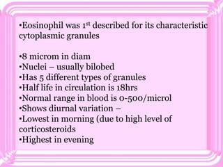
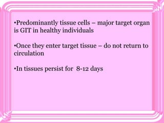
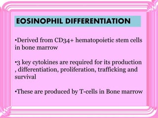
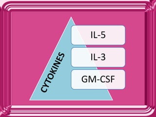
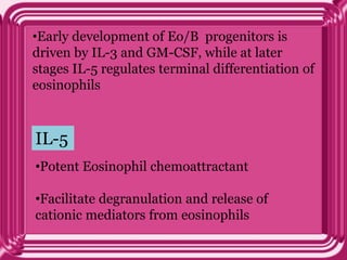
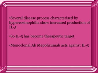
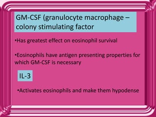
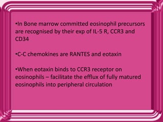
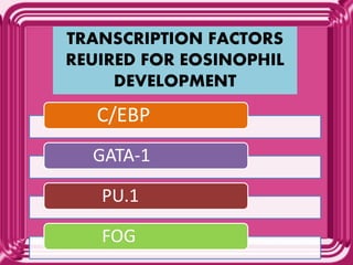
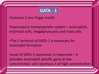
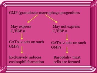
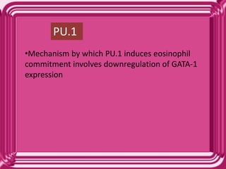
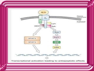
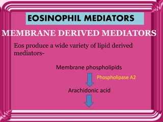
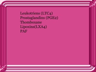
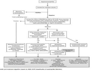
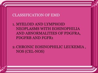
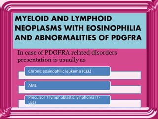
This document provides information on eosinophils:
- Eosinophils are white blood cells that are characterized by distinctive granules and a bilobed nucleus. They play a role in fighting parasites and allergic responses.
- Eosinophils develop from bone marrow stem cells and are regulated by cytokines like IL-5, IL-3 and GM-CSF. Transcription factors like C/EBP, GATA-1 and PU.1 are required for eosinophil development.
- Eosinophils traffic to tissues where they can persist for over a week. Their granules contain toxic proteins that are released during degranulation which is regulated by cytokines and chemokines.
- Eosin
Eosinophilia occurs when a large number of eosinophils are recruited to a specific site in your body or when the bone marrow produces too many eosinophils
I have listed out the LE cells structure and Microscopical examinaton of LE CELLS, Difference between tart cells and le cells, clinical symptoms and diagnostic procedure.
The destruction of red cells
Clinical signs & symptoms of haemolysis
Classification of Haemolytic anaemias
G6PD deficiency
Thalassaemias
http://www.usmlemcq.com/
ROLE OF NEUTROPHILS IN HEALTH & DISEASE.pptxjasmine918783
The slide contains information about neutrophils, its basic structure, formation, trafficking, functions, the role of neutrophils in acute inflammation, various disorders of neutrophils, etc.
RBC
FATE OF RBCS
ERYTHIOPOIESIS
HEMOGLOBIN
1) circular biconcave discs.
2) non nucleated
3) not contain cell organelles
4) are elastic and highly
deforming.
5) Life span 120 days
In red cell membrane there are very important proteins which maintain the shape of RBCs.
These are:
1) Spectrin
2) Ankyrin
3) Stromatin,
4) Actin
5) glycoprotein Elanin.
A congenital haemolytic anaemia i.e; hereditary spherocytosis results because of a significant deficiency of spectrin in the wall of RBCs cannot be maintained.
In RBCs membrane, blood group antigens are present.
SITES
Embryonic life (early wks) --- Yolk sac
Middle trimester ---- Liver, spleen, Lymph nodes
Last months & after birth --- Bone marrow
5 years --- B0ne marrow of all bones
Till 20 years --- proximal portions of humeri & tibiae
Onwards --- marrow of membranous bones such as vertebrae, sternum, ribs and ilia.
(trimester is 3 months duration in pregnancy)
The process of the origin, development and maturation of red blood cells.
It is an extremely active process
About 2.5 million erythrocytes are produced every second in order to replace those that are continuously destroyed by spleen and liver
Different growth and differentiation inducers control growth and differentiation of stem cells controlled by factors outside bone marrow
Above downward
Size of nucleus decrease
Size of hemoglobin increase
Cytoplasmic/ nucleus ratio increase
Amount of RNA responsible for basophilic stain decrease
In orthochromatic erythroblast stage , nucleus is expelled.
During reticulocyte stage, cell passes from bone marrow in to blood capillaries by the process of “Diapedesis”. Small amount of basophilic material.
Remaining basophilic material in the reticulocyte disappear with in 1 to 2 days and then it is called “Erythrocytes”.
Life span 120 days
After 120 days, taken by reticuloendothelial cells i.e; spleen & liver.
Spleen is the graveyard of RBCs.
Decrease in enzyme activity, ATP levels, and MCH
Decreased deformability
Metabolic processes slow down
Membrane becomes fragile
Destroyed in spleen as the red pulp space is narrow (3 μm) as diameter is very small. So, the spleen is called “Graveyard of RBCs”.
RBCs burst out
Hb is immediately phagocytized by macrophages pf the body. Iron and globin released.
EMBRYO
In 2nd month of intrauterine life:
There is Gower 1 & Gower 2 Hb.
Gower 1 contain:
2 zeta & 2 Epsilon
Gower 2 contain:
2 alpha & 2 Epsilon
3rd month onwards:
Fetal Hb:
Hb-F occurs in fetal red cells and disappears by 2 to 3 months after birth.
During I/U life, there is
HbF= 2alpha + 2 gamma chains.
Adult Haemoglobin
(HbA1):
2 alpha chains and 2 beta chains
HbA2:
is a minor component & is only 2.5% in normal adults.
After birth, there is Hb A2 (small amount), which is composed of 2 alpha & 2 delta chains.
Retinal Pigment epithelial cells perform a multitude of functions to protect the retina and maintain normal vision. Any kind of RPE malfunction leads to a variety of ocular diseases.
This PPT explains the various functions performed by RPE to maintain normal vision.
Specialized Tissues, Stem Cells and Tissue RenewalGarry D. Lasaga
This report will talk about the physiology of tissue renewal among different tissues and the important role played by stem cells in such mechanism as wells as the various key signaling mechanism involved.
Tom Selleck Health: A Comprehensive Look at the Iconic Actor’s Wellness Journeygreendigital
Tom Selleck, an enduring figure in Hollywood. has captivated audiences for decades with his rugged charm, iconic moustache. and memorable roles in television and film. From his breakout role as Thomas Magnum in Magnum P.I. to his current portrayal of Frank Reagan in Blue Bloods. Selleck's career has spanned over 50 years. But beyond his professional achievements. fans have often been curious about Tom Selleck Health. especially as he has aged in the public eye.
Follow us on: Pinterest
Introduction
Many have been interested in Tom Selleck health. not only because of his enduring presence on screen but also because of the challenges. and lifestyle choices he has faced and made over the years. This article delves into the various aspects of Tom Selleck health. exploring his fitness regimen, diet, mental health. and the challenges he has encountered as he ages. We'll look at how he maintains his well-being. the health issues he has faced, and his approach to ageing .
Early Life and Career
Childhood and Athletic Beginnings
Tom Selleck was born on January 29, 1945, in Detroit, Michigan, and grew up in Sherman Oaks, California. From an early age, he was involved in sports, particularly basketball. which played a significant role in his physical development. His athletic pursuits continued into college. where he attended the University of Southern California (USC) on a basketball scholarship. This early involvement in sports laid a strong foundation for his physical health and disciplined lifestyle.
Transition to Acting
Selleck's transition from an athlete to an actor came with its physical demands. His first significant role in "Magnum P.I." required him to perform various stunts and maintain a fit appearance. This role, which he played from 1980 to 1988. necessitated a rigorous fitness routine to meet the show's demands. setting the stage for his long-term commitment to health and wellness.
Fitness Regimen
Workout Routine
Tom Selleck health and fitness regimen has evolved. adapting to his changing roles and age. During his "Magnum, P.I." days. Selleck's workouts were intense and focused on building and maintaining muscle mass. His routine included weightlifting, cardiovascular exercises. and specific training for the stunts he performed on the show.
Selleck adjusted his fitness routine as he aged to suit his body's needs. Today, his workouts focus on maintaining flexibility, strength, and cardiovascular health. He incorporates low-impact exercises such as swimming, walking, and light weightlifting. This balanced approach helps him stay fit without putting undue strain on his joints and muscles.
Importance of Flexibility and Mobility
In recent years, Selleck has emphasized the importance of flexibility and mobility in his fitness regimen. Understanding the natural decline in muscle mass and joint flexibility with age. he includes stretching and yoga in his routine. These practices help prevent injuries, improve posture, and maintain mobilit
ARTIFICIAL INTELLIGENCE IN HEALTHCARE.pdfAnujkumaranit
Artificial intelligence (AI) refers to the simulation of human intelligence processes by machines, especially computer systems. It encompasses tasks such as learning, reasoning, problem-solving, perception, and language understanding. AI technologies are revolutionizing various fields, from healthcare to finance, by enabling machines to perform tasks that typically require human intelligence.
Recomendações da OMS sobre cuidados maternos e neonatais para uma experiência pós-natal positiva.
Em consonância com os ODS – Objetivos do Desenvolvimento Sustentável e a Estratégia Global para a Saúde das Mulheres, Crianças e Adolescentes, e aplicando uma abordagem baseada nos direitos humanos, os esforços de cuidados pós-natais devem expandir-se para além da cobertura e da simples sobrevivência, de modo a incluir cuidados de qualidade.
Estas diretrizes visam melhorar a qualidade dos cuidados pós-natais essenciais e de rotina prestados às mulheres e aos recém-nascidos, com o objetivo final de melhorar a saúde e o bem-estar materno e neonatal.
Uma “experiência pós-natal positiva” é um resultado importante para todas as mulheres que dão à luz e para os seus recém-nascidos, estabelecendo as bases para a melhoria da saúde e do bem-estar a curto e longo prazo. Uma experiência pós-natal positiva é definida como aquela em que as mulheres, pessoas que gestam, os recém-nascidos, os casais, os pais, os cuidadores e as famílias recebem informação consistente, garantia e apoio de profissionais de saúde motivados; e onde um sistema de saúde flexível e com recursos reconheça as necessidades das mulheres e dos bebês e respeite o seu contexto cultural.
Estas diretrizes consolidadas apresentam algumas recomendações novas e já bem fundamentadas sobre cuidados pós-natais de rotina para mulheres e neonatos que recebem cuidados no pós-parto em unidades de saúde ou na comunidade, independentemente dos recursos disponíveis.
É fornecido um conjunto abrangente de recomendações para cuidados durante o período puerperal, com ênfase nos cuidados essenciais que todas as mulheres e recém-nascidos devem receber, e com a devida atenção à qualidade dos cuidados; isto é, a entrega e a experiência do cuidado recebido. Estas diretrizes atualizam e ampliam as recomendações da OMS de 2014 sobre cuidados pós-natais da mãe e do recém-nascido e complementam as atuais diretrizes da OMS sobre a gestão de complicações pós-natais.
O estabelecimento da amamentação e o manejo das principais intercorrências é contemplada.
Recomendamos muito.
Vamos discutir essas recomendações no nosso curso de pós-graduação em Aleitamento no Instituto Ciclos.
Esta publicação só está disponível em inglês até o momento.
Prof. Marcus Renato de Carvalho
www.agostodourado.com
Lung Cancer: Artificial Intelligence, Synergetics, Complex System Analysis, S...Oleg Kshivets
RESULTS: Overall life span (LS) was 2252.1±1742.5 days and cumulative 5-year survival (5YS) reached 73.2%, 10 years – 64.8%, 20 years – 42.5%. 513 LCP lived more than 5 years (LS=3124.6±1525.6 days), 148 LCP – more than 10 years (LS=5054.4±1504.1 days).199 LCP died because of LC (LS=562.7±374.5 days). 5YS of LCP after bi/lobectomies was significantly superior in comparison with LCP after pneumonectomies (78.1% vs.63.7%, P=0.00001 by log-rank test). AT significantly improved 5YS (66.3% vs. 34.8%) (P=0.00000 by log-rank test) only for LCP with N1-2. Cox modeling displayed that 5YS of LCP significantly depended on: phase transition (PT) early-invasive LC in terms of synergetics, PT N0—N12, cell ratio factors (ratio between cancer cells- CC and blood cells subpopulations), G1-3, histology, glucose, AT, blood cell circuit, prothrombin index, heparin tolerance, recalcification time (P=0.000-0.038). Neural networks, genetic algorithm selection and bootstrap simulation revealed relationships between 5YS and PT early-invasive LC (rank=1), PT N0—N12 (rank=2), thrombocytes/CC (3), erythrocytes/CC (4), eosinophils/CC (5), healthy cells/CC (6), lymphocytes/CC (7), segmented neutrophils/CC (8), stick neutrophils/CC (9), monocytes/CC (10); leucocytes/CC (11). Correct prediction of 5YS was 100% by neural networks computing (area under ROC curve=1.0; error=0.0).
CONCLUSIONS: 5YS of LCP after radical procedures significantly depended on: 1) PT early-invasive cancer; 2) PT N0--N12; 3) cell ratio factors; 4) blood cell circuit; 5) biochemical factors; 6) hemostasis system; 7) AT; 8) LC characteristics; 9) LC cell dynamics; 10) surgery type: lobectomy/pneumonectomy; 11) anthropometric data. Optimal diagnosis and treatment strategies for LC are: 1) screening and early detection of LC; 2) availability of experienced thoracic surgeons because of complexity of radical procedures; 3) aggressive en block surgery and adequate lymph node dissection for completeness; 4) precise prediction; 5) adjuvant chemoimmunoradiotherapy for LCP with unfavorable prognosis.
New Drug Discovery and Development .....NEHA GUPTA
The "New Drug Discovery and Development" process involves the identification, design, testing, and manufacturing of novel pharmaceutical compounds with the aim of introducing new and improved treatments for various medical conditions. This comprehensive endeavor encompasses various stages, including target identification, preclinical studies, clinical trials, regulatory approval, and post-market surveillance. It involves multidisciplinary collaboration among scientists, researchers, clinicians, regulatory experts, and pharmaceutical companies to bring innovative therapies to market and address unmet medical needs.
micro teaching on communication m.sc nursing.pdfAnurag Sharma
Microteaching is a unique model of practice teaching. It is a viable instrument for the. desired change in the teaching behavior or the behavior potential which, in specified types of real. classroom situations, tends to facilitate the achievement of specified types of objectives.
Basavarajeeyam is an important text for ayurvedic physician belonging to andhra pradehs. It is a popular compendium in various parts of our country as well as in andhra pradesh. The content of the text was presented in sanskrit and telugu language (Bilingual). One of the most famous book in ayurvedic pharmaceutics and therapeutics. This book contains 25 chapters called as prakaranas. Many rasaoushadis were explained, pioneer of dhatu druti, nadi pareeksha, mutra pareeksha etc. Belongs to the period of 15-16 century. New diseases like upadamsha, phiranga rogas are explained.
MANAGEMENT OF ATRIOVENTRICULAR CONDUCTION BLOCK.pdfJim Jacob Roy
Cardiac conduction defects can occur due to various causes.
Atrioventricular conduction blocks ( AV blocks ) are classified into 3 types.
This document describes the acute management of AV block.
TEST BANK for Operations Management, 14th Edition by William J. Stevenson, Ve...kevinkariuki227
TEST BANK for Operations Management, 14th Edition by William J. Stevenson, Verified Chapters 1 - 19, Complete Newest Version.pdf
2. •Eosinophil was 1st described for its characteristic
cytoplasmic granules
•8 microm in diam
•Nuclei – usually bilobed
•Has 5 different types of granules
•Half life in circulation is 18hrs
•Normal range in blood is 0-500/microl
•Shows diurnal variation –
•Lowest in morning (due to high level of
corticosteroids
•Highest in evening
3. •Predominantly tissue cells – major target organ
is GIT in healthy individuals
•Once they enter target tissue – do not return to
circulation
•In tissues persist for 8-12 days
4. EOSINOPHIL DIFFERENTIATION
•Derived from CD34+ hematopoietic stem cells
in bone marrow
•3 key cytokines are required for its production
, differentiation, proliferation, trafficking and
survival
•These are produced by T-cells in Bone marrow
6. •Early development of Eo/B progenitors is
driven by IL-3 and GM-CSF, while at later
stages IL-5 regulates terminal differentiation of
eosinophils
•Potent Eosinophil chemoattractant
•Facilitate degranulation and release of
cationic mediators from eosinophils
IL-5
7. •Several disease process characterised by
hypereosinophilia show increased production of
IL-5
•So IL-5 has become therapeutic target
•Monoclonal Ab Mepolizumab acts against IL-5
8. •Has greatest effect on eosinophil survival
•Eosinophils have antigen presenting properties for
which GM-CSF is necessary
GM-CSF (granulocyte macrophage –
colony stimulating factor
•Activates eosinophils and make them hypodense
IL-3
9. •In Bone marrow committed eosinophil precursors
are recognised by their exp of IL-5 R, CCR3 and
CD34
•C-C chemokines are RANTES and eotaxin
•When eotaxin binds to CCR3 receptor on
eosinophils – facilitate the efflux of fully matured
eosinophils into peripheral circulation
11. C/EBP (CCAAT/ enhancer
binding protein)
•C/EBP α -If deficient – defects in formation of
granulocytes
•C/EBP β - if deficient - immature eosinophils are
formed (as it is required for eosinophil maturation)
12. GATA - 1
•Contains 2 zinc finger motifs
•Expressed in hematopoietic system – eosinophils,
erythroid cells, megakaryocytes and mast cells
•The C-terminal of GATA-1 is necessary for
eosionphil formation
•Level of GATA-1 expression is important – it
activates eosinophil specific gene at low
concentration and represses it at high concentration
13. GMP (granulocte-macrophage progenitors
May express
C/EBP α
May not express
C/EBP α
GATA-2 acts on such
GMPs
GATA-2 acts on such
GMPs
Exclusively induces
eosinophil formation
Basophils/ mast
cells are formed
14. PU.1
•Mechanism by which PU.1 induces eosinophil
commitment involves downregulation of GATA-1
expression
15. FOG
•Acts as a repressor of eosinophil lineage
•Expression of FOG in eosinophil leads to a loss of
eosinophil markers and acquisition of multipotent
lineage
16. • 2 additional trascription factor in eosinophil
development are –
1. IFN consensus sequence binding protein (Icsbp)
2. Id protein –
Exp of Id1 protein – inhibits eosinophil development
Exp of Id2 protein – accelerates final maturation of
eosinophil
17. EOSINOPHIL PRODUCTION
AND SURVIVAL IN
PERIPHERAL TISSUES
•Eosinophil development and maturation
can occur in peripheral / extramedullary
sites
Eo/B progenitors are released into blood
stream directly from bone marrow
18. goes to specific target organs on response to cytokines
and chemokines
• local tissues such as epithelial cells, endothelial
cells and fibroblast produce IL-5 and GM-CSF
•Eosinophils themselves produce IL-5 and GM-CSF
that leads to autocrine activation
This is responsible for eosinophil maturation
and survival in peripheral tissues
19. ROLE OF IL-5 IN EOSINOPHIL
SURVIVAL
•IL-5 consist of 2 subunits - α subunit and βc subunit
•βc subunit is shared by IL-3 R and GM-CSF R
•IL-5 stimulation through βc subunit leads to
phosphorylation of tyrosine kinases Jak2 , Lyn and
Syk
•Jak 2 signals through STAT1 pathway
•Lyn and Syk signals through Ras-Raf-MEK-ERK
pathway
20. •They finally enhances exp of antiapoptotic protein
Bcl-x in eosinophils and decreases proapoptotic
factors like Bax
•So increase eosinophil survival
21.
22. EOSINOPHIL HETEROGENEITY
3 different population of eosinophils is
known
1. Normodense eosinophils- constitutes
>90% of blood eosinophils
2.Hypodense eosinophils – raised in
Hypereosinophilia
3.Primed eosinophils
23. HYPODENSE EOSINOPHILS –
• Morphology – vacuolated , contain more
lipid bodies, express less MBP (major basic
protein) and possess small size crystalloid
granules
• Has greater cell volume
• Appears more activated , release more LTC4
and shows increased cyttoxicity towards
helminths
24. EOSINOPHIL TISSUE
ACCUMULATION
• Recruitment of eosinophils across vascular
endothelium occurs in 4 steps-
1. TETHERING – of eos to luminal surface of
vascular endothelium
2.ROLLING – along luminal surface of activated
endothelium in a reversible manner
3.FIRM ADHESION – of eos to endothelium
4.TRANSMIGRATION – through endothelial
cells into target tissues
25. SELECTINS α 4 INTEGRINS
FOR TETHERING AND ROLLING
β 2 INTEGRINSFOR FIRM ADHESION
C-C CHEMOKINES
FOR TRANSMIGRATION
26.
27. EOSINOPHIL MEDIATORS
MEMBRANE DERIVED MEDIATORS
Eos produce a wide variety of lipid derived
mediators-
Membrane phospholipids
Arachidonic acid
Phospholipase A2
29. GRANULE DERIVED PROTEINS
•Eosinophil contain 5 different population of
phospholipid bilayer membrane bound
granules-
CRYSTALLOID GRANULES
•Contain crystalline electron dense cores
surrounded by electron lucent materx
•Take up acidic dyes as they are cationic in nature
•Present in mature eosinophils
•In immature eosinophils – coreless granules may
be seen
30. •Approx 200 such granules / cell present
•CD63 – marker of these granules
•The cores contain – MBP
•Matrix contain EPO, ECP, EDN
PRIMARY GRANULES
•Coreless granules
•Enriched with CHARCOT LEYDEN CRYSTAL
(CLC) proteins
•Present in immature and mature eosinophils
•Less abundant than crystalloid granules
31. SMALL GRANULES
•Coreless granules
•Contain acid phosphatase, arylsulfatase ,
catalase and cytb558
LIPID BODIES
•5 LB/mature eosinophils
•Increased in idiopathic hypereosinophilia
35. MAJOR BASIC PROTEIN
•Synthesized during promyelocytic stage of
eosinophil development as pre pro MBP -------
pro MBP ---------MBP
•Once eos have reached full maturity – MBP is
no longer synthesized
•EPO, ECN, EDN continue to be synthesized
even in mature Eosinophil
36. CHARCOT LEYDEN CRYSTAL (CLC)
PROTEIN
•Produced at very high levels
•Designated as galectin-10
•Needle shaped structures that are colorless
37. DEGRANULATION MECHANISM
•Defined as exocytic fusion of granules with
plasma membrane during receptor mediated
secretion
Outer leaflet of lipid bilayer membrane surrounding
the granule encounters inner leaflet of plasma
membrane of the cell
38. After docking, granule and plasma membrane fuse
together and form a reversible structure known as
FUSION PORE
Depending on intensity of stimulus, the fusion
pore may either
Retreat- leading to
separation of granule from
plasma membrane
Expand and allow complete
integration of granule
membrane into plasma
membrane as a continous sheet
44. Normal range of peripheral blood eosinophils
is 0-500/microl
Mild eosinophilia 500-1500/microl
Moderate eosinophilia 1500-5000/microl
Severe eosinophilia >5000/microl
Hypereosinophilia (HE) >1500/microl
45. TISSUE HYPEREOSINOPHILIA
•In bone marrow and GIT normally increased
numbers of eosinophils may be seen
•Other than BM and GIT - eosinophils are
undetectable in other organs
In BM – HE is applied when >20% of all
nucleated cells are eosinophils
•Exact definition for tissue HE is
extramedullary organs is not available
49. MECHANISM RESULTING IN HE
ARE TWO-FOLD
Neoplastic HE
Eosinophils are either a part
(In Myeloid neoplasms
associated with eosinophilia
Or the major
representative(in CEL) of a
malignant clone
Reactive/non
neoplastic HE
Non clonal
eosinophilic
proliferation
50. NEOPLASTIC HE
•c/o myeloid neoplasms associated with HE
1. CML
2.CEL
3.JAK2 mutated MPN
4.Certain variant of AML
5. Some cases of advanced Systemic
mastocytosis
51. 1. Lymphoid neoplasms associated with
eosinophilia
2.Solid tumors associted with eosinophilia
3.Reactive conditions associated with
eosinophilia
4.Some cases of mastocytosis
REACTIVE /NON NEOPLASTIC
HE
it includes
54. 1. Lymphoid neoplasms associated with
eosinophilia
2.Solid tumors associated with eosinophilia
3.Reactive conditions associated with
eosinophilia
4.Some cases of mastocytosis
•Here eosinophils are non neoplastic cells
•It includes-
57. LPD ASSOCIATED WITH
HYPEREOSINOPHILIA ARE –
1. Hodgkin lymphoma (B-cell neoplasm)
2.Lymphoid neoplasms derived from mature
CD4+ Tcells
3.Lymphoid neoplasms derived from CD8+
Tcells
4.Immature B-cells and T-cells
58. PERIPHERAL T CELL LYMPHOMAS
(PTCL) ASSOCIATED WITH
HYPEREOSINOPHILIA
MYCOSIS FUNGOIDES AND SEZARY SYNDROME
ANGIOIMMUNOBLASTIC T CELL LYMPHOMA
ADULT T CELL LEUKEMIA/LYMPHOMA
SYSTEMIC ANAPLASTIC LARGE CELL LYMPHOMA
PRIMARY CUTANEOUS CD30+ LPD
ENTEROPATHY ASSOCIATED T CELL LYMPHOMA
PTCL,NOS
59. NON MALIGNANT T -CELL
LYMPHOMA ASSOCIATED WITH
HYPEREOSINOPHILIA
LYMPHOCYTIC VARIANT HYPEREOSINOPHILIC
SYNDROME (HES)
LYMPHOID DYSCRASIAS OF UNKNOWN
SIGNIFICANCE
61. PERIPHERAL T CELL LYMPHOMA
(PTCL)
•Most T cell malignancy associated with HE
derive from mature CD4+ T cells – that can
produce eosinophilopoietic factors
•PTCL are malignancies of mature T cells
typically involving peripheral tissues
Classified according to their site of involvement
NODAL - Primary in lymph
node
Extranodal sites (spleen
sin GIT)
62. PERIPHERAL T CELL LYMPHOMAS
(PTCL) ASSOCIATED WITH
HYPEREOSINOPHILIA
MYCOSIS FUNGOIDES AND SEZARY SYNDROME
ANGIOIMMUNOBLASTIC T CELL LYMPHOMA
ADULT T CELL LEUKEMIA/LYMPHOMA
SYSTEMIC ANAPLASTIC LARGE CELL LYMPHOMA
PRIMARY CUTANEOUS CD30+ LPD
ENTEROPATHY ASSOCIATED T CELL LYMPHOMA
PTCL,NOS
63. CUTANEOUS T- CELL LYMPHOMA
(CTCL)
•CTCL are the most common PTCL associated
with HE
•Most common CTCL associated with HE are –
mycosis fungoides and sezary syndrome
MYCOSIS FUNGOIDES (MF) AND SEZARY
SYNDROME (SS)
•MF – presence of epidermotropic clonal T cells
showing convoluted cerebriform nuclei
•These T cells are mature CD4+ memory T cells
64. •SS – aggressive leukemic variant of CTCL
•Epidermotropism –ve
•Sezary cells in circulation +ve (derived from TH2
cells)
•Disseminated LN involvement +ve
•Can arise de-novo or as a progression of
preexisting MF
•SS is more commonly ass with eosinophilia than
MF
•Blood eosinophilia is >700/microl
•Peripheral blood eosinophilia in MF and SS –
poor prognosis
65. •Eosinophil mediated end organ damage is
uncommon
•Mechanism of eosinophilia –
Sezary cells produce IL-4 this induces
exp of eotaxin by surrounding cells
chemoattractant for CCR3+ eosinophils
66. ANGIOIMMUNOBLASTIC T CELL
LYMPHOMA (AITL)
•Neoplastic cells are mature CD4+ T cells
•Eosinophilia > 500/microl
•Has no impact on prognosis
67. ADULT T- CELL
LEUKEMIA/LYMPHOMA (ATLL)
•Associated with HTLV1
•Neoplastic cells are mature CD4+ T cells –
multilobated nucleus- aka FLOWER CELLS
•Peripheral blood eosinophilia significantly
associated with skin and spleen involvement
•HTLV-1 encoded transactivator – tax may
contribute to eosinophilopoietic cytokine
production by the ATLL cells
•Ass with poor prognosis
75. •Imatinib sensitive fusion genes in a subset of patients
with eosinophilia has changed the way we approach
the diagnosis and treatment of this group of patients
•Associated with 28 fusion genes encoding an aberrant
tyrosine kinase and involving either-
PDGFRA
(4q)
PDGFRB
(5q)
FGFR1
(8p)
76. •Patients with fusion genes involving
PDGFRA/B are responsive to imatinib with a
very low rates of primary and secondary
resistance
•In contrast , fusion genes involving FGFR1
are imatinib resistant and associated with
aggressive clinical course
•All 3 disorders can present as chronic MPN
but frequency of manifestation as lymphoid
neoplasm varies
77. CLASSIFICATION OF EMD
1. MYELOID AND LYMPHOID
NEOPLASMS WITH EOSINOPHILIA
AND ABNORMALITIES OF PDGFRA,
PDGFRB AND FGFR1
2.CHRONIC EOSINOPHILIC LEUKEMIA ,
NOS (CEL-NOS)
78. MYELOID AND LYMPHOID
NEOPLASMS WITH EOSINOPHILIA
AND ABNORMALITIES OF PDGFRA
In case of PDGFRA related disorders
presentation is usually as
Chronic eosinophilic leukemia (CEL)
AML
Precursor T lymphoblastic lymphoma (T-
LBL)
79. •Most common MPN associated with PDGFRA
rearrangement is associated with –
•FIP1L1-PDGFRA
•This is formed by a cryptic deletion of CHIC2 at
4q12 in exon 12
•The breakpoint in FIP1L1 is variable
•This fusion results in an activated tyrosine kinase
which is inhibited by imatinib
•Mutation can be detected by FISH or PCR
•Some are negative for fusion gene – instead can
overexpress PDGFRA – also candidates for imatinib
80. •M>F
•MC in 4th decade
•Manifestation are due to eosinophilic tissue
infiltration and release of proinflammatory
mediators and toxic granule products
•Most feared complication - ENDOMYOCARDIAL
FIBROSIS WITH ENSUING RESTRICTIVE
CARDIOMYOPATHY
•So prior to initiating imatinib serum troponin and
echocardiogram must be done
81. •Peripheral blood – HALLMARK IS EOSINOPHILIA
•Mostly mature eosinophils with only small
numbers of eosinophil myelocytes or
promyelocytes
•Anemia and thrombocytopenia – frequently seen
82. Increased eosinophil size
Sparse granules with clear
areas of cytoplasm
Cytoplasmic vacuolation
Smaller size granules
Immature purplish granules in
romanowsky stain
Nuclear hypo/hyper
segmentation
83. Bone marrow findings
Hypercellular with increased
eosinophils and precursors
Charcot leyden crystals +
Increased CD25+ spindle
shaped atypical mast cells
84. •Serum tryptase and Vit B12 levels increased
•FIP1L1-PDGFRA related disease is more
sensitive to imatinib than is BCR-ABL
•Effective dose is lower in these cases
compared to BCR-ABL
•Rarely imatinib resistance develops – due to
hypermethylation of PTEN gene
85. •Eosinophilia can develop in patients with
systemic mastocytosis (SM-Eo) and increased
mast cells are seen in FIP1L1-PDGFRA related
eosinophilia
•So confusion can arise between the two
diseases
•Diagnostic criteria for SM-Eo is –
• Persistent eosinophilia of at least 6 months
duration
• Absolute eosinophil count >1500/microl
• WHO diagnostic criteria for SM
89. MYELOID NEOPLASM WITH
EOSINOPHILA AND
ABNORMALITIES OF PDGFRB
•20 PDGFRB fusion partners have been reported
•Most common – ETV6-PDGFRB – t (5;12)
•Result in activated tyrosine inase – sensitive to
imatinib
•Commonly manifested as CMML with
eosinophilia
91. MYELOID AND LYMPHOID
NEOPLASM WITH
EOSINOPHILA AND
ABNORMALITIES OF FGFR1
•In FGFR1 related diseases the manifestations
are most commonly lymphomatous with most
common being T-LBL
•Lymphadenopathy is common
•LN biopsy – Lymphoblatic lymphoma – most
often of T-cell or mixed myeloid/T-cell lineage
92. Various fusion genes - associated with FGFR1
are
1. t(8;13) – MC translocation - associated
with T-LBL and lymphadenopathy
2.t(8;22) – (BCR;FGFR1) – associated with
leucocytosis and basophilia
3.t(8;9) – tonsillar involvement and
monocytosis
4.T(8;6) – associated with polycythemia
Prognosis is poor
Not sensitive to imatinib
95. IDIOPATHIC HYPEREOSINOPHILIC
SYNDROME
1. Eosinophil count >1500/microl persisting for
at least 6 months
2.Reactive eosinophilia is excluded
3.AML, MDS, MPN, MDS/MPN and systemic
mastocytosis is excluded
4.A cytoine producing Immunophenotypically
aberrant T-cell population is excluded
5. Evidence of Tissue damage as a result of HE
is present