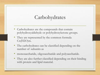
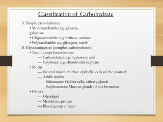
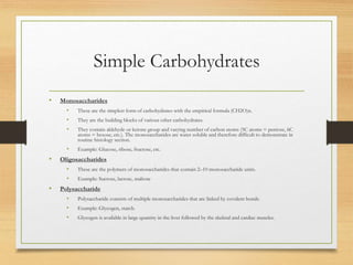
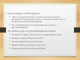
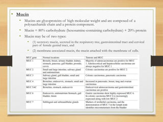
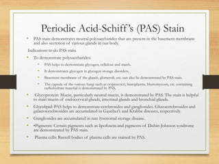
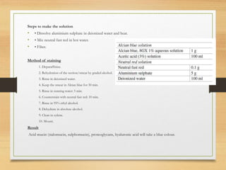
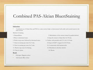
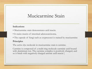
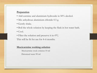
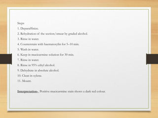
Special Stain for
Carbohydrate
Dr. Meenakshi Chaudhary
Demonstrator (Deptt. of Pathology)
SHKM GMC, Nuh
Carbohydrates
• Carbohydrates are the compounds that contain
polyhydroxyaldehyde or polyhydroxyketone groups.
• They are represented by the common formula
Cn(H2O)m.
• The carbohydrates can be classified depending on the
number of subunits as
• monosaccharide, oligosaccharide and polysaccharide.
• They are also further classified depending on their binding
with protein and lipid material.
A. Simple carbohydrates
• Monosaccharide: e.g. glucose,
galactose
• Oligosaccharide: e.g. maltose, sucrose
• Polysaccharide: e.g. glycogen, starch
B. Glycoconjugates (complex carbohydrates)
• Acid mucopolysaccharides
–– Carboxylated: e.g. hyaluronic acid
–– Sulphated: e.g. chondroitin sulphate
• Mucin
–– Neutral mucin: Surface epithelial cells of the stomach
–– Acidic mucin
Sialomucin: Goblet cells, salivary glands
Sulphomucin: Mucous glands of the bronchus
• Others
–– Glycolipid
–– Membrane protein
–– Blood group antigen
Classification of Carbohydrate
Simple Carbohydrates
• Monosaccharides
• These are the simplest form of carbohydrates with the empirical formula (CH2O)n.
• They are the building blocks of various other carbohydrates.
• They contain aldehyde or ketone group and varying number of carbon atoms (5C atoms = pentose, 6C
atoms = hexose, etc.). The monosaccharides are water soluble and therefore difficult to demonstrate in
routine histology section.
• Example: Glucose, ribose, fructose, etc.
• Oligosaccharides
• These are the polymers of monosaccharides that contain 2–10 monosaccharide units.
• Example: Sucrose, lactose, maltose
• Polysaccharide
• Polysaccharide consists of multiple monosaccharides that are linked by covalent bonds.
• Example: Glycogen, starch.
• Glycogen is available in large quantity in the liver followed by the skeletal and cardiac muscles.
• Glycoconjugates or Proteoglycans
ļ‚§ These are primarily groups of proteins that are extensively
glycosylated. The proteoglycans have a central core protein that is
covalently linked with polysaccharide.
ļ‚§ The carbohydrate part of the proteoglycan is known as
glycosaminoglycans.
• The different types of glycosaminoglycans include:
ļ‚§ Chondroitin sulphate: Present in the cartilage, ligament, bone
ļ‚§ Dermatan sulphate: Present in the skin
ļ‚§ Heparan sulphate: Present in the aorta
ļ‚§ Heparin: Present in granules of mast cell
ļ‚§ Hyaluronic acid: Present in synovial fluid
• Mucin
• Mucins are glycoproteins of high molecular weight and are composed of a
polysaccharide chain and a protein component.
• Mucin = 80% carbohydrate (hexosamine-containing carbohydrate) + 20% protein
• Mucin may be of two types:
• (1) secretory mucin, secreted in the respiratory tree, gastrointestinal tract and cervical
part of female genital tract, and
• (2) membrane-associated mucin, the mucin attached with the membrane of cells.
Periodic Acid-Schiff’s (PAS) Stain
• PAS stain demonstrates neutral polysaccharides that are present in the basement membrane
and also secretion of various glands in our body.
Indications to do PAS stain
• To demonstrate polysaccharides:
• PAS helps to demonstrate glycogen, cellulose and starch.
• It demonstrates glycogen in glycogen storage disorders.
• Basement membrane of the glands, glomeruli, etc. can also be demonstrated by PAS stain.
• The capsule of the various fungi such as cryptococci, histoplasma, blastomycosis, etc. containing
carbohydrate material is demonstrated by PAS,
• Glycoprotein: Mucin, particularly neutral mucin, is demonstrated by PAS. The stain is helpful
to stain mucin of endocervical glands, intestinal glands and bronchial glands.
• Glycolipid: PAS helps to demonstrate cerebrosides and gangliosides. Glucocerebrosides and
galactocerebrosides are accumulated in Gaucher’s and Krabbe diseases, respectively.
• Gangliosides are accumulated in rare lysosomal storage disease.
• •Pigments: Certain pigments such as lipofuscin and pigments of Dubin-Johnson syndrome
are demonstrated by PAS stain.
• Plasma cells: Russell bodies of plasma cells are stained by PAS.
• Principle
The hydroxyl group (OH) of the carbohydrate molecule is oxidized
to aldehyde (CHO) group by periodic acid.
• These aldehyde groups react with Schiff’s reagent to form a magenta-
coloured compound.
Preparation
• Dissolve basic fuchsin (1 g) in 200 ml of boiling distilled water.
• Cool the solution.
• Add 1 N hydrochloric acid and mix well
• Add potassium metabisulphite (2 g).
• Add activated charcoal (2 g).
• Keep the solution in the dark.
Steps
• 1. Deparaffinize.
• 2. Pass through graded lower concentration of alcohol and section/smear to bring in water.
• 3. Oxidize with periodic acid (1%) for 5–10 min.
• 4. Clean with water.
• 5. Keep in Schiff’s reagent for 20–30 min.
• 6. Clean in running tap water for 5 min.
• 7. Counterstain with haematoxylin.
• 8. Wash in tap water for blueing.
• 9. Dehydrate in absolute alcohol.
• 10. Clear in xylene.
• 11. Mount.
Result
• Glycogen and glycoprotein: Magenta colour
• Materials Positive for PAS Reaction Glycogen, starch,
mucin, reticulin, basement membrane, capsule of
fungi, etc.
Testing Schiff ’s reagent
• Add drops of Schiff’s reagent to formalin.
• Active Schiff’s reagent will quickly change the colour
of formalin to pink.
Alcian Blue
• Alcian blue stains acid mucin (in acidic pH 2.5), such as sialomucin and sulphomucin.
• It stains mucin of the salivary glands, prostate and large intestine.
• Alcian blue also stains proteoglycans of cartilaginous material.
Basic Principle
Alcian blue is a group of water-soluble polyvalent basic dye.
The dye is made of a copper-containing phthalocyanine ring with a copper atom in its
centre. Th phthalocyanine ring is also attached with four isothiouronium groups that are
positively charged (
This positively charged Alcian blue dye complex has an attraction with anionic sites of
the mucin. Copper imparts the blue colour of the dye-mucin complex.
Steps to make the solution
• • Dissolve aluminium sulphate in deionized water and heat.
• • Mix neutral fast red in hot water.
• • Filter.
Method of staining
1. Deparaffinize.
2. Rehydration of the section/smear by graded alcohol.
3. Rinse in deionized water.
4. Keep the smear in Alcian blue for 30 min.
5. Rinse in running water: 5 min.
6. Counterstain with neutral fast red: 10 min.
7. Rinse in 95% ethyl alcohol.
8. Dehydrate in absolute alcohol.
9. Clean in xylene.
10. Mount.
Result
Acid mucin (sialomucin, sulphomucin), proteoglycans, hyaluronic acid will take a blue colour.
Combined PAS-Alcian BluenStaining
Indications
• Combined use of Alcian blue and PAS in a same section helps to demonstrate both acidic and neutral mucin in the
same section
Method of staining
1. Deparaffinize. 2. Rehydration of the section/smear by graded alcohol.
3. Rinse in deionized water. 4. Keep the smear in Alcian blue for 30 min.
5. Wash in tap water followed by deionized water. 6. Oxidize with periodic acid (1%) for 5–10 min.
7. Clean in running tap water for 5 min. 8. Keep in Schiff ’s reagent for 20–30 min.
9. Clean in running tap water for 5 min. 10. Counterstain with haematoxylin.
11. Wash in tap water for blueing. 12. Dehydrate in absolute alcohol.
13. Clear in xylene. 14. Mount.
Result
• Glycogen: Magenta colour
• Acid mucin: Blue colour
Mucicarmine Stain
Indications
• Mucicarmine stain demonstrates acid mucin.
• It stains mucin of intestinal adenocarcinoma.
• The capsule of fungi such as cryptococci is stained by mucicarmine
Principles
The active dye molecule in mucicarmine stain is carmine.
Carmine is composed of a multi-ring molecule carminic acid bound
with aluminium ion. The carmine complex is positively charged, and
so it binds with negatively charged anionic acid mucin (
Preparation
• Add carmine and aluminium hydroxide in 50% alcohol.
• Mix anhydrous aluminium chloride 0.5 g.
• Gently shake.
• Boil the whole solution by keeping the flask in hot water bath.
• Cool.
• Filter the solution and preserve it in 4°C.
This will be fit for use for 4–6 months.
Mucicarmine working solution
Mucicarmine stock solution 10 ml
Deionized water 90 ml
Steps
1. Deparaffinize.
2. Rehydration of the section/smear by graded alcohol.
3. Rinse in water.
4. Counterstain with haematoxylin for 5–10 min.
5. Wash in water.
6. Keep in mucicarmine solution for 30 min.
7. Rinse in water.
8. Rinse in 95% ethyl alcohol.
9. Dehydrate in absolute alcohol.
10. Clean in xylene.
11. Mount.
Interpretation- Positive mucicarmine stain shows a dark red colour.
Thankyou

Special Stain for Carbohydrate.pptx

  • 1.
    Special Stain for Carbohydrate Dr.Meenakshi Chaudhary Demonstrator (Deptt. of Pathology) SHKM GMC, Nuh
  • 2.
    Carbohydrates • Carbohydrates arethe compounds that contain polyhydroxyaldehyde or polyhydroxyketone groups. • They are represented by the common formula Cn(H2O)m. • The carbohydrates can be classified depending on the number of subunits as • monosaccharide, oligosaccharide and polysaccharide. • They are also further classified depending on their binding with protein and lipid material.
  • 3.
    A. Simple carbohydrates •Monosaccharide: e.g. glucose, galactose • Oligosaccharide: e.g. maltose, sucrose • Polysaccharide: e.g. glycogen, starch B. Glycoconjugates (complex carbohydrates) • Acid mucopolysaccharides –– Carboxylated: e.g. hyaluronic acid –– Sulphated: e.g. chondroitin sulphate • Mucin –– Neutral mucin: Surface epithelial cells of the stomach –– Acidic mucin Sialomucin: Goblet cells, salivary glands Sulphomucin: Mucous glands of the bronchus • Others –– Glycolipid –– Membrane protein –– Blood group antigen Classification of Carbohydrate
  • 4.
    Simple Carbohydrates • Monosaccharides •These are the simplest form of carbohydrates with the empirical formula (CH2O)n. • They are the building blocks of various other carbohydrates. • They contain aldehyde or ketone group and varying number of carbon atoms (5C atoms = pentose, 6C atoms = hexose, etc.). The monosaccharides are water soluble and therefore difficult to demonstrate in routine histology section. • Example: Glucose, ribose, fructose, etc. • Oligosaccharides • These are the polymers of monosaccharides that contain 2–10 monosaccharide units. • Example: Sucrose, lactose, maltose • Polysaccharide • Polysaccharide consists of multiple monosaccharides that are linked by covalent bonds. • Example: Glycogen, starch. • Glycogen is available in large quantity in the liver followed by the skeletal and cardiac muscles.
  • 5.
    • Glycoconjugates orProteoglycans ļ‚§ These are primarily groups of proteins that are extensively glycosylated. The proteoglycans have a central core protein that is covalently linked with polysaccharide. ļ‚§ The carbohydrate part of the proteoglycan is known as glycosaminoglycans. • The different types of glycosaminoglycans include: ļ‚§ Chondroitin sulphate: Present in the cartilage, ligament, bone ļ‚§ Dermatan sulphate: Present in the skin ļ‚§ Heparan sulphate: Present in the aorta ļ‚§ Heparin: Present in granules of mast cell ļ‚§ Hyaluronic acid: Present in synovial fluid
  • 6.
    • Mucin • Mucinsare glycoproteins of high molecular weight and are composed of a polysaccharide chain and a protein component. • Mucin = 80% carbohydrate (hexosamine-containing carbohydrate) + 20% protein • Mucin may be of two types: • (1) secretory mucin, secreted in the respiratory tree, gastrointestinal tract and cervical part of female genital tract, and • (2) membrane-associated mucin, the mucin attached with the membrane of cells.
  • 8.
    Periodic Acid-Schiff’s (PAS)Stain • PAS stain demonstrates neutral polysaccharides that are present in the basement membrane and also secretion of various glands in our body. Indications to do PAS stain • To demonstrate polysaccharides: • PAS helps to demonstrate glycogen, cellulose and starch. • It demonstrates glycogen in glycogen storage disorders. • Basement membrane of the glands, glomeruli, etc. can also be demonstrated by PAS stain. • The capsule of the various fungi such as cryptococci, histoplasma, blastomycosis, etc. containing carbohydrate material is demonstrated by PAS, • Glycoprotein: Mucin, particularly neutral mucin, is demonstrated by PAS. The stain is helpful to stain mucin of endocervical glands, intestinal glands and bronchial glands. • Glycolipid: PAS helps to demonstrate cerebrosides and gangliosides. Glucocerebrosides and galactocerebrosides are accumulated in Gaucher’s and Krabbe diseases, respectively. • Gangliosides are accumulated in rare lysosomal storage disease. • •Pigments: Certain pigments such as lipofuscin and pigments of Dubin-Johnson syndrome are demonstrated by PAS stain. • Plasma cells: Russell bodies of plasma cells are stained by PAS.
  • 9.
    • Principle The hydroxylgroup (OH) of the carbohydrate molecule is oxidized to aldehyde (CHO) group by periodic acid. • These aldehyde groups react with Schiff’s reagent to form a magenta- coloured compound. Preparation • Dissolve basic fuchsin (1 g) in 200 ml of boiling distilled water. • Cool the solution. • Add 1 N hydrochloric acid and mix well • Add potassium metabisulphite (2 g). • Add activated charcoal (2 g). • Keep the solution in the dark.
  • 10.
    Steps • 1. Deparaffinize. •2. Pass through graded lower concentration of alcohol and section/smear to bring in water. • 3. Oxidize with periodic acid (1%) for 5–10 min. • 4. Clean with water. • 5. Keep in Schiff’s reagent for 20–30 min. • 6. Clean in running tap water for 5 min. • 7. Counterstain with haematoxylin. • 8. Wash in tap water for blueing. • 9. Dehydrate in absolute alcohol. • 10. Clear in xylene. • 11. Mount. Result • Glycogen and glycoprotein: Magenta colour
  • 11.
    • Materials Positivefor PAS Reaction Glycogen, starch, mucin, reticulin, basement membrane, capsule of fungi, etc. Testing Schiff ’s reagent • Add drops of Schiff’s reagent to formalin. • Active Schiff’s reagent will quickly change the colour of formalin to pink.
  • 12.
    Alcian Blue • Alcianblue stains acid mucin (in acidic pH 2.5), such as sialomucin and sulphomucin. • It stains mucin of the salivary glands, prostate and large intestine. • Alcian blue also stains proteoglycans of cartilaginous material. Basic Principle Alcian blue is a group of water-soluble polyvalent basic dye. The dye is made of a copper-containing phthalocyanine ring with a copper atom in its centre. Th phthalocyanine ring is also attached with four isothiouronium groups that are positively charged ( This positively charged Alcian blue dye complex has an attraction with anionic sites of the mucin. Copper imparts the blue colour of the dye-mucin complex.
  • 13.
    Steps to makethe solution • • Dissolve aluminium sulphate in deionized water and heat. • • Mix neutral fast red in hot water. • • Filter. Method of staining 1. Deparaffinize. 2. Rehydration of the section/smear by graded alcohol. 3. Rinse in deionized water. 4. Keep the smear in Alcian blue for 30 min. 5. Rinse in running water: 5 min. 6. Counterstain with neutral fast red: 10 min. 7. Rinse in 95% ethyl alcohol. 8. Dehydrate in absolute alcohol. 9. Clean in xylene. 10. Mount. Result Acid mucin (sialomucin, sulphomucin), proteoglycans, hyaluronic acid will take a blue colour.
  • 14.
    Combined PAS-Alcian BluenStaining Indications •Combined use of Alcian blue and PAS in a same section helps to demonstrate both acidic and neutral mucin in the same section Method of staining 1. Deparaffinize. 2. Rehydration of the section/smear by graded alcohol. 3. Rinse in deionized water. 4. Keep the smear in Alcian blue for 30 min. 5. Wash in tap water followed by deionized water. 6. Oxidize with periodic acid (1%) for 5–10 min. 7. Clean in running tap water for 5 min. 8. Keep in Schiff ’s reagent for 20–30 min. 9. Clean in running tap water for 5 min. 10. Counterstain with haematoxylin. 11. Wash in tap water for blueing. 12. Dehydrate in absolute alcohol. 13. Clear in xylene. 14. Mount. Result • Glycogen: Magenta colour • Acid mucin: Blue colour
  • 15.
    Mucicarmine Stain Indications • Mucicarminestain demonstrates acid mucin. • It stains mucin of intestinal adenocarcinoma. • The capsule of fungi such as cryptococci is stained by mucicarmine Principles The active dye molecule in mucicarmine stain is carmine. Carmine is composed of a multi-ring molecule carminic acid bound with aluminium ion. The carmine complex is positively charged, and so it binds with negatively charged anionic acid mucin (
  • 16.
    Preparation • Add carmineand aluminium hydroxide in 50% alcohol. • Mix anhydrous aluminium chloride 0.5 g. • Gently shake. • Boil the whole solution by keeping the flask in hot water bath. • Cool. • Filter the solution and preserve it in 4°C. This will be fit for use for 4–6 months. Mucicarmine working solution Mucicarmine stock solution 10 ml Deionized water 90 ml
  • 17.
    Steps 1. Deparaffinize. 2. Rehydrationof the section/smear by graded alcohol. 3. Rinse in water. 4. Counterstain with haematoxylin for 5–10 min. 5. Wash in water. 6. Keep in mucicarmine solution for 30 min. 7. Rinse in water. 8. Rinse in 95% ethyl alcohol. 9. Dehydrate in absolute alcohol. 10. Clean in xylene. 11. Mount. Interpretation- Positive mucicarmine stain shows a dark red colour.
  • 18.