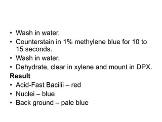
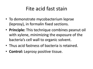
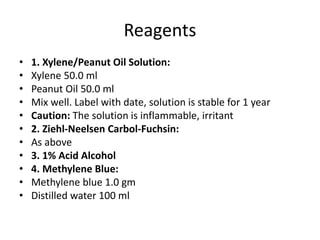
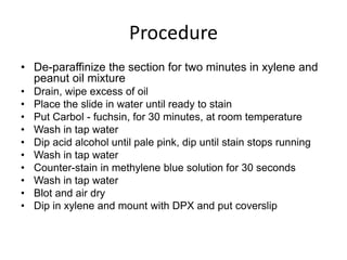
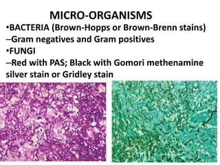
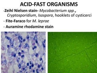
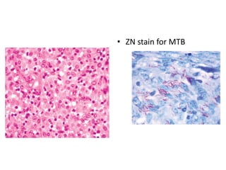
SPECIAL (MICRO-ORGANISM) STAINS IN
HISTOPATHOLOGY
Babatunde M Duduyemi
OUTLINE
• Introduction
• Types of stains
• Classification
• Examples
• Summary
Introduction
• Traditional methods for the demonstration of
microorganisms in tissue sections can only be based
upon staining characteristics and simple morphology
because the organisms are fixed and dead.
• The most effective means of isolating and identifying
individual species of most organisms is to study their
growth patterns and morphology in vitro.
• The study of these criteria has formed the basis for the
identification and classification of microorganisms
Objectives
• After reading this lesson, you will be able
to:
• describe Ziehl-Neelsen stain
• demonstrate Fite acid fast stain
• explain periodic acid Schiff, Mucicarmine
stain, Giemsa etc
Introduction…..
• Of the hundreds of “special” stains
known only a relatively small minority
are of real diagnostic importance
• The advent of immunohistochemistry
has rendered many of them obsolete.
CLASSIFICATION OF STAINS
• Natural dyes (organic substances, plant,
insect)
• Synthetic dyes (coal tar)
Theory of stains
1. Physical (adsorption, absorption,
solubility, osmotic pressure, capillary
attraction)
2. Chemical (acidic, basic, amphoteric,
neutral)
CLASSIFICATION OF STAINS (CONTD.)
• Most histological dyes are classified either
as acid or as basic dyes.
• An acid dye exists as an anion (negatively
charged) in solution, while a basic dye
exists as a cation (positive charge).
• For instance, in the haematoxylin-eosin
stain (H&E), the haematoxylin-metal
complex acts as a basic dye. The eosin acts
as an acid dye.
Gram stain
• Some of the bacteria like mycobacterium do
not get stained with Gram stain due to their
lipid capsule.
• Ziehl-Neelsen stain is applied to identify
mycobacteria which are stained pink to red in
colour.
Gram stain……
• This reaction may depend on the difference in
permeability of cytoplasmic membrane.
• During staining a dye iodine complex is
formed within the cell- insoluble in water.
• The complex diffuses freely from gram
negative organisms but diffuses less in Gram
positive bacteria due to poor membrane
permeability.
Preparation of reagents
Crystal violet solution
• Crystal violet 1.0gm
• Absolute alcohol 20ml
• Ammonium oxalate(1%) 80ml
Basic fuchsin
• Basic fuchsin 1.0gm
• Distilled water 100ml
Gram’s iodine
• Iodine crystal 1.0gm
• Potassium iodide 2.0gm
• Distilled water 300ml
procedure
• Deparaffinize the section and bring it to water
• Put crystal violet for one minute
• Add Gram’s iodine for 30 seconds
• Differentiate by dipping the section once or
twice in alcohol
• Wash with water and counterstain with
safranin for 45 seconds
• Wash with water, air dry and mount.
RESULT
• Gram positive bacteria stain blue black.
• Gram negative bacteria stain red or pink.
• Some of the bacteria like mycobacterium
do not get stained with Gram stain due to
their lipid capsule.
• Ziehl-Neelsen stain is applied to identify
mycobacteria which are stained pink to red
in colour
Ziehl-Neelsen stain
• To demonstrate Mycobacterium tuberculosis
in paraffin sections.
• Principle: When Mycobacterium tuberculosis
are stained with a strong reagent like basic
fuchsin in aqueous 5% phenol at high
temperature the bacilli resist decolourization
by strong acids (20% sulphuric acid).
• Any decolourized non AFB is counterstained
with methylene blue or malachite green.
Reagents
• 1. Ziehl-Neelsen’s carbol fuchsin
• Basic fuchsin 1gm
• Absolute alcohol 10ml
• 5% phenol (aqueous) 100ml
• Dissolve basic fuchsin in alcohol, and then add 5%
phenol.
• 2. Methylene blue solution
• Methylene blue 1gm
• Distilled water 100ml
Procedure
• De-wax the sections in xylene and bring to
water.
• Flood sections with carbol fuchsin and heat to
steaming by intermittent flaming for 10 to 15
minutes or stain in coplin jar at 56°-60°C
(oven or water bath) for 30 minutes.
• Wash in water to remove excess of stain.
• Differentiate with 20% sulphuric acid or in 3%
hydrochloric acid in 70% alcohol until the
tissue is very pale pink colour. Then washed
in water (for 5 to 10 minutes).
• Wash in water.
• Counterstain in 1% methylene blue for 10 to
15 seconds.
• Wash in water.
• Dehydrate, clear in xylene and mount in DPX.
Result
• Acid-Fast Bacilii – red
• Nuclei – blue
• Back ground – pale blue
Fite acid fast stain
• To demonstrate mycobacterium leprae
(leprosy), in formalin fixed sections.
• Principle: This technique combines peanut oil
with xylene, minimizing the exposure of the
bacteria’s cell wall to organic solvent.
• Thus acid fastness of bacteria is retained.
• Control: Leprosy positive tissue.
Reagents
• 1. Xylene/Peanut Oil Solution:
• Xylene 50.0 ml
• Peanut Oil 50.0 ml
• Mix well. Label with date, solution is stable for 1 year
• Caution: The solution is inflammable, irritant
• 2. Ziehl-Neelsen Carbol-Fuchsin:
• As above
• 3. 1% Acid Alcohol
• 4. Methylene Blue:
• Methylene blue 1.0 gm
• Distilled water 100 ml
Procedure
• De-paraffinize the section for two minutes in xylene and
peanut oil mixture
• Drain, wipe excess of oil
• Place the slide in water until ready to stain
• Put Carbol - fuchsin, for 30 minutes, at room temperature
• Wash in tap water
• Dip acid alcohol until pale pink, dip until stain stops running
• Wash in tap water
• Counter-stain in methylene blue solution for 30 seconds
• Wash in tap water
• Blot and air dry
• Dip in xylene and mount with DPX and put coverslip
• Results
• Acid-fast bacilli – red
• Background – blue
• Note: Mineral oil may be substituted for
peanut oil.
PAS
• Periodic acid causes oxidation of 1:2 glycol
groups in the tissues to di-aldehydes.
• The di-aldehyde reacts with fuchsin –
sulfurous acid solution(Schiff’s) to form a
magenta coloured compound.
• Aim: To demonstrate glycogen, epithelial
mucin, fungi, amoeba and basement
membrane.
• Control: Liver and intestine
Reagents
• Periodic acid 1%
• Distilled water 100 ml
• Schiff’s reagent
• Basic fuchsin 1 gm
• Distilled water 200 ml
• 1N hydrochloric acid 20 ml
• Sodium or Potassium metabitesulfite1gm
• Activated charcoal 2gm
• Dissolve basic fuchsin in boiling distilled water
• Shake for 5 minutes and cool to 50°C
• Filter and add 1N solution.
• Cool further and add sodium or potassium metabisulfite. Keep for 18
hours in dark.
• Add activated charcoal, shake well, filter and store the solution at
40°C.
Procedure
• Bring the sections to water.
• Dip the slide in Periodic acid solution for 5-10 minutes.
• Wash in tap water and rinse in distilled water.
• Put Schiff’s reagent on the section for 20 minutes.
• Wash thoroughly in running water.
• Counerstain with Hematoxylin, dehydrate, clear and mount in DPX.
Result
• Glycogen (except non-sulfated acid
mucopolysaccharide), basement
membrane, fungi, parasites and other
positive substances – magenta
• Nucleus – blue or violet
GIEMSA stain
• Used to demonstrate bacteria, haematologic element,
bone marrow elements, blood parasites
Reagents
• Giemsa stain
• Stock solution:
• Giemsa powder 1.0 gm
• Methyl alcohol 66 ml
• Glycerin 66 ml
• Add glycerin to Giemsa powder and put in oven at
60oC for 30 minutes to 2 hours (until the stain
dissolves). Then add methyl alcohol.
Procedure
• Deparaffinize and take the section to
water.
• Place the slide in working Giemsa
solution.
• Wash with tap water
• Differentiate with 0.5% acetic acid solution
• Dehydrate and mount.
Result
• Nuclei – blue
• Malarial parasite – purple
• Collagen and other elements – pink
• As a histology stain, detects mast cells and
microorganisms, such as Giardia or
Helicobacter pylori which stains dark blue with
Giemsa and black with silver stains.
Note
• Sections stained at room temperature for
longer period show better results than
sections stained at higher temperature for
shorter period.
• Differentiation with acetic acid removes only
blue dye hence gives better red intensity.
• Giemsa reagent improves with age
Mucicarmine stain
• To demonstrate mucin, glycogen in
tissues.
• Principle: Aluminium is believed to form a
chelation complex with carmine changing
the molecule to a positive charge allowing
it to bind with the acid substrates of low
density such as mucus.
Result
• Mucin – deep rose
• Nuclei – black
• Other tissue elements – yellow (if metanil
yellow is used)
• Colorless (if metanil yellow is not used)
• Capsule of cryptococci (fungus) – deep rose
• Control - small intestine
GROCOTT-GOMORI’S METHANAMINE SILVER
• To demonstrate fungi and bacteria particularly to
stain carbohydrate.
• Cell wall of fungi like Pneumocystis jiroveci is
outlined by black stain.
Result
• Basement membrane black
• Fungi cell wall black
• Background pink or green (depends on
counterstain nuclear fast red or light green)
• Control kidney or skin
GIEMSA STAIN
Giemsa stain for H. pylori
MICRO-ORGANISMS
•BACTERIA (Brown-Hopps or Brown-Brenn stains)
–Gram negatives and Gram positives
•FUNGI
–Red with PAS; Black with Gomori methenamine
silver stain or Gridley stain
ACID-FAST ORGANISMS
-Zeihl Nielsen stain- Mycobacterium spp.,
Cryptosporidium, Isospora, hooklets of cysticerci
- Fite-Faraco for M. leprae
- Auramine rhodamine stain
Mycobacterium avium complex
• ZN stain for MTB
WARTHIN-STARRY SILVER STAIN- Helicobacter
pylori, spirochetes, Klebsiella rhinoscleromatis
VIRAL STAINS
HBV SURFACE ANTIGEN
Demonstrated with either
Shikata orcein- brownish granular
particles in the hepatocyte
cytoplasm.
Victorian Blue- blue.
Summary
• Ziehl-Neelson stain is used to demonstrate
mycobacterium tuberculosis in paraffin sections
• Methylene blue or malachite green is used as counter
stain
• Fite acid fast stain is used to demonstrate
mycobacterium leprae in formalin fixed sections and
this technique combines peanut with xylene
• Periodic acid Schiff stain is used to demonstrate
glycogen, epithelial mucin, fungi and amoeba
• Liver and intestine is used as control in periodic acid
Schiff stain
• Giema stain is used to demonstrate bacteria and blood
parasites
• In Giema stain nuclei appears blue,
malarial parasite appears purple and
collagen and other elements appear pink
• Mucin in tissue is demonstrated by
mucicarmine stain
• Grocott-Gomori’s Methanamine silver stain
is used to demonstrate fungi and bacteria
particularly to stain carbohydrate
Practice questions
• 1. Mycobacterium Tuberculosis in paraffin section is demonstrated
by.....................
• 2. ..................... or ..................... is used as counter stain
• 3. Periodic acid Schiff stain is used to demonstrate .....................,
.....................& .....................
• 4. In periodic acid Schiff stain the nucleus appears ..................... &
glycogen, fungi, parasites appear .....................
• 5. Mucin in tissue is demonstrated by ..................... stain
• 6. ..................... stain is used to demonstrate fungi and bacteria
• 7. In Grocott-Gromori’s methanamine silver stain the fungal cell
wall and basement membrane appears ............. & background
appears...............
• 8. The control used in Grocott-Gromori’s Methanamine silver stain
is .................... or .....................
Practice questions
• What are the stains which may be used to
demonstrate fungi?
• What is the principle of silver methanamine
staining?
• What is the difference in Acid Fast staining for
Mycobacterium tuberculosis and leprae?
• What are the uses of Giemsa stain?
• What control is used in PAS stain
THANK YOU FOR LISTENING
• Successful people have
libraries, others have
large screen TVs

microorganism stain.pptx

  • 1.
    SPECIAL (MICRO-ORGANISM) STAINSIN HISTOPATHOLOGY Babatunde M Duduyemi
  • 2.
    OUTLINE • Introduction • Typesof stains • Classification • Examples • Summary
  • 3.
    Introduction • Traditional methodsfor the demonstration of microorganisms in tissue sections can only be based upon staining characteristics and simple morphology because the organisms are fixed and dead. • The most effective means of isolating and identifying individual species of most organisms is to study their growth patterns and morphology in vitro. • The study of these criteria has formed the basis for the identification and classification of microorganisms
  • 4.
    Objectives • After readingthis lesson, you will be able to: • describe Ziehl-Neelsen stain • demonstrate Fite acid fast stain • explain periodic acid Schiff, Mucicarmine stain, Giemsa etc
  • 5.
    Introduction….. • Of thehundreds of “special” stains known only a relatively small minority are of real diagnostic importance • The advent of immunohistochemistry has rendered many of them obsolete.
  • 6.
    CLASSIFICATION OF STAINS •Natural dyes (organic substances, plant, insect) • Synthetic dyes (coal tar) Theory of stains 1. Physical (adsorption, absorption, solubility, osmotic pressure, capillary attraction) 2. Chemical (acidic, basic, amphoteric, neutral)
  • 7.
    CLASSIFICATION OF STAINS(CONTD.) • Most histological dyes are classified either as acid or as basic dyes. • An acid dye exists as an anion (negatively charged) in solution, while a basic dye exists as a cation (positive charge). • For instance, in the haematoxylin-eosin stain (H&E), the haematoxylin-metal complex acts as a basic dye. The eosin acts as an acid dye.
  • 8.
    Gram stain • Someof the bacteria like mycobacterium do not get stained with Gram stain due to their lipid capsule. • Ziehl-Neelsen stain is applied to identify mycobacteria which are stained pink to red in colour.
  • 9.
    Gram stain…… • Thisreaction may depend on the difference in permeability of cytoplasmic membrane. • During staining a dye iodine complex is formed within the cell- insoluble in water. • The complex diffuses freely from gram negative organisms but diffuses less in Gram positive bacteria due to poor membrane permeability.
  • 10.
    Preparation of reagents Crystalviolet solution • Crystal violet 1.0gm • Absolute alcohol 20ml • Ammonium oxalate(1%) 80ml Basic fuchsin • Basic fuchsin 1.0gm • Distilled water 100ml Gram’s iodine • Iodine crystal 1.0gm • Potassium iodide 2.0gm • Distilled water 300ml
  • 11.
    procedure • Deparaffinize thesection and bring it to water • Put crystal violet for one minute • Add Gram’s iodine for 30 seconds • Differentiate by dipping the section once or twice in alcohol • Wash with water and counterstain with safranin for 45 seconds • Wash with water, air dry and mount.
  • 12.
    RESULT • Gram positivebacteria stain blue black. • Gram negative bacteria stain red or pink. • Some of the bacteria like mycobacterium do not get stained with Gram stain due to their lipid capsule. • Ziehl-Neelsen stain is applied to identify mycobacteria which are stained pink to red in colour
  • 13.
    Ziehl-Neelsen stain • Todemonstrate Mycobacterium tuberculosis in paraffin sections. • Principle: When Mycobacterium tuberculosis are stained with a strong reagent like basic fuchsin in aqueous 5% phenol at high temperature the bacilli resist decolourization by strong acids (20% sulphuric acid). • Any decolourized non AFB is counterstained with methylene blue or malachite green.
  • 14.
    Reagents • 1. Ziehl-Neelsen’scarbol fuchsin • Basic fuchsin 1gm • Absolute alcohol 10ml • 5% phenol (aqueous) 100ml • Dissolve basic fuchsin in alcohol, and then add 5% phenol. • 2. Methylene blue solution • Methylene blue 1gm • Distilled water 100ml
  • 15.
    Procedure • De-wax thesections in xylene and bring to water. • Flood sections with carbol fuchsin and heat to steaming by intermittent flaming for 10 to 15 minutes or stain in coplin jar at 56°-60°C (oven or water bath) for 30 minutes. • Wash in water to remove excess of stain. • Differentiate with 20% sulphuric acid or in 3% hydrochloric acid in 70% alcohol until the tissue is very pale pink colour. Then washed in water (for 5 to 10 minutes).
  • 16.
    • Wash inwater. • Counterstain in 1% methylene blue for 10 to 15 seconds. • Wash in water. • Dehydrate, clear in xylene and mount in DPX. Result • Acid-Fast Bacilii – red • Nuclei – blue • Back ground – pale blue
  • 17.
    Fite acid faststain • To demonstrate mycobacterium leprae (leprosy), in formalin fixed sections. • Principle: This technique combines peanut oil with xylene, minimizing the exposure of the bacteria’s cell wall to organic solvent. • Thus acid fastness of bacteria is retained. • Control: Leprosy positive tissue.
  • 18.
    Reagents • 1. Xylene/PeanutOil Solution: • Xylene 50.0 ml • Peanut Oil 50.0 ml • Mix well. Label with date, solution is stable for 1 year • Caution: The solution is inflammable, irritant • 2. Ziehl-Neelsen Carbol-Fuchsin: • As above • 3. 1% Acid Alcohol • 4. Methylene Blue: • Methylene blue 1.0 gm • Distilled water 100 ml
  • 19.
    Procedure • De-paraffinize thesection for two minutes in xylene and peanut oil mixture • Drain, wipe excess of oil • Place the slide in water until ready to stain • Put Carbol - fuchsin, for 30 minutes, at room temperature • Wash in tap water • Dip acid alcohol until pale pink, dip until stain stops running • Wash in tap water • Counter-stain in methylene blue solution for 30 seconds • Wash in tap water • Blot and air dry • Dip in xylene and mount with DPX and put coverslip
  • 20.
    • Results • Acid-fastbacilli – red • Background – blue • Note: Mineral oil may be substituted for peanut oil.
  • 21.
    PAS • Periodic acidcauses oxidation of 1:2 glycol groups in the tissues to di-aldehydes. • The di-aldehyde reacts with fuchsin – sulfurous acid solution(Schiff’s) to form a magenta coloured compound. • Aim: To demonstrate glycogen, epithelial mucin, fungi, amoeba and basement membrane. • Control: Liver and intestine
  • 22.
    Reagents • Periodic acid1% • Distilled water 100 ml • Schiff’s reagent • Basic fuchsin 1 gm • Distilled water 200 ml • 1N hydrochloric acid 20 ml • Sodium or Potassium metabitesulfite1gm • Activated charcoal 2gm
  • 23.
    • Dissolve basicfuchsin in boiling distilled water • Shake for 5 minutes and cool to 50°C • Filter and add 1N solution. • Cool further and add sodium or potassium metabisulfite. Keep for 18 hours in dark. • Add activated charcoal, shake well, filter and store the solution at 40°C. Procedure • Bring the sections to water. • Dip the slide in Periodic acid solution for 5-10 minutes. • Wash in tap water and rinse in distilled water. • Put Schiff’s reagent on the section for 20 minutes. • Wash thoroughly in running water. • Counerstain with Hematoxylin, dehydrate, clear and mount in DPX.
  • 24.
    Result • Glycogen (exceptnon-sulfated acid mucopolysaccharide), basement membrane, fungi, parasites and other positive substances – magenta • Nucleus – blue or violet
  • 25.
    GIEMSA stain • Usedto demonstrate bacteria, haematologic element, bone marrow elements, blood parasites Reagents • Giemsa stain • Stock solution: • Giemsa powder 1.0 gm • Methyl alcohol 66 ml • Glycerin 66 ml • Add glycerin to Giemsa powder and put in oven at 60oC for 30 minutes to 2 hours (until the stain dissolves). Then add methyl alcohol.
  • 26.
    Procedure • Deparaffinize andtake the section to water. • Place the slide in working Giemsa solution. • Wash with tap water • Differentiate with 0.5% acetic acid solution • Dehydrate and mount.
  • 27.
    Result • Nuclei –blue • Malarial parasite – purple • Collagen and other elements – pink • As a histology stain, detects mast cells and microorganisms, such as Giardia or Helicobacter pylori which stains dark blue with Giemsa and black with silver stains.
  • 28.
    Note • Sections stainedat room temperature for longer period show better results than sections stained at higher temperature for shorter period. • Differentiation with acetic acid removes only blue dye hence gives better red intensity. • Giemsa reagent improves with age
  • 29.
    Mucicarmine stain • Todemonstrate mucin, glycogen in tissues. • Principle: Aluminium is believed to form a chelation complex with carmine changing the molecule to a positive charge allowing it to bind with the acid substrates of low density such as mucus.
  • 30.
    Result • Mucin –deep rose • Nuclei – black • Other tissue elements – yellow (if metanil yellow is used) • Colorless (if metanil yellow is not used) • Capsule of cryptococci (fungus) – deep rose • Control - small intestine
  • 31.
    GROCOTT-GOMORI’S METHANAMINE SILVER •To demonstrate fungi and bacteria particularly to stain carbohydrate. • Cell wall of fungi like Pneumocystis jiroveci is outlined by black stain. Result • Basement membrane black • Fungi cell wall black • Background pink or green (depends on counterstain nuclear fast red or light green) • Control kidney or skin
  • 32.
  • 33.
  • 34.
    MICRO-ORGANISMS •BACTERIA (Brown-Hopps orBrown-Brenn stains) –Gram negatives and Gram positives •FUNGI –Red with PAS; Black with Gomori methenamine silver stain or Gridley stain
  • 35.
    ACID-FAST ORGANISMS -Zeihl Nielsenstain- Mycobacterium spp., Cryptosporidium, Isospora, hooklets of cysticerci - Fite-Faraco for M. leprae - Auramine rhodamine stain
  • 36.
  • 37.
    WARTHIN-STARRY SILVER STAIN-Helicobacter pylori, spirochetes, Klebsiella rhinoscleromatis
  • 38.
    VIRAL STAINS HBV SURFACEANTIGEN Demonstrated with either Shikata orcein- brownish granular particles in the hepatocyte cytoplasm. Victorian Blue- blue.
  • 39.
    Summary • Ziehl-Neelson stainis used to demonstrate mycobacterium tuberculosis in paraffin sections • Methylene blue or malachite green is used as counter stain • Fite acid fast stain is used to demonstrate mycobacterium leprae in formalin fixed sections and this technique combines peanut with xylene • Periodic acid Schiff stain is used to demonstrate glycogen, epithelial mucin, fungi and amoeba • Liver and intestine is used as control in periodic acid Schiff stain • Giema stain is used to demonstrate bacteria and blood parasites
  • 40.
    • In Giemastain nuclei appears blue, malarial parasite appears purple and collagen and other elements appear pink • Mucin in tissue is demonstrated by mucicarmine stain • Grocott-Gomori’s Methanamine silver stain is used to demonstrate fungi and bacteria particularly to stain carbohydrate
  • 41.
    Practice questions • 1.Mycobacterium Tuberculosis in paraffin section is demonstrated by..................... • 2. ..................... or ..................... is used as counter stain • 3. Periodic acid Schiff stain is used to demonstrate ....................., .....................& ..................... • 4. In periodic acid Schiff stain the nucleus appears ..................... & glycogen, fungi, parasites appear ..................... • 5. Mucin in tissue is demonstrated by ..................... stain • 6. ..................... stain is used to demonstrate fungi and bacteria • 7. In Grocott-Gromori’s methanamine silver stain the fungal cell wall and basement membrane appears ............. & background appears............... • 8. The control used in Grocott-Gromori’s Methanamine silver stain is .................... or .....................
  • 42.
    Practice questions • Whatare the stains which may be used to demonstrate fungi? • What is the principle of silver methanamine staining? • What is the difference in Acid Fast staining for Mycobacterium tuberculosis and leprae? • What are the uses of Giemsa stain? • What control is used in PAS stain
  • 43.
    THANK YOU FORLISTENING • Successful people have libraries, others have large screen TVs