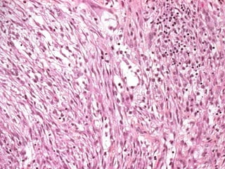
Soft tissue tumors are defined as mesenchymal proliferations that occur in extraskeletal, nonepithelial tissues. They are classified based on the tissue they originate from, such as muscle, fat, fibrous tissue, vessels, and nerves. The most common soft tissue tumors are lipomas, which are benign fatty tumors. Liposarcomas are malignant fatty tumors that can recur if not adequately excised. Rhabdomyosarcoma is the most common soft tissue sarcoma in children that arises in skeletal muscle tissue and is usually treated with surgery and chemotherapy.
non-skeletal mesodermal tissues: adipose tissue, fibrous tissue, muscle, blood vessels and peripheral nerves (despite neuroectodermal origin)
benign, malignant and intermediate (low-grade malignant – locally aggressive, can recur, no metastatic potential)
originate from primitive mesenchymal stem cells
classification according to differentiation lines (e.g. liposarcoma is not a tumor arising from adipose tissue but exhibiting lipoblastic differentiation)
pathology of round cell tumours of osseo articular system like ewings sarcoma, mesenchymal chondrosarcoma,small cell osteosarcoma, plasma cell neoplasms and other hematopoietic malignancies. how immunochemistry os playing pivotal role in differential diagnosis.
Histopathological Grossing of Kidney Tumors with the common gross differentials encountered,
reference - TATA memorial grossing techniques , Rosai and ackerman surgical pathology , Fletcher , Springer histopathology Specimen
Soft tissue pathology is rapidly changing
Novel molecular findings
Tumors previously known under one rubric are reclassified with relative frequency
Lesions that for decades were thought to be reactive now are discovered to possess gene rearrangements
Features of previously unknown or incompletely described tumors are coalesced and synthesized into new entities
Relative rarity of soft tissue tumors only adds to the challenge
of keeping abreast of all of these advances
Presentation about lipoma and liposarcoma, origin, cause, description, diagnosis, treatment with pictures that help the better understanding of the topic.
HOT NEW PRODUCT! BIG SALES FAST SHIPPING NOW FROM CHINA!! EU KU DB BK substit...GL Anaacs
Contact us if you are interested:
Email / Skype : kefaya1771@gmail.com
Threema: PXHY5PDH
New BATCH Ku !!! MUCH IN DEMAND FAST SALE EVERY BATCH HAPPY GOOD EFFECT BIG BATCH !
Contact me on Threema or skype to start big business!!
Hot-sale products:
NEW HOT EUTYLONE WHITE CRYSTAL!!
5cl-adba precursor (semi finished )
5cl-adba raw materials
ADBB precursor (semi finished )
ADBB raw materials
APVP powder
5fadb/4f-adb
Jwh018 / Jwh210
Eutylone crystal
Protonitazene (hydrochloride) CAS: 119276-01-6
Flubrotizolam CAS: 57801-95-3
Metonitazene CAS: 14680-51-4
Payment terms: Western Union,MoneyGram,Bitcoin or USDT.
Deliver Time: Usually 7-15days
Shipping method: FedEx, TNT, DHL,UPS etc.Our deliveries are 100% safe, fast, reliable and discreet.
Samples will be sent for your evaluation!If you are interested in, please contact me, let's talk details.
We specializes in exporting high quality Research chemical, medical intermediate, Pharmaceutical chemicals and so on. Products are exported to USA, Canada, France, Korea, Japan,Russia, Southeast Asia and other countries.
Flu Vaccine Alert in Bangalore Karnatakaaddon Scans
As flu season approaches, health officials in Bangalore, Karnataka, are urging residents to get their flu vaccinations. The seasonal flu, while common, can lead to severe health complications, particularly for vulnerable populations such as young children, the elderly, and those with underlying health conditions.
Dr. Vidisha Kumari, a leading epidemiologist in Bangalore, emphasizes the importance of getting vaccinated. "The flu vaccine is our best defense against the influenza virus. It not only protects individuals but also helps prevent the spread of the virus in our communities," he says.
This year, the flu season is expected to coincide with a potential increase in other respiratory illnesses. The Karnataka Health Department has launched an awareness campaign highlighting the significance of flu vaccinations. They have set up multiple vaccination centers across Bangalore, making it convenient for residents to receive their shots.
To encourage widespread vaccination, the government is also collaborating with local schools, workplaces, and community centers to facilitate vaccination drives. Special attention is being given to ensuring that the vaccine is accessible to all, including marginalized communities who may have limited access to healthcare.
Residents are reminded that the flu vaccine is safe and effective. Common side effects are mild and may include soreness at the injection site, mild fever, or muscle aches. These side effects are generally short-lived and far less severe than the flu itself.
Healthcare providers are also stressing the importance of continuing COVID-19 precautions. Wearing masks, practicing good hand hygiene, and maintaining social distancing are still crucial, especially in crowded places.
Protect yourself and your loved ones by getting vaccinated. Together, we can help keep Bangalore healthy and safe this flu season. For more information on vaccination centers and schedules, residents can visit the Karnataka Health Department’s official website or follow their social media pages.
Stay informed, stay safe, and get your flu shot today!
Knee anatomy and clinical tests 2024.pdfvimalpl1234
This includes all relevant anatomy and clinical tests compiled from standard textbooks, Campbell,netter etc..It is comprehensive and best suited for orthopaedicians and orthopaedic residents.
NVBDCP.pptx Nation vector borne disease control programSapna Thakur
NVBDCP was launched in 2003-2004 . Vector-Borne Disease: Disease that results from an infection transmitted to humans and other animals by blood-feeding arthropods, such as mosquitoes, ticks, and fleas. Examples of vector-borne diseases include Dengue fever, West Nile Virus, Lyme disease, and malaria.
New Directions in Targeted Therapeutic Approaches for Older Adults With Mantl...i3 Health
i3 Health is pleased to make the speaker slides from this activity available for use as a non-accredited self-study or teaching resource.
This slide deck presented by Dr. Kami Maddocks, Professor-Clinical in the Division of Hematology and
Associate Division Director for Ambulatory Operations
The Ohio State University Comprehensive Cancer Center, will provide insight into new directions in targeted therapeutic approaches for older adults with mantle cell lymphoma.
STATEMENT OF NEED
Mantle cell lymphoma (MCL) is a rare, aggressive B-cell non-Hodgkin lymphoma (NHL) accounting for 5% to 7% of all lymphomas. Its prognosis ranges from indolent disease that does not require treatment for years to very aggressive disease, which is associated with poor survival (Silkenstedt et al, 2021). Typically, MCL is diagnosed at advanced stage and in older patients who cannot tolerate intensive therapy (NCCN, 2022). Although recent advances have slightly increased remission rates, recurrence and relapse remain very common, leading to a median overall survival between 3 and 6 years (LLS, 2021). Though there are several effective options, progress is still needed towards establishing an accepted frontline approach for MCL (Castellino et al, 2022). Treatment selection and management of MCL are complicated by the heterogeneity of prognosis, advanced age and comorbidities of patients, and lack of an established standard approach for treatment, making it vital that clinicians be familiar with the latest research and advances in this area. In this activity chaired by Michael Wang, MD, Professor in the Department of Lymphoma & Myeloma at MD Anderson Cancer Center, expert faculty will discuss prognostic factors informing treatment, the promising results of recent trials in new therapeutic approaches, and the implications of treatment resistance in therapeutic selection for MCL.
Target Audience
Hematology/oncology fellows, attending faculty, and other health care professionals involved in the treatment of patients with mantle cell lymphoma (MCL).
Learning Objectives
1.) Identify clinical and biological prognostic factors that can guide treatment decision making for older adults with MCL
2.) Evaluate emerging data on targeted therapeutic approaches for treatment-naive and relapsed/refractory MCL and their applicability to older adults
3.) Assess mechanisms of resistance to targeted therapies for MCL and their implications for treatment selection
Lung Cancer: Artificial Intelligence, Synergetics, Complex System Analysis, S...Oleg Kshivets
RESULTS: Overall life span (LS) was 2252.1±1742.5 days and cumulative 5-year survival (5YS) reached 73.2%, 10 years – 64.8%, 20 years – 42.5%. 513 LCP lived more than 5 years (LS=3124.6±1525.6 days), 148 LCP – more than 10 years (LS=5054.4±1504.1 days).199 LCP died because of LC (LS=562.7±374.5 days). 5YS of LCP after bi/lobectomies was significantly superior in comparison with LCP after pneumonectomies (78.1% vs.63.7%, P=0.00001 by log-rank test). AT significantly improved 5YS (66.3% vs. 34.8%) (P=0.00000 by log-rank test) only for LCP with N1-2. Cox modeling displayed that 5YS of LCP significantly depended on: phase transition (PT) early-invasive LC in terms of synergetics, PT N0—N12, cell ratio factors (ratio between cancer cells- CC and blood cells subpopulations), G1-3, histology, glucose, AT, blood cell circuit, prothrombin index, heparin tolerance, recalcification time (P=0.000-0.038). Neural networks, genetic algorithm selection and bootstrap simulation revealed relationships between 5YS and PT early-invasive LC (rank=1), PT N0—N12 (rank=2), thrombocytes/CC (3), erythrocytes/CC (4), eosinophils/CC (5), healthy cells/CC (6), lymphocytes/CC (7), segmented neutrophils/CC (8), stick neutrophils/CC (9), monocytes/CC (10); leucocytes/CC (11). Correct prediction of 5YS was 100% by neural networks computing (area under ROC curve=1.0; error=0.0).
CONCLUSIONS: 5YS of LCP after radical procedures significantly depended on: 1) PT early-invasive cancer; 2) PT N0--N12; 3) cell ratio factors; 4) blood cell circuit; 5) biochemical factors; 6) hemostasis system; 7) AT; 8) LC characteristics; 9) LC cell dynamics; 10) surgery type: lobectomy/pneumonectomy; 11) anthropometric data. Optimal diagnosis and treatment strategies for LC are: 1) screening and early detection of LC; 2) availability of experienced thoracic surgeons because of complexity of radical procedures; 3) aggressive en block surgery and adequate lymph node dissection for completeness; 4) precise prediction; 5) adjuvant chemoimmunoradiotherapy for LCP with unfavorable prognosis.
Tom Selleck Health: A Comprehensive Look at the Iconic Actor’s Wellness Journeygreendigital
Tom Selleck, an enduring figure in Hollywood. has captivated audiences for decades with his rugged charm, iconic moustache. and memorable roles in television and film. From his breakout role as Thomas Magnum in Magnum P.I. to his current portrayal of Frank Reagan in Blue Bloods. Selleck's career has spanned over 50 years. But beyond his professional achievements. fans have often been curious about Tom Selleck Health. especially as he has aged in the public eye.
Follow us on: Pinterest
Introduction
Many have been interested in Tom Selleck health. not only because of his enduring presence on screen but also because of the challenges. and lifestyle choices he has faced and made over the years. This article delves into the various aspects of Tom Selleck health. exploring his fitness regimen, diet, mental health. and the challenges he has encountered as he ages. We'll look at how he maintains his well-being. the health issues he has faced, and his approach to ageing .
Early Life and Career
Childhood and Athletic Beginnings
Tom Selleck was born on January 29, 1945, in Detroit, Michigan, and grew up in Sherman Oaks, California. From an early age, he was involved in sports, particularly basketball. which played a significant role in his physical development. His athletic pursuits continued into college. where he attended the University of Southern California (USC) on a basketball scholarship. This early involvement in sports laid a strong foundation for his physical health and disciplined lifestyle.
Transition to Acting
Selleck's transition from an athlete to an actor came with its physical demands. His first significant role in "Magnum P.I." required him to perform various stunts and maintain a fit appearance. This role, which he played from 1980 to 1988. necessitated a rigorous fitness routine to meet the show's demands. setting the stage for his long-term commitment to health and wellness.
Fitness Regimen
Workout Routine
Tom Selleck health and fitness regimen has evolved. adapting to his changing roles and age. During his "Magnum, P.I." days. Selleck's workouts were intense and focused on building and maintaining muscle mass. His routine included weightlifting, cardiovascular exercises. and specific training for the stunts he performed on the show.
Selleck adjusted his fitness routine as he aged to suit his body's needs. Today, his workouts focus on maintaining flexibility, strength, and cardiovascular health. He incorporates low-impact exercises such as swimming, walking, and light weightlifting. This balanced approach helps him stay fit without putting undue strain on his joints and muscles.
Importance of Flexibility and Mobility
In recent years, Selleck has emphasized the importance of flexibility and mobility in his fitness regimen. Understanding the natural decline in muscle mass and joint flexibility with age. he includes stretching and yoga in his routine. These practices help prevent injuries, improve posture, and maintain mobilit
2. OBJECTIVESOBJECTIVES
To define soft tissuesTo define soft tissues
Decribe the types of soft tissueDecribe the types of soft tissue
tumorstumors
3. SOFT TISSUE TUMORS
Soft tissue tumors are defined asSoft tissue tumors are defined as
mesenchymal proliferations that occur inmesenchymal proliferations that occur in
the extraskeletal, nonepithelial tissues ofthe extraskeletal, nonepithelial tissues of
the body.the body.
4. SOFT TISSUE TUMORS
They are classified according to theThey are classified according to the
tissue they originate such as muscle,tissue they originate such as muscle,
fat, fibrous tissue, vessels, andfat, fibrous tissue, vessels, and
nerves.nerves.
5. SOFT TISSUE TUMORS
The cause of most soft tissue tumors isThe cause of most soft tissue tumors is
unknown.unknown.
The majority of soft tissue tumors occurThe majority of soft tissue tumors occur
sporadically, but a small minority is associatedsporadically, but a small minority is associated
with genetic syndromeswith genetic syndromes
1.1. neurofibromatosis type 1 (neurofibroma,neurofibromatosis type 1 (neurofibroma,
malignant schwannoma),malignant schwannoma),
2.2. Gardner syndrome (fibromatosis),Gardner syndrome (fibromatosis),
3.3. Li-Fraumeni syndrome (soft tissue sarcoma),Li-Fraumeni syndrome (soft tissue sarcoma),
4.4. Osler-Weber-Rendu syndrome (telangiectasia)Osler-Weber-Rendu syndrome (telangiectasia)
6. SOFT TISSUE TUMORS
There are documented associations,There are documented associations,
between radiation therapy and rarebetween radiation therapy and rare
instances in which chemical burns,instances in which chemical burns,
thermal burns, or trauma were associatedthermal burns, or trauma were associated
with subsequent development of awith subsequent development of a
sarcoma.sarcoma.
7. SOFT TISSUE TUMORS
Exposure to phenoxyherbicides andExposure to phenoxyherbicides and
chlorophenols has also been implicated inchlorophenols has also been implicated in
some cases.some cases.
Kaposi sarcoma is causally associatedKaposi sarcoma is causally associated
with the human herpesvirus 8; (viruses arewith the human herpesvirus 8; (viruses are
probably not important in theprobably not important in the
pathogenesis of most sarcomas)pathogenesis of most sarcomas)
8. SOFT TISSUE TUMORS
Soft tissue tumors may arise in any location,Soft tissue tumors may arise in any location,
approximately 40% occur in the lowerapproximately 40% occur in the lower
extremities, especially the thigh;extremities, especially the thigh;
20% in the upper extremities;20% in the upper extremities;
10% in the head and neck;10% in the head and neck;
and 30% in the trunk and retroperitoneum.and 30% in the trunk and retroperitoneum.
Regarding sarcomas, males are affected moreRegarding sarcomas, males are affected more
frequently than females , and the incidencefrequently than females , and the incidence
generally increases with age.generally increases with age.
9. SOFT TISSUE TUMORS
Tumors of adipose tissueTumors of adipose tissue
LipomasLipomas
LiposarcomaLiposarcoma
Tumors and tumor-like lesions ofTumors and tumor-like lesions of
fibrous tissuefibrous tissue
Nodular fasciitis,Nodular fasciitis,
FibromatosesFibromatoses
Superficial fibromatosesSuperficial fibromatoses
Deep fibromatosesDeep fibromatoses
FibrosarcomaFibrosarcoma
Fibrohistiocytic tumors
Fibrous histiocytomaFibrous histiocytoma
Dermatofibrosarcoma protuberansDermatofibrosarcoma protuberans
Malignant fibrous histiocytomaMalignant fibrous histiocytoma
Tumors of skeletal muscleTumors of skeletal muscle
RhabdomyomaRhabdomyoma
RhabdomyosarcomaRhabdomyosarcoma
Vascular tumorsVascular tumors
HemangiomaHemangioma
LymphangiomaLymphangioma
HemangioendotheliomaHemangioendothelioma
HemangiopericytomaHemangiopericytoma
AngiosarcomaAngiosarcoma
Peripheral nerve tumorsPeripheral nerve tumors
NeurofibromaNeurofibroma
SchwannomaSchwannoma
Granular cell tumorGranular cell tumor
Malignant peripheral nerveMalignant peripheral nerve
sheath tumorssheath tumors
Tumors of uncertain histogenesisTumors of uncertain histogenesis
Synovial sarcomaSynovial sarcoma
Alveolar soft part sarcomaAlveolar soft part sarcoma
Epithelioid sarcomaEpithelioid sarcoma
10. FATTY TUMORSFATTY TUMORS
LIPOMAS
Benign tumors of fat, known as lipomas, are the
most common soft tissue tumor of adulthood.
They are subclassified according to particular
morphologic features as conventional lipoma,
fibrolipoma, angiolipoma, spindle cell lipoma,
myelolipoma, and pleomorphic lipoma.
Lipomas are soft, mobile, and painless (except
angiolipoma) and are usually cured by simple
excision
11. FATTY TUMORSFATTY TUMORS
MorphologyMorphology
The conventional lipoma, the mostThe conventional lipoma, the most
common subtype, is a well-encapsulatedcommon subtype, is a well-encapsulated
mass of mature adipocytes that variesmass of mature adipocytes that varies
considerably in size.considerably in size.
It arises in the subcutis of the proximalIt arises in the subcutis of the proximal
extremities and trunk, most frequentlyextremities and trunk, most frequently
during mid-adulthood.during mid-adulthood.
Histologically, they consist of mature whiteHistologically, they consist of mature white
fat cells with no pleomorphism.fat cells with no pleomorphism.
12.
13.
14. FATTY TUMORSFATTY TUMORS
LIPOSARCOMALIPOSARCOMA
Liposarcomas are one of the most commonLiposarcomas are one of the most common
sarcomas of adulthood and appear in those insarcomas of adulthood and appear in those in
their forties to sixties,their forties to sixties,
they are uncommon in children.they are uncommon in children.
They usually arise in the deep soft tissues of theThey usually arise in the deep soft tissues of the
proximal extremities and retroperitoneum andproximal extremities and retroperitoneum and
developing into large tumors.developing into large tumors.
All types of liposarcoma recur locally and oftenAll types of liposarcoma recur locally and often
repeatedly unless adequately excised.repeatedly unless adequately excised.
15. FATTY TUMORSFATTY TUMORS
MorphologyMorphology
Histologically, liposarcomas can be dividedHistologically, liposarcomas can be divided
into( atypical lipomatous tumor) well-into( atypical lipomatous tumor) well-
differentiated, myxoid, round cell, anddifferentiated, myxoid, round cell, and
pleomorphic variants.pleomorphic variants.
Myxoid type is the most common.Myxoid type is the most common.
The cells in well-differentiated liposarcomas areThe cells in well-differentiated liposarcomas are
readily recognized as lipocytes.readily recognized as lipocytes.
In the other variants, most of the tumor cells areIn the other variants, most of the tumor cells are
not obviously adipogenic, but some cellsnot obviously adipogenic, but some cells
indicative of fatty differentiation are almostindicative of fatty differentiation are almost
always present.always present.
These cells are known asThese cells are known as lipoblastslipoblasts; they mimic; they mimic
fetal fat cells and contain round clearfetal fat cells and contain round clear
cytoplasmic vacuoles of lipid that scallop thecytoplasmic vacuoles of lipid that scallop the
20. TUMORS OF SKELETALTUMORS OF SKELETAL
MUSCLEMUSCLE
Skeletal muscle neoplasms, in contrast toSkeletal muscle neoplasms, in contrast to
other groups of tumors, are almost allother groups of tumors, are almost all
malignant.malignant.
The benign variant, rhabdomyoma, isThe benign variant, rhabdomyoma, is
distinctly rare.distinctly rare.
21. TUMORS OF SKELETALTUMORS OF SKELETAL
MUSCLEMUSCLE
RHABDOMYOSARCOMARHABDOMYOSARCOMA
Rhabdomyosarcoma,Rhabdomyosarcoma, the most common softthe most common soft
tissue sarcoma of childhood and adolescence,tissue sarcoma of childhood and adolescence,
usually appears before age 20.usually appears before age 20.
They may arise in any anatomic location, butThey may arise in any anatomic location, but
mostly the head and neck or genitourinary tract.mostly the head and neck or genitourinary tract.
Only in the extremities they appear in relation toOnly in the extremities they appear in relation to
skeletal muscle.skeletal muscle.
24. Common Histiotypes of RhabdomyosarcomaCommon Histiotypes of Rhabdomyosarcoma
60% HN-GU 20% Extremities
Botryoid 10% Vagina-Grapelike
25. TUMORS OF SKELETALTUMORS OF SKELETAL
MUSCLEMUSCLE
MorphologyMorphology
Rhabdomyosarcoma is histologically subclassified intoRhabdomyosarcoma is histologically subclassified into
thethe embryonal, alveolar, and pleomorphic variants.variants.
The rhabdomyoblast-the diagnostic cell in all types-The rhabdomyoblast-the diagnostic cell in all types-
contains eccentric eosinophilic granular cytoplasm rich incontains eccentric eosinophilic granular cytoplasm rich in
thick and thin filaments.thick and thin filaments.
The rhabdomyoblasts may be round or elongate; theThe rhabdomyoblasts may be round or elongate; the
latter are known aslatter are known as tadpole or strap cellstadpole or strap cells and mayand may
contain cross-striations visible by light microscopy.contain cross-striations visible by light microscopy.
Ultrastructurally, rhabdomyoblasts contain sarcomeres,Ultrastructurally, rhabdomyoblasts contain sarcomeres,
and immunohistochemically they stain with antibodies toand immunohistochemically they stain with antibodies to
the myogenic markersthe myogenic markers desmin, MYOD1, and myogenin.desmin, MYOD1, and myogenin.
26.
27. Rhabdomyosarcoma composed of malignant small roundRhabdomyosarcoma composed of malignant small round
cells. The rhabdomyoblasts are large and round and havecells. The rhabdomyoblasts are large and round and have
abundant eosinophilic cytoplasm; no cross-striations areabundant eosinophilic cytoplasm; no cross-striations are
evidentevident
30. TUMORS OF SKELETALTUMORS OF SKELETAL
MUSCLEMUSCLE
Embryonal rhabdomyosarcomaEmbryonal rhabdomyosarcoma is theis the
most common type, accounting for 66% ofmost common type, accounting for 66% of
rhabdomyosarcomas.rhabdomyosarcomas.
It includes theIt includes the sarcoma botryoidessarcoma botryoides
described in and spindle cell variants.described in and spindle cell variants.
The tumor occurs in children under age 10The tumor occurs in children under age 10
years and typically arises in the nasalyears and typically arises in the nasal
cavity, orbit, middle ear, prostate, andcavity, orbit, middle ear, prostate, and
paratesticular region.paratesticular region.
33. TUMORS OF SKELETALTUMORS OF SKELETAL
MUSCLEMUSCLE
Alveolar rhabdomyosarcomaAlveolar rhabdomyosarcoma is mostis most
common in early to mid-adolescence andcommon in early to mid-adolescence and
usually arises in the deep musculature ofusually arises in the deep musculature of
the extremities.the extremities.
Histologically the tumor is traversed by aHistologically the tumor is traversed by a
network of fibrous septae that divide thenetwork of fibrous septae that divide the
cells into clusters or aggregates; as thecells into clusters or aggregates; as the
central cells degenerate and drop out, withcentral cells degenerate and drop out, with
resemblance to pulmonary alveolae.resemblance to pulmonary alveolae.
36. TUMORS OF SKELETALTUMORS OF SKELETAL
MUSCLEMUSCLE
Pleomorphic rhabdomyosarcomaPleomorphic rhabdomyosarcoma isis
characterized by numerous large,characterized by numerous large,
sometimes multinucleated, bizarresometimes multinucleated, bizarre
eosinophilic tumor cells.eosinophilic tumor cells.
This variant is rare, has a tendency toThis variant is rare, has a tendency to
arise in the deep soft tissue of adults and,arise in the deep soft tissue of adults and,
as noted earlier, can resemble malignantas noted earlier, can resemble malignant
fibrous histiocytoma histologically.fibrous histiocytoma histologically.
37. TUMORS OF SKELETALTUMORS OF SKELETAL
MUSCLEMUSCLE
Rhabdomyosarcomas are aggressiveRhabdomyosarcomas are aggressive
neoplasms and are usually treated with aneoplasms and are usually treated with a
combination of surgery and chemotherapy withcombination of surgery and chemotherapy with
or without radiation.or without radiation.
The histologic variant and location of the tumorThe histologic variant and location of the tumor
influence survival.influence survival.
The botryoid subtype has the best prognosis,The botryoid subtype has the best prognosis,
followed by the embryonal, pleomorphic, andfollowed by the embryonal, pleomorphic, and
alveolar variants.alveolar variants.
Overall, approximately 65% of children are curedOverall, approximately 65% of children are cured
of their disease, but adults fare less well.of their disease, but adults fare less well.
38. TUMORS OF SMOOTHTUMORS OF SMOOTH
MUSCLEMUSCLE
LEIOMYOMALEIOMYOMA
Leiomyomas, the benign smooth muscleLeiomyomas, the benign smooth muscle
tumors, often arise in the uterus where theytumors, often arise in the uterus where they
represent the most common neoplasmrepresent the most common neoplasm inin
womenwomen
Leiomyomas may also arise in the erector piliLeiomyomas may also arise in the erector pili
muscles found in the skin, nipples, scrotum, andmuscles found in the skin, nipples, scrotum, and
labia (genital leiomyomas) and less frequentlylabia (genital leiomyomas) and less frequently
develop in the deep soft tissues.develop in the deep soft tissues.
39. TUMORS OF SMOOTHTUMORS OF SMOOTH
MUSCLEMUSCLE
They are usually not larger than 1 to 2 cm inThey are usually not larger than 1 to 2 cm in
greatest dimension and are composed ofgreatest dimension and are composed of
fascicles of spindle cells that tend to intersectfascicles of spindle cells that tend to intersect
each other at right angles.each other at right angles.
The tumor cells have blunt-ended, elongatedThe tumor cells have blunt-ended, elongated
nuclei and show minimal atypia and few mitoticnuclei and show minimal atypia and few mitotic
figures.figures.
Solitary lesions are easily cured; however, theySolitary lesions are easily cured; however, they
may be so numerous that complete surgicalmay be so numerous that complete surgical
removal is impractical.removal is impractical.
40.
41.
42.
43. TUMORS OF SMOOTHTUMORS OF SMOOTH
MUSCLEMUSCLE
LEIOMYOSARCOMA
Leiomyosarcomas account for 10% toLeiomyosarcomas account for 10% to
20% of soft tissue sarcomas.20% of soft tissue sarcomas.
They occur in adults and afflict womenThey occur in adults and afflict women
more frequently than men.more frequently than men.
Most develop in the skin and deep softMost develop in the skin and deep soft
tissues of the extremities andtissues of the extremities and
retroperitoneum.retroperitoneum.
44. TUMORS OF SMOOTHTUMORS OF SMOOTH
MUSCLEMUSCLE
MorphologyMorphology
Leiomyosarcomas present as pain-less firmLeiomyosarcomas present as pain-less firm
masses.masses.
Retroperitoneal tumors may be large and bulkyRetroperitoneal tumors may be large and bulky
and cause abdominal symptoms.and cause abdominal symptoms.
Histologically, they are characterized byHistologically, they are characterized by
malignant spindle cells that have cigar-shapedmalignant spindle cells that have cigar-shaped
nuclei arranged in interweaving fascicles.nuclei arranged in interweaving fascicles.
Morphologic variants include tumors with aMorphologic variants include tumors with a
prominent myxoid stroma and others withprominent myxoid stroma and others with
epithelioid cells.epithelioid cells.
Immunohistochemically, they stain withImmunohistochemically, they stain with
antibodies toantibodies to vimentin, actin, smooth musclevimentin, actin, smooth muscle
actin, and desmin.actin, and desmin.
45. TUMORS OF SMOOTHTUMORS OF SMOOTH
MUSCLEMUSCLE
Treatment depends on the size, location,Treatment depends on the size, location,
and grade of the tumor.and grade of the tumor.
Superficial or cutaneous leiomyosarcomasSuperficial or cutaneous leiomyosarcomas
are usually small and have a goodare usually small and have a good
prognosis,prognosis,
large, retroperitoneal ones cannot belarge, retroperitoneal ones cannot be
entirely excised, and cause death by bothentirely excised, and cause death by both
local extension and metastatic spread.local extension and metastatic spread.
50. FIBROHISTIOCYTIC TUMORSFIBROHISTIOCYTIC TUMORS
BENIGN FIBROUS HISTIOCYTOMABENIGN FIBROUS HISTIOCYTOMA
(DERMATOFIBROMA)(DERMATOFIBROMA)
Benign fibrous histiocytoma is a relativelyBenign fibrous histiocytoma is a relatively
common lesion that usually occurs in thecommon lesion that usually occurs in the
dermis and subcutis.dermis and subcutis.
It is painless and slow growing and mostIt is painless and slow growing and most
often presents in mid-adult life as a firm,often presents in mid-adult life as a firm,
small (up to 1 cm) mobile nodule.small (up to 1 cm) mobile nodule.
51. FIBROHISTIOCYTIC TUMORSFIBROHISTIOCYTIC TUMORS
MorphologyMorphology
Most benign fibrous histiocytomas consistMost benign fibrous histiocytomas consist
of a proliferation of bland spindle cellsof a proliferation of bland spindle cells
arranged in a storiform pattern.arranged in a storiform pattern.
These tumors have infiltrative margins;These tumors have infiltrative margins;
common secondary findings include thecommon secondary findings include the
presence of foam cells, hemosiderinpresence of foam cells, hemosiderin
deposits, multinucleated giant cells, anddeposits, multinucleated giant cells, and
hyperplasia of the overlying epidermishyperplasia of the overlying epidermis
52.
53.
54. FIBROHISTIOCYTIC TUMORSFIBROHISTIOCYTIC TUMORS
MALIGNANT FIBROUS HISTIOCYTOMAMALIGNANT FIBROUS HISTIOCYTOMA
Malignant fibrous histiocytomaMalignant fibrous histiocytoma refers to a grouprefers to a group
of related soft tissue sarcomas characterized byof related soft tissue sarcomas characterized by
considerable cytologic pleomorphism, theconsiderable cytologic pleomorphism, the
presence of bizarre multinucleate cells, andpresence of bizarre multinucleate cells, and
storiform architecture.storiform architecture.
Malignant fibrous histiocytoma usually arises inMalignant fibrous histiocytoma usually arises in
the musculature of the proximal extremities andthe musculature of the proximal extremities and
the retroperitoneum.the retroperitoneum.
Cutaneous variants have also been calledCutaneous variants have also been called
atypical fibroxanthomasatypical fibroxanthomas..
55. FIBROHISTIOCYTIC TUMORSFIBROHISTIOCYTIC TUMORS
Morphology.Morphology.
These tumors are usually large (5 to 20 cm),These tumors are usually large (5 to 20 cm),
gray-white unencapsulated masses but oftengray-white unencapsulated masses but often
appear deceptively circumscribed.appear deceptively circumscribed.
Malignant fibrous histiocytomas have beenMalignant fibrous histiocytomas have been
categorized intocategorized into storiform-pleomorphic,storiform-pleomorphic,
myxoid, inflammatory, giant cell,myxoid, inflammatory, giant cell, andand
angiomatoidangiomatoid variants based on their histologicvariants based on their histologic
features.features.
TheThe storiform-pleomorphic type is the moststoriform-pleomorphic type is the most
commoncommon and as the name indicates isand as the name indicates is
composed of malignant spindle cells oriented incomposed of malignant spindle cells oriented in
a storiform pattern with scattered, large rounda storiform pattern with scattered, large round
pleomorphic cells .pleomorphic cells .
56. FIBROHISTIOCYTIC TUMORSFIBROHISTIOCYTIC TUMORS
Most variants of malignant fibrous histiocytoma,Most variants of malignant fibrous histiocytoma,
except for the angiomatoid type, are aggressive,except for the angiomatoid type, are aggressive,
recur unless widely excised, and have arecur unless widely excised, and have a
metastatic rate of 30% to 50%.metastatic rate of 30% to 50%.
However, cutaneous tumors rarely disseminate;However, cutaneous tumors rarely disseminate;
the angiomatoid variant is also indolent and inthe angiomatoid variant is also indolent and in
contrast to the other types occurs in adolescentscontrast to the other types occurs in adolescents
and young adults.and young adults.
57. Malignant fibrous histiocytoma revealing fascicles ofMalignant fibrous histiocytoma revealing fascicles of
plump spindle cells in a swirling (storiform) pattern,plump spindle cells in a swirling (storiform) pattern,
typical but not pathognomonic of this neoplasmtypical but not pathognomonic of this neoplasm
58.
59.
60. REACTIVE PSEUDOSARCOMATOUSREACTIVE PSEUDOSARCOMATOUS
PROLIFERATIONSPROLIFERATIONS
Reactive pseudosarcomatous proliferations are non-Reactive pseudosarcomatous proliferations are non-
neoplastic lesions that either develop in response toneoplastic lesions that either develop in response to
some form of local trauma (physical or ischemic) or aresome form of local trauma (physical or ischemic) or are
idiopathic.idiopathic.
They are composed of plump reactive fibroblasts orThey are composed of plump reactive fibroblasts or
related mesenchymal cells.related mesenchymal cells.
They are alarming because they develop suddenly andThey are alarming because they develop suddenly and
grow rapidly;grow rapidly;
histologically, they cause concern because they mimichistologically, they cause concern because they mimic
sarcomas owing to their hyper cellularity, mitotic activity,sarcomas owing to their hyper cellularity, mitotic activity,
and a primitive appearance.and a primitive appearance.
Representative of this family of lesions areRepresentative of this family of lesions are nodularnodular
fasciitisfasciitis andand myositis ossificans.myositis ossificans.
61. Nodular FasciitisNodular Fasciitis
Nodular fasciitis, also known asNodular fasciitis, also known as infiltrativeinfiltrative oror
pseudosarcomatous fasciitis,pseudosarcomatous fasciitis, is the mostis the most
common of the reactive pseudosarcomas.common of the reactive pseudosarcomas.
It most often occurs in adults on the volar aspectIt most often occurs in adults on the volar aspect
of the forearm, followed in order of frequency byof the forearm, followed in order of frequency by
the chest and back.the chest and back.
Patients typically present with a several-weekPatients typically present with a several-week
history of a solitary, rapidly growing, andhistory of a solitary, rapidly growing, and
sometimes painful mass.sometimes painful mass.
Trauma is noted in only 10% to 15% of cases.Trauma is noted in only 10% to 15% of cases.
62. Nodular FasciitisNodular Fasciitis
MorphologyMorphology
Nodular fasciitis lesions arise in the deep
dermis, subcutis, or muscle.
Grossly the lesion is several centimeters in
greatest dimension, is nodular in configuration,
and has poorly defined margins.
Nodular fasciitis is richly cellular and consists of
plump, immature-appearing fibroblasts arranged
randomly (simulating cells growing in tissue
culture) or in short intersecting fascicles
63.
64. Nodular fasciitis with plump, randomly
oriented spindle cells surrounded by myxoid
stroma
65. Myositis OssificansMyositis Ossificans
Myositis ossificans is distinguished from the otherMyositis ossificans is distinguished from the other
fibroblastic proliferations by the presence offibroblastic proliferations by the presence of metaplasticmetaplastic
bone.bone.
It usually develops in athletic adolescents and youngIt usually develops in athletic adolescents and young
adults and follows an episode of trauma in more thanadults and follows an episode of trauma in more than
50% of cases.50% of cases.
The lesion typically arises in the musculature of theThe lesion typically arises in the musculature of the
proximal extremities.proximal extremities.
The clinical findings are related to its stage ofThe clinical findings are related to its stage of
development; in the early phase, the involved area isdevelopment; in the early phase, the involved area is
swollen and painful, and within several weeks, itswollen and painful, and within several weeks, it
becomes more circumscribed and firm.becomes more circumscribed and firm.
Eventually, it evolves into a painless, hard, well-Eventually, it evolves into a painless, hard, well-
demarcated mass.demarcated mass.
66. MorphologyMorphology
Grossly, the usual lesions are 3 to 6 cm in greatestGrossly, the usual lesions are 3 to 6 cm in greatest
dimension.dimension.
Most are well delineated and have soft, glisteningMost are well delineated and have soft, glistening
centers and a firm, gritty periphery.centers and a firm, gritty periphery.
The microscopic findings vary according to the age ofThe microscopic findings vary according to the age of
the lesion; in the earliest phase, the lesion is the mostthe lesion; in the earliest phase, the lesion is the most
cellular and consists of plump, elongated fibroblast-likecellular and consists of plump, elongated fibroblast-like
cells simulating nodular fasciitiscells simulating nodular fasciitis
Morphologic zonation begins within 3 weeks; the centerMorphologic zonation begins within 3 weeks; the center
retains its population of fibroblasts; however, it mergesretains its population of fibroblasts; however, it merges
with an adjacent intermediate zone that containswith an adjacent intermediate zone that contains
osteoblasts, which deposit ill-defined trabeculae ofosteoblasts, which deposit ill-defined trabeculae of
woven bone.woven bone.
The most peripheral zone contains well-formed,The most peripheral zone contains well-formed,
mineralized trabeculae that closely resemble cancellousmineralized trabeculae that closely resemble cancellous
bone.bone.
69. Superficial Fibromatosis (Palmar,
Plantar, and Penile Fibromatoses)
Palmar, plantar, and penile fibromatoses, morePalmar, plantar, and penile fibromatoses, more
bothersome than serious lesions, constitute a smallbothersome than serious lesions, constitute a small
group of superficial fibromatoses.group of superficial fibromatoses.
They are characterized by nodular or poorly definedThey are characterized by nodular or poorly defined
broad fascicles of mature-appearing fibroblastsbroad fascicles of mature-appearing fibroblasts
surrounded by abundant dense collagen.surrounded by abundant dense collagen.
Immunohistochemical and ultrastructural studies indicateImmunohistochemical and ultrastructural studies indicate
that many of these cells arethat many of these cells are myofibroblastsmyofibroblasts..
All forms of superficial fibromatosis affect males moreAll forms of superficial fibromatosis affect males more
frequently than females.frequently than females.
In about 20% to 25% of cases, the palmar and plantarIn about 20% to 25% of cases, the palmar and plantar
fibromatoses stabilize and do not progress, in somefibromatoses stabilize and do not progress, in some
instances resolving spontaneously. Some recur afterinstances resolving spontaneously. Some recur after
excision, particularly the plantar variant.excision, particularly the plantar variant.
70. Superficial Fibromatosis (Palmar,
Plantar, and Penile Fibromatoses)
In the palmar variantIn the palmar variant (Dupuytren contracture),(Dupuytren contracture), there isthere is
irregular or nodular thickening of the palmar fascia eitherirregular or nodular thickening of the palmar fascia either
unilaterally or bilaterally (50%).unilaterally or bilaterally (50%).
Over a span of years, attachment to the overlying skinOver a span of years, attachment to the overlying skin
causes puckering and dimpling. At the same time, acauses puckering and dimpling. At the same time, a
slowly progressive flexion contracture develops, mainlyslowly progressive flexion contracture develops, mainly
of the fourth and fifth fingers of the hand.of the fourth and fifth fingers of the hand.
Essentially similar changes are seen withEssentially similar changes are seen with plantarplantar
fibromatosis,fibromatosis, except that flexion contractures areexcept that flexion contractures are
uncommon and bilateral involvement is infrequent.uncommon and bilateral involvement is infrequent.
71. Superficial Fibromatosis (Palmar,
Plantar, and Penile Fibromatoses)
InIn penile fibromatosis (Peyronie disease),penile fibromatosis (Peyronie disease),
a palpable induration or mass appearsa palpable induration or mass appears
usually on the dorsolateral aspect of theusually on the dorsolateral aspect of the
penis. It may cause eventually abnormalpenis. It may cause eventually abnormal
curvature of the shaft or constriction of thecurvature of the shaft or constriction of the
urethra, or both.urethra, or both.
72. Deep-Seated FibromatosisDeep-Seated Fibromatosis
(Desmoid Tumors)(Desmoid Tumors)
Deep-seated fibromatoses lie in the borderlandDeep-seated fibromatoses lie in the borderland
between nonaggressive fibrous tumors and low-between nonaggressive fibrous tumors and low-
grade fibrosarcomas.grade fibrosarcomas.
They commonly present as large, infiltrativeThey commonly present as large, infiltrative
masses that frequently recur after incompletemasses that frequently recur after incomplete
excision;excision;
They are composed of banal well-differentiatedThey are composed of banal well-differentiated
fibroblasts that do not metastasize.fibroblasts that do not metastasize.
They may occur at any age but are mostThey may occur at any age but are most
frequent in the teens to thirties.frequent in the teens to thirties.
73. Deep-Seated Fibromatosis
(Desmoid Tumors)
Desmoids are divided intoDesmoids are divided into extra-abdominal, abdominal,extra-abdominal, abdominal,
and intra-abdominal,and intra-abdominal, but all have essentially similarbut all have essentially similar
gross and microscopic features.gross and microscopic features.
Extra-abdominal desmoids occur in men and womenExtra-abdominal desmoids occur in men and women
with equal frequency and arise principally in thewith equal frequency and arise principally in the
musculature of the shoulder, chest wall, back, and thigh.musculature of the shoulder, chest wall, back, and thigh.
Abdominal desmoids generally arise in theAbdominal desmoids generally arise in the
musculoaponeurotic structures of the anterior abdominalmusculoaponeurotic structures of the anterior abdominal
wall in women during or after pregnancy.wall in women during or after pregnancy.
Intra-abdominal desmoids tend to occur in the mesenteryIntra-abdominal desmoids tend to occur in the mesentery
or pelvic walls, often in patients having familialor pelvic walls, often in patients having familial
adenomatous polyposis (Gardner syndrome)adenomatous polyposis (Gardner syndrome)
74. Deep-Seated FibromatosisDeep-Seated Fibromatosis
(Desmoid Tumors)(Desmoid Tumors)
Morphology.Morphology.
These tumors occur as gray-white, firm, poorlyThese tumors occur as gray-white, firm, poorly
demarcated masses varying from 1 to 15 cm indemarcated masses varying from 1 to 15 cm in
greatest diameter.greatest diameter.
They are rubbery and tough and infiltrateThey are rubbery and tough and infiltrate
surrounding structures.surrounding structures.
Histologically deep-seated fibromatosis isHistologically deep-seated fibromatosis is
composed of plump fibroblasts arranged incomposed of plump fibroblasts arranged in
broad sweeping fascicles that infiltrate to thebroad sweeping fascicles that infiltrate to the
adjacent tissueadjacent tissue
Mitoses are usually infrequent.Mitoses are usually infrequent.
Regenerating muscle cells, when trapped withinRegenerating muscle cells, when trapped within
these lesions, may take on the appearance ofthese lesions, may take on the appearance of
multinucleated giant cells.multinucleated giant cells.
76. FIBROSARCOMAFIBROSARCOMA
Fibrosarcomas are rare but may occur anywhereFibrosarcomas are rare but may occur anywhere
in the body, most commonly in thein the body, most commonly in the
retroperitoneum, the thigh, the knee, and theretroperitoneum, the thigh, the knee, and the
distal extremities.distal extremities.
Many tumors previously consideredMany tumors previously considered
fibrosarcoma have been reclassified asfibrosarcoma have been reclassified as
aggressive fibromatosis (desmoid), malignantaggressive fibromatosis (desmoid), malignant
fibrous histiocytoma, malignant peripheral nervefibrous histiocytoma, malignant peripheral nerve
sheath tumors, or synovial sarcomas.sheath tumors, or synovial sarcomas.
77. FIBROSARCOMAFIBROSARCOMA
MorphologyMorphology
Typically, these neoplasms are unencapsulated,Typically, these neoplasms are unencapsulated,
infiltrative, soft, fish-flesh masses often having areas ofinfiltrative, soft, fish-flesh masses often having areas of
hemorrhage and necrosis.hemorrhage and necrosis.
Better-differentiated lesions may appear deceptivelyBetter-differentiated lesions may appear deceptively
encapsulated.encapsulated.
Histologic examination shows all degrees ofHistologic examination shows all degrees of
differentiation, from slowly growing tumors that closelydifferentiation, from slowly growing tumors that closely
resemble cellular fibromatosis sometimes havingresemble cellular fibromatosis sometimes having
spindled cells growing in a herringbone fashion to highlyspindled cells growing in a herringbone fashion to highly
cellular neoplasms dominated by architectural disarray,cellular neoplasms dominated by architectural disarray,
pleomorphism, frequent mitoses, and areas of necrosis.pleomorphism, frequent mitoses, and areas of necrosis.
78. FIBROSARCOMAFIBROSARCOMA
They are aggressive tumors, however,They are aggressive tumors, however,
recurring in more than 50% of the casesrecurring in more than 50% of the cases
and metastasizing in more than 25%.and metastasizing in more than 25%.
79.
80.
81. Fibrosarcoma composed of malignantFibrosarcoma composed of malignant
spindle cells arranged in a herringbonespindle cells arranged in a herringbone
patternpattern
82.
83. SYNOVIAL SARCOMASYNOVIAL SARCOMA
Synovial sarcomaSynovial sarcoma is so named because it wasis so named because it was
once believed to recapitulate synovium, but theonce believed to recapitulate synovium, but the
cell of origin is still unclear.cell of origin is still unclear.
In addition, although the termIn addition, although the term synovial sarcomasynovial sarcoma
implies an origin from the joint linings, less thanimplies an origin from the joint linings, less than
10% are intra-articular.10% are intra-articular.
Synovial sarcomas account for approximatelySynovial sarcomas account for approximately
10% of all soft tissue sarcomas and rank as the10% of all soft tissue sarcomas and rank as the
fourth most common sarcoma.fourth most common sarcoma.
84. SYNOVIAL SARCOMASYNOVIAL SARCOMA
Most occur in patients in their twenties to forties.Most occur in patients in their twenties to forties.
The majority develop in the deep soft tissue inThe majority develop in the deep soft tissue in
the vicinity of the large joints of the extremities,the vicinity of the large joints of the extremities,
and about 60% to 70% involve the lowerand about 60% to 70% involve the lower
extremities, especially around the knee andextremities, especially around the knee and
thigh.thigh.
Patients usually present with a deep-seatedPatients usually present with a deep-seated
mass that has been noted for several years.mass that has been noted for several years.
Uncommonly, these tumors occur in the headUncommonly, these tumors occur in the head
and neck or the different viscera.and neck or the different viscera.
85. SYNOVIAL SARCOMASYNOVIAL SARCOMA
MorphologyMorphology
The histologic hallmark of biphasic synovial
sarcoma is the dual line of differentiation of the
tumor cells (i.e., epithelial-like and spindle cells).
Despite the mimicry of synovium, the tumor
cells do not have the features of synoviocytes.
The epithelial cells are cuboidal to columnar and
form glands or grow in solid cords or
aggregates.
86.
87.
88.
89.
90. SYNOVIAL SARCOMASYNOVIAL SARCOMA
The spindle cells are arranged in denselyThe spindle cells are arranged in densely
cellular fascicles that surround thecellular fascicles that surround the
epithelial cellsepithelial cells
Many synovial sarcomas areMany synovial sarcomas are monophasicmonophasic
in that they are composed of only spindledin that they are composed of only spindled
cells or, rarely, epithelial cells.cells or, rarely, epithelial cells.
91. SYNOVIAL SARCOMASYNOVIAL SARCOMA
Lesions composed of spindled cells only,Lesions composed of spindled cells only,
are easily mistaken for fibrosarcomas orare easily mistaken for fibrosarcomas or
malignant peripheral nerve sheath tumors.malignant peripheral nerve sheath tumors.
Immunohistochemistry is helpful inImmunohistochemistry is helpful in
identifying these tumors, since the tumoridentifying these tumors, since the tumor
cells yield positive reactions for keratincells yield positive reactions for keratin
and epithelial membrane antigen,and epithelial membrane antigen,
differentiating these tumors from mostdifferentiating these tumors from most
other sarcomas.other sarcomas.
92. SUMMARYSUMMARY
The most common soft tissue tumor is lipoma.The most common soft tissue tumor is lipoma.
The most common soft tissue sarcomaThe most common soft tissue sarcoma
(malignant soft tissue tumor) in the(malignant soft tissue tumor) in the
retroperitoneal region is liposarcoma.retroperitoneal region is liposarcoma.
Malignant fibrous histiocytoma is the mostMalignant fibrous histiocytoma is the most
common sarcoma of adulthood.common sarcoma of adulthood.
Malignant fibrous histiocytoma is the mostMalignant fibrous histiocytoma is the most
common sarcoma that arises after radiationcommon sarcoma that arises after radiation
therapy.therapy.
The most common malignant soft tissue tumorThe most common malignant soft tissue tumor
of chidhood is rhabdomyosarcoma.of chidhood is rhabdomyosarcoma.
93. SUMMARYSUMMARY
Embryonal rhabdomyosarcoma is theEmbryonal rhabdomyosarcoma is the
most common type of RMS and has themost common type of RMS and has the
best clinical outcome.best clinical outcome.
Alveolar RMS has the worst prognosis andAlveolar RMS has the worst prognosis and
is likely to metastatize to LN.is likely to metastatize to LN.
Alveolar type RMS is the most commonAlveolar type RMS is the most common
RMS encountered in adulthood.RMS encountered in adulthood.
Dermatofibrosarcoma protuberans is theDermatofibrosarcoma protuberans is the
borderline fibrohistiocytic tumor.borderline fibrohistiocytic tumor.
94. THANK YOU FORTHANK YOU FOR
YOURYOUR INTEREST ,INTEREST ,
YOUR ENTHUSIASM ,YOUR ENTHUSIASM ,
YOUR QUESTIONS,YOUR QUESTIONS,
AND YOUR WILLINGNESS TOAND YOUR WILLINGNESS TO
INVEST SO MUCH EFFORTINVEST SO MUCH EFFORT
Good luck in the clinical yearsGood luck in the clinical years
FERDA ÖZKANFERDA ÖZKAN