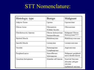
This document discusses soft tissue tumors, including:
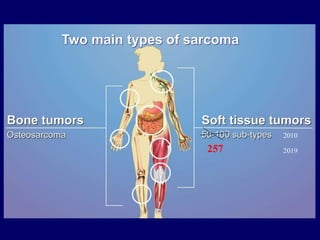
1. Soft tissue tumors can arise from various mesodermal tissues and there are over 257 subtypes classified by the WHO.
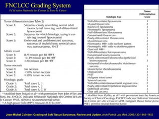
2. Benign tumors are generally superficial, grow slowly, and are not infiltrative, while malignant tumors are generally deeper, larger, grow rapidly, and are infiltrative.
3. The diagnosis of soft tissue tumors involves histological morphology, immunohistochemistry to identify cell markers, cytogenetic analysis to identify translocations, and molecular analysis.