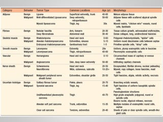
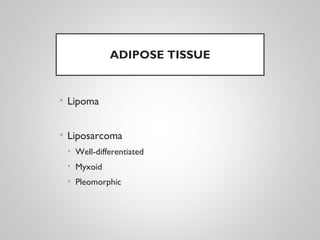
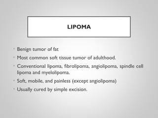
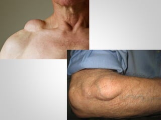
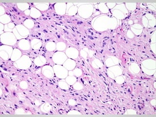
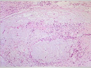
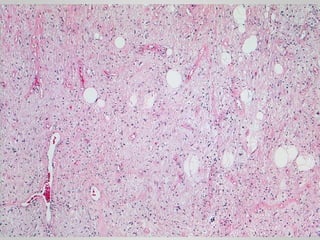
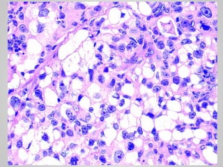
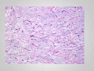
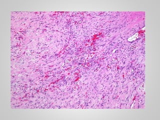
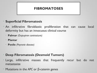
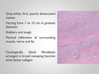
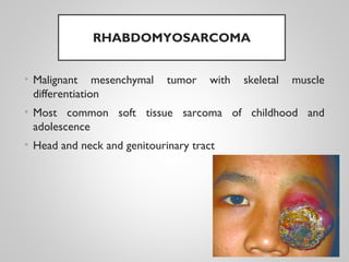
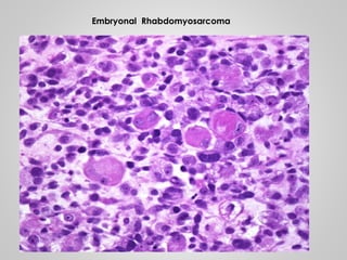
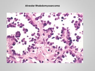
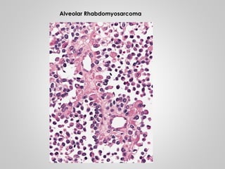
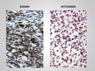
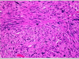
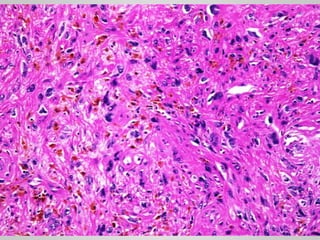
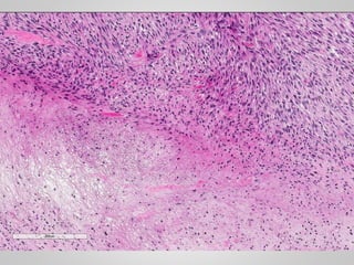
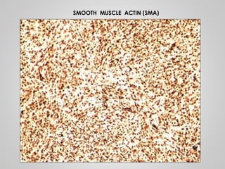
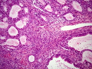
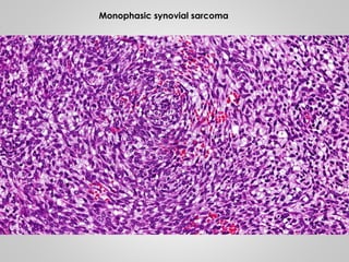
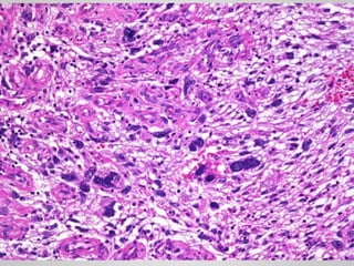
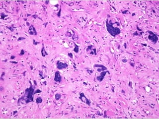
SOFT TISSUE TUMORS
BY: DR. USMAN SHAMS
INTRODUCTION
• Soft tissue refers to non-epithelial tissue excluding the
skeleton, joints, central nervous system, hematopoietic and
lymphoid tissues
• The area of soft tissue pathology is restricted to
neoplasms.
• Less than 1% of all cancers
• 2% of all cancer mortality
PATHOGENESIS
• Sporadic with no known predisposing cause.
• Germline mutations in tumor suppressor genes
• Neurofibromatosis 1
• Gardner syndrome
• Li-Fraumeni syndrome
• Osler-Weber Rendu syndrome
• Known environmental exposures such as radiation, burns
or toxins.
• No known precursor lesion
CHROMOSOMAL ABNORMALITIES
CLASSIFICATION
• Classification of soft tissue tumors continues to evolve as
new molecular genetic abnormalities are identified.
• Pathologic classification integrates morphology (e.g.,
muscle differentiation), immunohistochemistry and
molecular diagnostics
ADIPOSE TISSUE
• Lipoma
• Liposarcoma
• Well-differentiated
• Myxoid
• Pleomorphic
LIPOMA
• Benign tumor of fat
• Most common soft tissue tumor of adulthood.
• Conventional lipoma, fibrolipoma, angiolipoma, spindle cell
lipoma and myelolipoma.
• Soft, mobile, and painless (except angiolipoma)
• Usually cured by simple excision.
LIPOSARCOMA
• One of the most common sarcomas of adulthood
• People in their 50s to 60s
• Deep soft tissues of the extremities and in the
retroperitoneum
• Amplification of MDM2 gene
• Well-differentiated liposarcoma
• Adipocytes with scattered atypical spindle cells
• Myxoid liposarcoma
• Abundant basophilic extracellular matrix, arborizing
capillaries and primitive cells at various stages of adipocyte
differentiation
• Pleomorphic liposarcoma
• Sheets of anaplastic cells, bizarre nuclei and variable
amounts of immature adipocytes (lipoblasts).
FIBROUS TUMORS
• Nodular fasciitis
• Fibromatosis
NODULAR FASCIITIS
• Self-limited
• Fibroblastic and myofibroblastic proliferation
• Young adults
• Upper extremity
• History of trauma
• Rapid growth over a period of several weeks or months
• Typically no larger than 5 cm
• Spontaneously regresses and if excised, it rarely recurs.
• Circumscribed, or slightly infiltrative
• Arranged randomly or in short fascicles reminiscent of
tissue culture fibroblasts
• The cells vary in size and shape (spindle to stellate) and have
conspicuous nucleoli; mitotic figures are abundant.
• Lymphocytes and extravasated red blood cells are common.
FIBROMATOSES
• Superficial Fibromatosis
• An infiltrative fibroblastic proliferation that can cause local
deformity but has an innocuous clinical course
• Palmar (Dupuytren contracture)
• Plantar
• Penile (Peyronie disease)
• Deep Fibromatosis (Desmoid Tumors)
• Large, infiltrative masses that frequently recur but do not
metastasize
• Mutations in the APC or -catenin genesβ
Palmar Fibromatosis (Dupuytren contracture)
• Gray-white, firm, poorly demarcated
masses
• Varying from 1 to 15 cm in greatest
diameter
• Rubbery and tough
• Marked infiltration of surrounding
muscle, nerve and fat.
• Cytologically bland fibroblasts
arranged in broad sweeping fascicles
amid dense collagen
SKELETAL MUSCLE TUMORS
• Rhabdomyoma
• Rhabdomyosarcoma
RHABDOMYOSARCOMA
• Malignant mesenchymal tumor with skeletal muscle
differentiation
• Most common soft tissue sarcoma of childhood and
adolescence
• Head and neck and genitourinary tract
• Embryonal (60%)
Primitive spindle cells, “strap cells”
• Alveolar (20%)
Uniform round discohesive cells between septa
• Pleomorphic (20%)
Embryonal Rhabdomyosarcoma
Alveolar Rhabdomyosarcoma
DESMIN MYOGENIN
SMOOTH MUSCLE TUMORS
• Leiomyoma
• Leiomyosarcoma
LEIOMYOMA
• Uterine Leiomyoma
• Pilar Leiomyoma (Erector pili muscle in skin)
• Hereditary leiomyomatosis and renal cell cancer syndrome
LEIOMYOSARCOMA
• 10% to 20% of soft tissue sarcomas
• Adults
• Women more frequently than men
• Deep soft tissues of the extremities and retroperitoneum
• Great vessels, especially the inferior vena cava
• Painless firm masses
• Retroperitoneal tumors - abdominal symptoms
SMOOTH MUSCLE ACTIN (SMA)
TUMORS OF UNCERTAIN ORIGIN
• Synovial sarcoma
• Undifferentiated pleomorphic sarcoma
SYNOVIAL SARCOMA
• Misnomer
• 10% of all soft tissue sarcomas
• 20s to 40s age
• Deep-seated mass
• t(x;18)(p11;q11)
• Monophasic synovial sarcoma
• Uniform spindle cells with scant cytoplasm and dense chromatin
growing in short, tightly packed fascicles
• Biphasic synovial sarcoma
• Spindle cell component + gland-like structures
• Epithelial markers (e.g., keratins)
Monophasic synovial sarcoma
UNDIFFERENTIATED PLEOMORPHIC
SARCOMA
Soft tissue tumours
Soft tissue tumours
Soft tissue tumours

Soft tissue tumours

  • 1.
    SOFT TISSUE TUMORS BY:DR. USMAN SHAMS
  • 2.
    INTRODUCTION • Soft tissuerefers to non-epithelial tissue excluding the skeleton, joints, central nervous system, hematopoietic and lymphoid tissues • The area of soft tissue pathology is restricted to neoplasms. • Less than 1% of all cancers • 2% of all cancer mortality
  • 3.
    PATHOGENESIS • Sporadic withno known predisposing cause. • Germline mutations in tumor suppressor genes • Neurofibromatosis 1 • Gardner syndrome • Li-Fraumeni syndrome • Osler-Weber Rendu syndrome • Known environmental exposures such as radiation, burns or toxins. • No known precursor lesion
  • 4.
  • 5.
    CLASSIFICATION • Classification ofsoft tissue tumors continues to evolve as new molecular genetic abnormalities are identified. • Pathologic classification integrates morphology (e.g., muscle differentiation), immunohistochemistry and molecular diagnostics
  • 7.
    ADIPOSE TISSUE • Lipoma •Liposarcoma • Well-differentiated • Myxoid • Pleomorphic
  • 8.
    LIPOMA • Benign tumorof fat • Most common soft tissue tumor of adulthood. • Conventional lipoma, fibrolipoma, angiolipoma, spindle cell lipoma and myelolipoma. • Soft, mobile, and painless (except angiolipoma) • Usually cured by simple excision.
  • 12.
    LIPOSARCOMA • One ofthe most common sarcomas of adulthood • People in their 50s to 60s • Deep soft tissues of the extremities and in the retroperitoneum • Amplification of MDM2 gene
  • 13.
    • Well-differentiated liposarcoma •Adipocytes with scattered atypical spindle cells • Myxoid liposarcoma • Abundant basophilic extracellular matrix, arborizing capillaries and primitive cells at various stages of adipocyte differentiation • Pleomorphic liposarcoma • Sheets of anaplastic cells, bizarre nuclei and variable amounts of immature adipocytes (lipoblasts).
  • 20.
    FIBROUS TUMORS • Nodularfasciitis • Fibromatosis
  • 21.
    NODULAR FASCIITIS • Self-limited •Fibroblastic and myofibroblastic proliferation • Young adults • Upper extremity • History of trauma • Rapid growth over a period of several weeks or months • Typically no larger than 5 cm • Spontaneously regresses and if excised, it rarely recurs.
  • 22.
    • Circumscribed, orslightly infiltrative • Arranged randomly or in short fascicles reminiscent of tissue culture fibroblasts • The cells vary in size and shape (spindle to stellate) and have conspicuous nucleoli; mitotic figures are abundant. • Lymphocytes and extravasated red blood cells are common.
  • 26.
    FIBROMATOSES • Superficial Fibromatosis •An infiltrative fibroblastic proliferation that can cause local deformity but has an innocuous clinical course • Palmar (Dupuytren contracture) • Plantar • Penile (Peyronie disease) • Deep Fibromatosis (Desmoid Tumors) • Large, infiltrative masses that frequently recur but do not metastasize • Mutations in the APC or -catenin genesβ
  • 27.
  • 28.
    • Gray-white, firm,poorly demarcated masses • Varying from 1 to 15 cm in greatest diameter • Rubbery and tough • Marked infiltration of surrounding muscle, nerve and fat. • Cytologically bland fibroblasts arranged in broad sweeping fascicles amid dense collagen
  • 30.
    SKELETAL MUSCLE TUMORS •Rhabdomyoma • Rhabdomyosarcoma
  • 31.
    RHABDOMYOSARCOMA • Malignant mesenchymaltumor with skeletal muscle differentiation • Most common soft tissue sarcoma of childhood and adolescence • Head and neck and genitourinary tract
  • 32.
    • Embryonal (60%) Primitivespindle cells, “strap cells” • Alveolar (20%) Uniform round discohesive cells between septa • Pleomorphic (20%)
  • 33.
  • 34.
  • 35.
  • 36.
  • 37.
  • 38.
    SMOOTH MUSCLE TUMORS •Leiomyoma • Leiomyosarcoma
  • 39.
    LEIOMYOMA • Uterine Leiomyoma •Pilar Leiomyoma (Erector pili muscle in skin) • Hereditary leiomyomatosis and renal cell cancer syndrome
  • 42.
    LEIOMYOSARCOMA • 10% to20% of soft tissue sarcomas • Adults • Women more frequently than men • Deep soft tissues of the extremities and retroperitoneum • Great vessels, especially the inferior vena cava • Painless firm masses • Retroperitoneal tumors - abdominal symptoms
  • 46.
  • 47.
    TUMORS OF UNCERTAINORIGIN • Synovial sarcoma • Undifferentiated pleomorphic sarcoma
  • 48.
    SYNOVIAL SARCOMA • Misnomer •10% of all soft tissue sarcomas • 20s to 40s age • Deep-seated mass • t(x;18)(p11;q11) • Monophasic synovial sarcoma • Uniform spindle cells with scant cytoplasm and dense chromatin growing in short, tightly packed fascicles • Biphasic synovial sarcoma • Spindle cell component + gland-like structures • Epithelial markers (e.g., keratins)
  • 50.
  • 51.

Editor's Notes

  • #15 Well-differentiated liposarcoma
  • #17 Myxoid liposarcoma
  • #19 Pleomorphic liposarcoma
  • #50 Biphasic synovial sarcoma