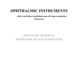
This document provides an overview of various ophthalmic instruments used in eye examinations, including:
1. Direct and indirect ophthalmoscopes are used to examine the interior of the eye. Indirect ophthalmoscopy uses a condensing lens to form an erect, magnified image of the retina.
2. The slit lamp examination uses different illumination techniques like diffuse, direct, and retroillumination to examine the anterior segment of the eye including the eyelids, cornea, iris, and lens.
3. Indirect techniques like specular reflection and optical sectioning are used to examine corneal structures at high magnification.
4. A lensometer is used to measure the refractive power of correct
Accommodation/ Accommodation of Eye, Measurement of Accommodation of Eye (hea...Bikash Sapkota
CLICK HERE TO DOWNLOAD FULL PPT ❤❤ https://healthkura.com/measurement-of-accommodation-of-eye/ ❤❤
Dear viewers Check Out my other piece of works at ❤❤❤ https://healthkura.com ❤❤❤
Measurement of Accommodation of eye:
Amplitude, Facility,
Relative Accommodation, Fatigue, Lag,
Dynamic Retinoscopy
Presentation Layout:
-Introduction to accommodation of eye
-Mechanism
-Components
-Measurement of accommodation of eye
- Amplitude
- Facility
- Relative accommodation
- Lag
-Dynamic Retinoscopy
Accommodation
-dioptric adjustment of the crystalline lens of the eye
- to obtain clear vision for a given target of regard
-process by which the refractive power of eye is altered
- to ensure a clear retinal image
For further reading
-Clinical Procedures in Optometry by J.D. Bartlett, J.B. Eskridge, J.F. Amos
-Primary Care Optometry by Theodere Grosvenor
-Borish’s Clinical Refraction by W.J. Benjamin
-Clinical Procedures for Ocular examination by Carlson et al
-American Academy of Ophthalmology
-Optometric Clinical Practice Guideline by American Optometric Association
-Internet
Follow me to get in touch with optometric and ophthalmic updates
Detailed instumentaion and use of manual Lensometer and just a outline of automated lensometer.
I have used the picture of manual lensometer with out the parts describtion because i have explained orally by showing the picture..
Hope u all like it and may help you in learning better. :)
Course: Bioinformatics for Biomedical Research (2014).
Session: 3.2- Basic Aspects of Microarray Technology and Data Analysis.
Statistics and Bioinformatisc Unit (UEB) & High Technology Unit (UAT) from Vall d'Hebron Research Institute (www.vhir.org), Barcelona.
SLIT LAMP AND ITS DIFFERENT ILLUMINATION TECHNIQUES.pptxAbhishek Kashyap
This presentation explains in detail about different illumination techniques and filters used in slit lamp examination and the procedure to perform slit lamp examination.
Binocular Indirect Ophthalmoscopy is known to provide a wider view of the inside of the eye. It is one of the most commonly used ophthalmic instrument.
Slit lamp biomicroscopy and illumination techniquesLoknath Goswami
It is a presentation on slitlamp for beginner, shown the parts and different illumination techniques both for eye and contact lens and it have short history
The Roman Empire A Historical Colossus.pdfkaushalkr1407
The Roman Empire, a vast and enduring power, stands as one of history's most remarkable civilizations, leaving an indelible imprint on the world. It emerged from the Roman Republic, transitioning into an imperial powerhouse under the leadership of Augustus Caesar in 27 BCE. This transformation marked the beginning of an era defined by unprecedented territorial expansion, architectural marvels, and profound cultural influence.
The empire's roots lie in the city of Rome, founded, according to legend, by Romulus in 753 BCE. Over centuries, Rome evolved from a small settlement to a formidable republic, characterized by a complex political system with elected officials and checks on power. However, internal strife, class conflicts, and military ambitions paved the way for the end of the Republic. Julius Caesar’s dictatorship and subsequent assassination in 44 BCE created a power vacuum, leading to a civil war. Octavian, later Augustus, emerged victorious, heralding the Roman Empire’s birth.
Under Augustus, the empire experienced the Pax Romana, a 200-year period of relative peace and stability. Augustus reformed the military, established efficient administrative systems, and initiated grand construction projects. The empire's borders expanded, encompassing territories from Britain to Egypt and from Spain to the Euphrates. Roman legions, renowned for their discipline and engineering prowess, secured and maintained these vast territories, building roads, fortifications, and cities that facilitated control and integration.
The Roman Empire’s society was hierarchical, with a rigid class system. At the top were the patricians, wealthy elites who held significant political power. Below them were the plebeians, free citizens with limited political influence, and the vast numbers of slaves who formed the backbone of the economy. The family unit was central, governed by the paterfamilias, the male head who held absolute authority.
Culturally, the Romans were eclectic, absorbing and adapting elements from the civilizations they encountered, particularly the Greeks. Roman art, literature, and philosophy reflected this synthesis, creating a rich cultural tapestry. Latin, the Roman language, became the lingua franca of the Western world, influencing numerous modern languages.
Roman architecture and engineering achievements were monumental. They perfected the arch, vault, and dome, constructing enduring structures like the Colosseum, Pantheon, and aqueducts. These engineering marvels not only showcased Roman ingenuity but also served practical purposes, from public entertainment to water supply.
Students, digital devices and success - Andreas Schleicher - 27 May 2024..pptxEduSkills OECD
Andreas Schleicher presents at the OECD webinar ‘Digital devices in schools: detrimental distraction or secret to success?’ on 27 May 2024. The presentation was based on findings from PISA 2022 results and the webinar helped launch the PISA in Focus ‘Managing screen time: How to protect and equip students against distraction’ https://www.oecd-ilibrary.org/education/managing-screen-time_7c225af4-en and the OECD Education Policy Perspective ‘Students, digital devices and success’ can be found here - https://oe.cd/il/5yV
Read| The latest issue of The Challenger is here! We are thrilled to announce that our school paper has qualified for the NATIONAL SCHOOLS PRESS CONFERENCE (NSPC) 2024. Thank you for your unwavering support and trust. Dive into the stories that made us stand out!
How to Create Map Views in the Odoo 17 ERPCeline George
The map views are useful for providing a geographical representation of data. They allow users to visualize and analyze the data in a more intuitive manner.
How to Split Bills in the Odoo 17 POS ModuleCeline George
Bills have a main role in point of sale procedure. It will help to track sales, handling payments and giving receipts to customers. Bill splitting also has an important role in POS. For example, If some friends come together for dinner and if they want to divide the bill then it is possible by POS bill splitting. This slide will show how to split bills in odoo 17 POS.
Unit 8 - Information and Communication Technology (Paper I).pdfThiyagu K
This slides describes the basic concepts of ICT, basics of Email, Emerging Technology and Digital Initiatives in Education. This presentations aligns with the UGC Paper I syllabus.
Synthetic Fiber Construction in lab .pptxPavel ( NSTU)
Synthetic fiber production is a fascinating and complex field that blends chemistry, engineering, and environmental science. By understanding these aspects, students can gain a comprehensive view of synthetic fiber production, its impact on society and the environment, and the potential for future innovations. Synthetic fibers play a crucial role in modern society, impacting various aspects of daily life, industry, and the environment. ynthetic fibers are integral to modern life, offering a range of benefits from cost-effectiveness and versatility to innovative applications and performance characteristics. While they pose environmental challenges, ongoing research and development aim to create more sustainable and eco-friendly alternatives. Understanding the importance of synthetic fibers helps in appreciating their role in the economy, industry, and daily life, while also emphasizing the need for sustainable practices and innovation.
Ethnobotany and Ethnopharmacology:
Ethnobotany in herbal drug evaluation,
Impact of Ethnobotany in traditional medicine,
New development in herbals,
Bio-prospecting tools for drug discovery,
Role of Ethnopharmacology in drug evaluation,
Reverse Pharmacology.
Model Attribute Check Company Auto PropertyCeline George
In Odoo, the multi-company feature allows you to manage multiple companies within a single Odoo database instance. Each company can have its own configurations while still sharing common resources such as products, customers, and suppliers.
How to Make a Field invisible in Odoo 17Celine George
It is possible to hide or invisible some fields in odoo. Commonly using “invisible” attribute in the field definition to invisible the fields. This slide will show how to make a field invisible in odoo 17.
2024.06.01 Introducing a competency framework for languag learning materials ...Sandy Millin
http://sandymillin.wordpress.com/iateflwebinar2024
Published classroom materials form the basis of syllabuses, drive teacher professional development, and have a potentially huge influence on learners, teachers and education systems. All teachers also create their own materials, whether a few sentences on a blackboard, a highly-structured fully-realised online course, or anything in between. Despite this, the knowledge and skills needed to create effective language learning materials are rarely part of teacher training, and are mostly learnt by trial and error.
Knowledge and skills frameworks, generally called competency frameworks, for ELT teachers, trainers and managers have existed for a few years now. However, until I created one for my MA dissertation, there wasn’t one drawing together what we need to know and do to be able to effectively produce language learning materials.
This webinar will introduce you to my framework, highlighting the key competencies I identified from my research. It will also show how anybody involved in language teaching (any language, not just English!), teacher training, managing schools or developing language learning materials can benefit from using the framework.
4. Distant Direct Ophthalmoscopy
• Performed with the help of self illuminating ophthalmoscope
or a plane mirror with a hole in centre
• Distance – 20 to 25 cm
• Applications-
1. To diagnose opacity in the refractive media
2. To differentiate between a mole and a hole of the iris
3. To recognize detached retina or a tumour arising from the
fundus
7. • Image – erect, virtual,14-15 times magnified emmetropes
(more in myopes and less in hyperopes)
• Field of vision is always smaller than field of illumination.
• Factors affecting it are-
Directly proportional to size of pupil
Directly proportional to axial length
Inversely proportional to the distance between the observed
and the observer’s eye
8. PROCEDURE
• Performed in a semidark room with the patient seated and
looking straight ahead and observer standing to the side of the
eye to be examined.
• The observer should reflect beam of light from the
ophthalmoscope into the patient’s pupil.
• Once the red reflex is seen, the observer should move as close
to the patient’s eye as possible.
• Once the retina is focused, the details should be examined
systematically.
10. • Introduced by Nagel in 1864
• Indirect Ophthalmoscopy (IDO) involves making the eye
highly myopic by placing a high power convex lens in front of
the eye so that a real, inverted and laterally reversed image is
formed close to the principle focus of the lens, between the
lens and the observer
• The technique is called Indirect because the fundus is seen
through a condensing lens.
11. Optical system of binocular indirect
ophthalmoscope
• Binocularity is achieved by reducing the observer’s
interpupillary distance from about 60mm to approximately
15mm by prism/mirror
13. • Image- real, inverted, magnified.
image magnification=power of eye/power of condensing lens
• Magnification of image depends upon
diopteric power of convex lens
position of lens in relation to the eyeball
refractive state of eyeball
• Field of observation is always greater than field of illumination
14. SIZE OF THE IMAGE IN DIFFERENT REFRACTIVE
STATE (EMMETROPIC, HYPERMETROPIC AND
MYOPIC EYE)
15. Procedure
• The practitioner should first illuminate the patient’s pupil area
by pointing the head and hence the illumination towards the
patient’s eye.
• Interpose the condenser lens close to the eye about 2 cm, and
centre the lens on to the pupil. The lens should be held with the
more convex side towards the practitioner.
• Pull back the lens away from the patient’s eye, at the same time
taking care to keep the illumination centered on the pupil. Whilst
withdrawing the lens, the practitioner will find a distance that
provides an optimum field of view. This should be approximately
at the focus of the lens, i.e. 5cms from the pupil using a +20D
lens
• Having obtained an image filling the BIO lens, the fundus may
then be examined by moving around the patient if reclining, or by
redirecting the patient’s fixation if seated
16. Scleral indentation
• Done with the depressor placed on the patient’s lid
• It should be moved in a direction opposite to that in which
examiner wants the depression to appear
• It should be rolled gently and tangentially over the eye surface
• Superonasal quadrant is most sensitive to scleral depression
18. ADVANTAGES OF IDO
• Larger field of view
• Lesser distortion of retinal image
• Easier to examine if patients eye movements are present and
with high spherical or astigmatic refractive errors
• Useful in hazy media due to its bright light and optical
property
• Can be used intraoperatively
• Vitreous can be examined easily and various vitreous
abnormalities diagnosed through this
19. DISADVANTAGES
• Difficult to learn
• Less magnification, therefore details of a small lesion not
visualized properly
• Impossible with very small pupils
• More uncomfortable to the patient
20. FUNDUS DRAWING
• It is made on special Amsler’s
chart, which has 12 clock hours
marked and has 3 concentric
circles
• Vortex veins ampulla seen along
the equator
• Long ciliary vein- 3 and 9 o’clock
• Branching vessels
21. COLOUR CODING OF RETINAL DRAWING
red Optic disc, retinal artery, hemorrhage, attached
retina, retinal neovascularisation
blue Retinal vein, detached retina, retinal oedema
green Media opacities, vitreous hemorrhage
yellow Retinal an choroidal exudates
brown Pigmented lesion, choroidal dethachment
Red line with
blue
Retinal breaks
25. • The slit lamp facilitates an examination which looks at anterior
segment of the human eye, which includes the
– Eyelid
– Cornea
– Sclera
– Conjunctiva
– Iris
– Anterior chamber
– Natural crystalline lens and
– Anterior vitreous.
26. HISTORICAL LANDMARKS
• De Wecker 1863 devised a portable ophthalmomicroscope .
• Albert1891,developed a binocular microscope which provided
stereoscopic view.
• Gullstrand ,1911 introduced the illumination system which had
for the first time a slit diaphragm in it
– Therefore Gullstrand is credited with the invention of slit
lamp.
27. • Operational components of slit lamp biomicroscope essentially
consist of:
• Illumination system
• Observation system
• Mechanical system
31. METHODS OF ILLUMINATION
1- DIFFUSE ILLUMINATION
• Angle between microscope and illumination system should be
30-45 degree.
• Slit width should be widest.
• Diffusing filter is used.
• Magnification: low to medium
• Illumination: medium to high
Applications:
General view of anterior of eye: lids,
lashes, sclera, cornea ,iris, pupil,
Gross pathology and media opacities
Contact lens fitting.
Assessment of lacrimal reflex.
32. 2- DIRECT ILLUMINATION
• Involves placing the light source at an angle of about 40-50
degree from microscope.
• This arrangement permits both light beam and microscope to
be sharply focused on the ocular tissue being observed.
• It is particularly suitable for assessment of cataracts, scars,
nerves, vessels etc.
• It is also of great importance for
the determination of stabilization
of axis of toric contact lens.
33. PARALLELOPIPED
Constructed by narrowing the beam to 1-2mm in width to
illuminate a rectangular area of cornea.
Microscope is placed directly in front of patients cornea.
Light source is approximately 45 degree from straight ahead
position.
Applications:
• Used to detect and examine corneal structures and defects.
• Used to detect corneal striae that develop when corneal
edema occurs with hydrogel lens wear and in keratoconus.
• Corneal scars and infilterates appears brighter than
surrounding because they have more density.
• Cells and flare in anterior chamber can be graded.
34. CONICAL BEAM
• Produced by narrowing the vertical height of a parallelopiped to
produce a small circular or square spot of light.
• Light source is 45-60 degree temporally and directed into pupil.
• Biomicroscope: directly in front of eye.
• Magnification: high(16-25x)
• Intensity of light source to heighest setting.
• Focusing: Beam is focused between cornea and anterior lens surface
and dark zone between cornea and anterior lens observed.
• Principle is same as that of beam of sun light streaming through a
room ,illuminating airborne dust particles.
• This occurance is called tyndall phenomenon.
• Most useful when examining the transparency of anterior chamber
for evidence of floating cells and flare seen in anterior uveitis.
35. OPTICAL SECTION
• Optic section is a very thin parallelopiped and optically cuts a
very thin slice of the cornea.
• Axis of illuminating and viewing path intersect in the area of
anterior eye media to be examined.
• Angle - 45 degree.
• With narrow slit the depth and portion of different
objects(penetration depth of foreign bodies, shape of lens etc)
can be resolved more easily.
• Magnification- maximum
• Used to localize:
– Infiltrates
– Cataracts
– AC depth.
36.
37. SPECULAR REFLECTION
• microscope and slit beam should be at equal angles from normal to cornea.
• Angle of illuminator to microscope must be equal and opposite.
• Angle of light should be moved until a very bright reflex obtained from
corneal surface which is called zone of specular reflection.
• Irregularities ,deposits in these smooth surface will fail to reflect light and
these appear darker than surrounding.
• Under specular reflection anterior corneal
surface appears as white uniform surface
and corneal endothelium takes on a mosaic
pattern.
• Uses:
a) Evaluate general appearance of corneal
endothelium
b) Lens surfaces
c) Corneal epithelium
38. INDIRECT ILLUMINATION
• The beam is focused in an area adjacent to ocular tissue to be
observed.
• Main application:
Examination of objects in direct vicinity
of corneal areas of reduced transparency
e,g infiltrates, corneal scars, deposits,
epithelial and stromal defects
• Illumination:
Narrow to medium slit beam
Decentred beam
• Magnification: approx 12x
39. RETROILLUMINATION
• Light is reflected off the iris or fundus, while the microscope is
focused on the cornea.
• 2 types-
DIRECT RETROILLUMINATION
• Observer is in direct pathway of
light reflected from structures.
• Pathology is seen against an
illuminated background
40. INDIRECT RETROILLUMINATION
• Observer is at right angle to the
observed structure and, therefore,
not in line with light.
• Pathology is seen against dark
background.
41. SCLEROTIC SCATTER
• Light beam is focused at limbus.
• Because of the phenomenon of total internal reflection, rays of
light pass through the substance of cornea and illuminate the
opposite side of limbus.
• Any corneal opacity becomes visible because it scatters the
rays of light.
• Magnification- 6-10 times
43. • It is a device designed to measure the refractive power
prescription of a unknown lens.
• 2 types-
1. Manual-
• Manual lensometer gives the accurate
power of a lens and were used in optical
industries.
• A manual lensometer is portable and
can be carried anywhere
• But a person needs to have a better
idea to measure the power of a lens.
(specially in case of a toric lens)
44. 2-Automated lensometer
• It is a fully automatic well programmed
device mostly used in clinics.
• It is easy and faster and can print
prescription.
• It is less accurate when compared to
manual lensometer.
45. Steps to neutralize spectacle lenses in
manual lensometer
• Calibration:- Adjusting the device to perform smoothly in
order to get accurate power of the lenses.
• Eye piece should be focused manually.
• Power drum should level zero.
• All the knobs should be placed
rightly and the device should be
neat and clean to avoid errors.
46. ADJUSTMENT OF EYEPIECE
• It is very important to focus the eye piece so that the
observer’s eye is relaxed and to avoid errors.
• In adjusting the eye piece the blurred black protractor is
focused.
• At first the eye piece is rotated completely Anti-clock
wise, the protractor views blurred.
• Then slowly rotate the eye piece clock wise and stop at
once where it is sharp and clear.
• Now the eye piece is set and focused.
47. Placing the spectacle lenses on the device
• Placing the spectacles lens on the device with its front surface
facing towards the eye piece.
• There are 2 knobs on the lensometer:-
1-Lens stop (holds the lens/
frame in place)
2-Frame leveling knob (makes
sure that the frame is leveled
and helps in accurate axis
measurement)
48. TARGET
• It is green in colour and appears when the device is switch on.
• It shows the position of the optical center of the lens.
• There is a ring of round dots at the centre of the target.
• This represents the power orientation of the lens as it rotates
with the lens rotation
• These round dots become small
lines oriented in one direction in
case of a toric lens
49. ANALYZING THE SPHERICAL LENSES
• Move the lens such that the target is exactly at the centre.
• Rotate the power drum until target is clear and sharp (all the
dots at the center should be separate & sharp)
• Stop rotating the power drum when the target is sharp for the
first time.
• Always count the upper mark on the power scale for accuracy
of the spherical power of the lens.
50. ANALYZING THE CYLINDRICAL LENS
• The process of Neutralizing a toric lens when compared to
spherical lens is completely a different concept.
• The key and the most important thing to remember is the
central orientation of the dots.
51. Process of toric lens neutralization
• Step 1:-
• Move the lens so that the target is at the centre of the protractor.
• This gives the optical centre location.
52. • Step 2-
• Rotate the power drum so that the central lines (oriented in one
direction) are sharp
53. • Step 3 -
• Rotate the axis wheel so that one meridian of the target is
parallel to the orientation of the central lines.
• Note that only one meridian of the target will be sharp.
• As the 180° meridian is sharp in this picture, we will write the
power and the axis as 180°
• Ex: ---DC X 180s°
54. • Rotate the power drum so that the opposite meridian in the
target is sharp.
• Note that the central lines will change in direction and will be
oriented in the opposite direction.
• As the 90° meridian is sharp in this picture, we will write the
power and the axis as 90°
• Ex: ----DC X 90°
• Writing the power
• As the Lensometer gives the readings of a toric lens power in 2
cylinder format, we need to transpose the power and write in
sphero – cylindrical format
• The power which we got in the example shall be written as:
• -----DS/----DC X 180°