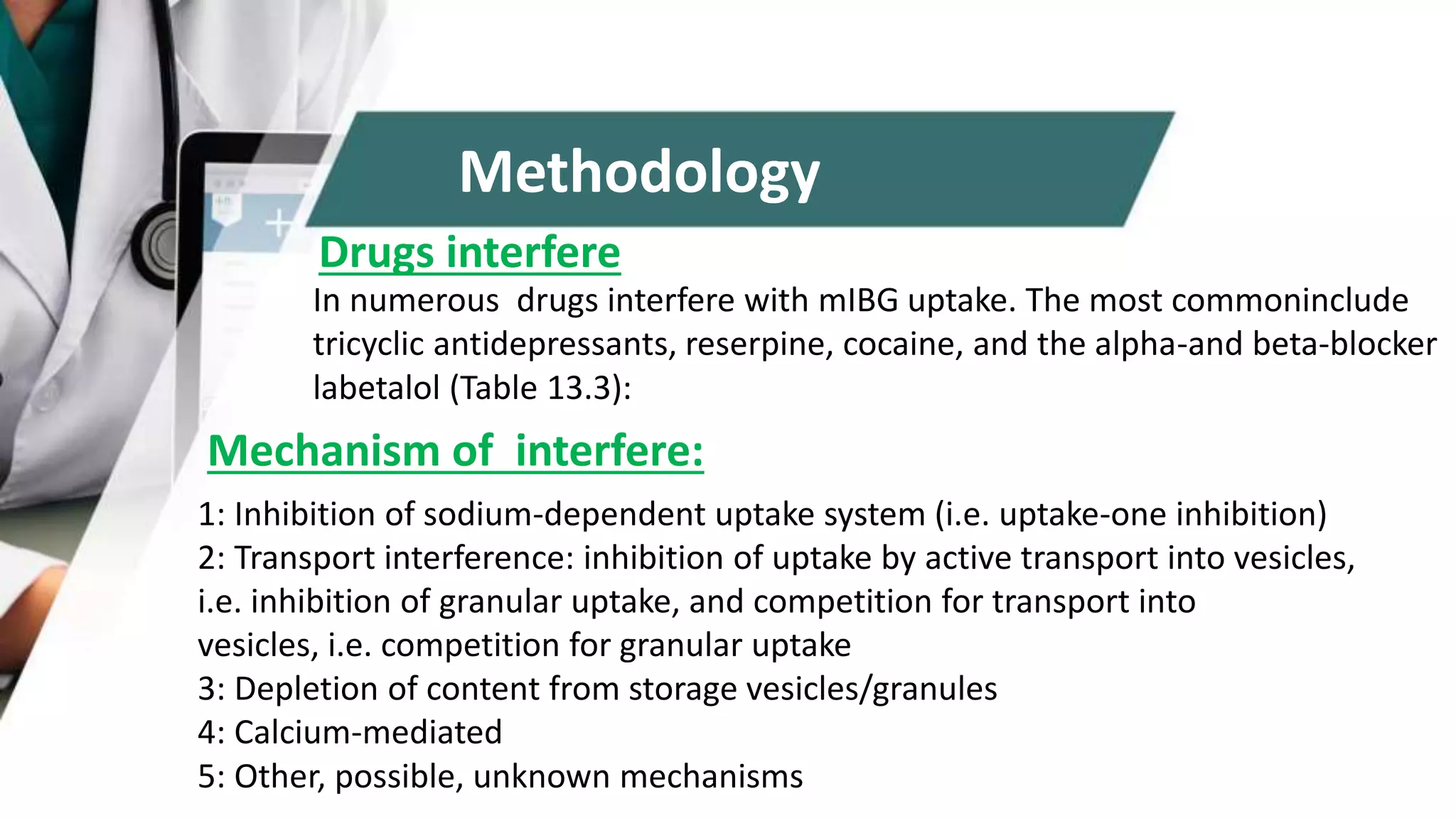
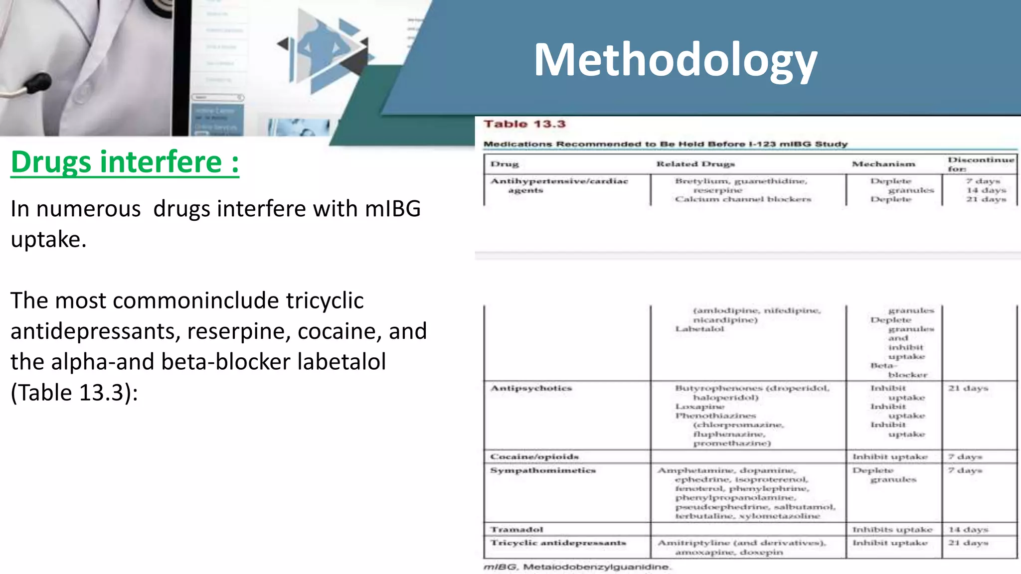
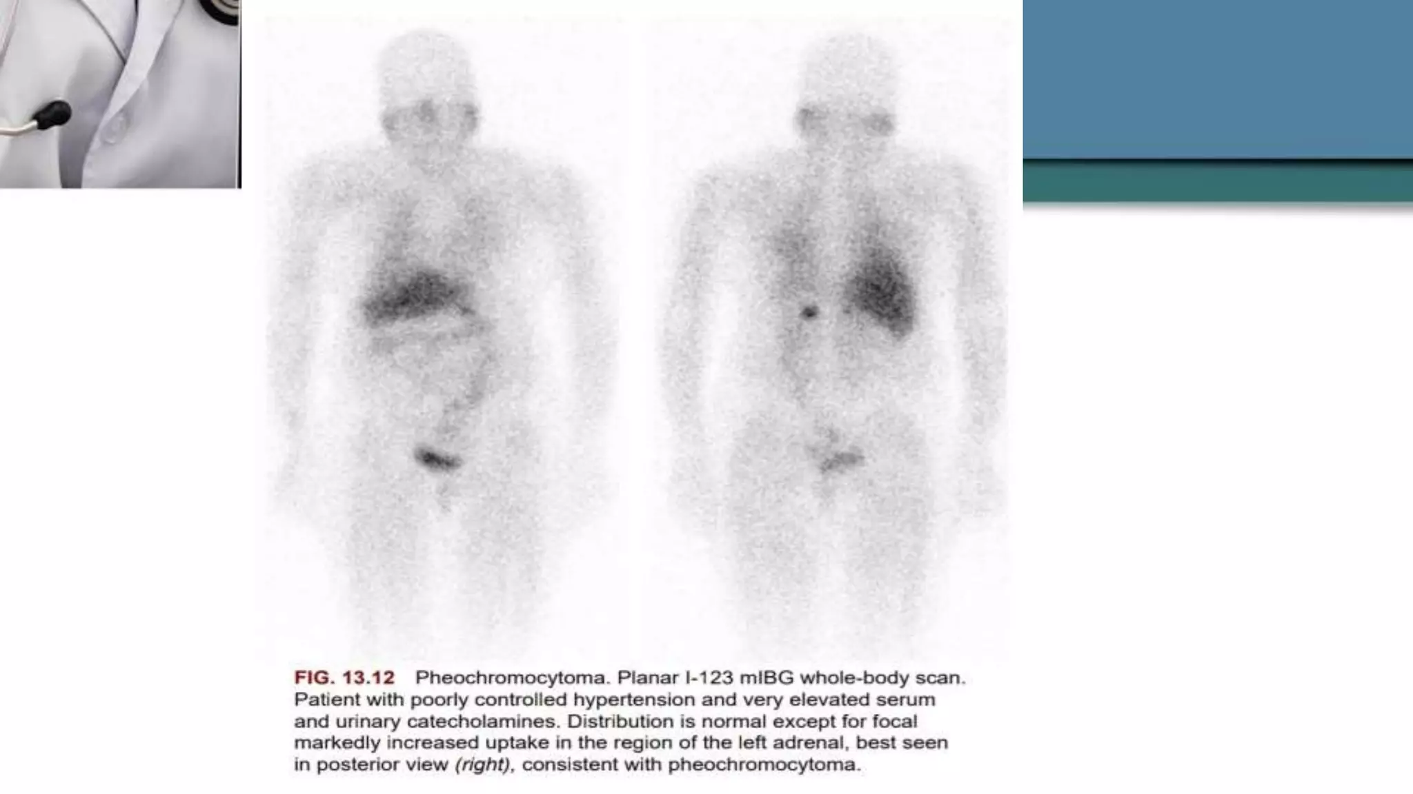
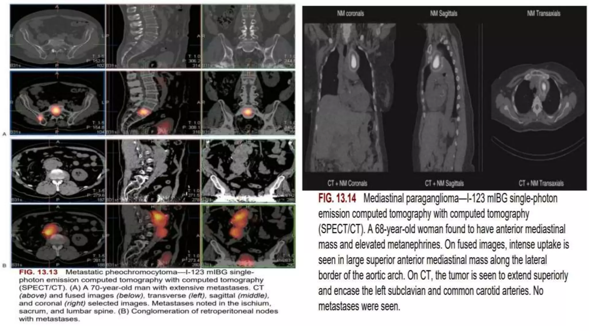
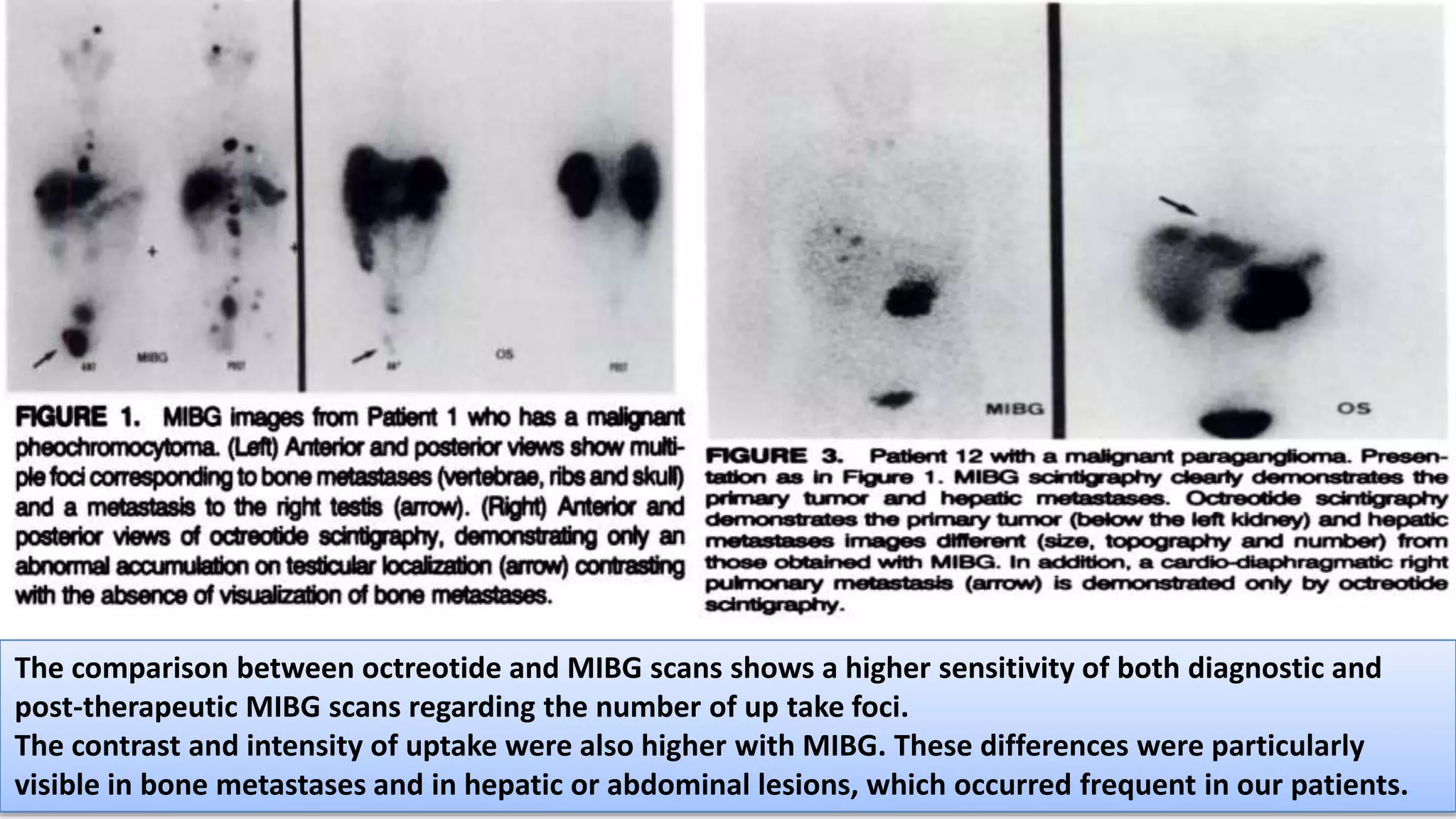
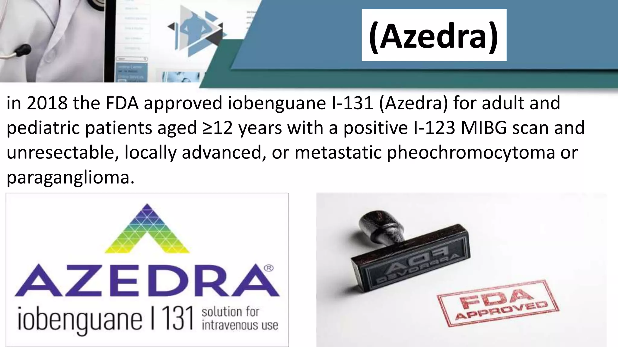
I-123 and I-131 mIBG Adrenergic
Tumor
Imaging and Therapy
Dr. Mustafa Al-Thabhawee
Tehran university of medical sciences
Research center for Nuclear medicine
National center of excellence
Shariati hospital
2020
Depending on nuclear medicine requisites 2021and MIBG
Guideline
Radiolabeled meta-iodo-benzyl-guanidine (MIBG) adrenal medullary scintigraphy has
been used clinically since the 1980s for diagnosis and staging of neural crest tumors
(e.g., pheochromocytomas, neuroblastomas and paragangliomas).
I-131-labeled MIBG was the original diagnostic agent; however, I-123-labeled MIBG is
now widely available and preferable because of its superior image quality with an
optimal 159-keV gamma-ray energy and lower patient radiation given a lack of β–
emissions and shorter half-life of 13 hours as opposed to I-131 with a 364-keV
gamma-ray energy, β– emissions, and 8-day half life. I-131 mIBG is reserved for
therapy.
Introduction
In numerous drugs interfere with mIBG uptake. The most commoninclude
tricyclic antidepressants, reserpine, cocaine, and the alpha-and beta-blocker
labetalol (Table 13.3):
Drugs interfere
Mechanism of interfere:
1: Inhibition of sodium-dependent uptake system (i.e. uptake-one inhibition)
2: Transport interference: inhibition of uptake by active transport into vesicles,
i.e. inhibition of granular uptake, and competition for transport into
vesicles, i.e. competition for granular uptake
3: Depletion of content from storage vesicles/granules
4: Calcium-mediated
5: Other, possible, unknown mechanisms
Methodology
In numerous drugs interfere with mIBG
uptake.
The most commoninclude tricyclic
antidepressants, reserpine, cocaine, and
the alpha-and beta-blocker labetalol
(Table 13.3):
Drugs interfere :
Methodology
Pretreatment with saturated potassium iodide (SSKI) or Lugol’s solution is recommended in the
package insert and in procedural guidelines to block thyroid uptake (Table 13.4)
BLock thyroid uptake
Before examination
The technologist, nurse or physician should give the patient (or parents if the patient is a child) a thorough explanation of
the preparation procedure and of the scintigraphic study.
Before examination
Patient History:
The patient should be clinically evaluated by the nuclear medicine physician who should consider any information that could be useful for
the interpretation of scintigraphic images:
1.Relevant history of suspected or known primary tumour
2.Intake of possibly interfering drugs
3.Absence or presence of symptoms
4.Laboratory test results (plasma and urinary catecholamine dosage, carcinoembryonic antigen, 5-hydroxyindoleacetic acid, neuron-specific
enolase, chromogranin A, calcitonin, etc.)
5.Results of any other imaging studies (CT, MRI, ultrasonography, plain radiographic imaging).
6.History of recent biopsy, surgery, chemotherapy, hor- mone therapy, radiation therapy.
After injection
Patients should be encouraged to drink large volumes of fluids following mIBG injection and
should void immediately prior to the study.
Side effects
1. Adverse effects of mIBG (tachycardia, pallor, vomiting, abdominal pain), that are not
related to allergy but to the pharmacological effects of the molecule, are very rare when
slow injection is used.
2.Injection via a central venous catheter must be avoided if possible (imaging artefacts,
potential adverse effects).
After injection
and
side effects of injection
Imaging protocol
I-123 MIBG avidly localizes in organs with high
adrenergic innervation, including the heart,
salivary glands, kidneys, and liver.
Variable activity is seen in the lungs, gallbladder,
salivary glands, and nasal mucosa.
Mild to moderate adrenal uptake often occurs with
planar I-123 MIBG imaging and is nearly always
seen with SPECT.
No uptake occurs in the normal skeleton.
It is cleared through the colon and kidneys.
Uptake and Distribution
The uptake of radiolabelled mIBG in different organs depends on catecholamine excretion and/or adrenergic innervation.
After intravenous injection approximately 50% of the administered radioactivity appears in the urine by 24 h, and 70–
90% of the residual activity is recovered within 48 h.
Since mIBG is excreted in the urine, the bladder and urinary tract show intense activity. mIBG is normally taken up mainly
by the liver; lower uptake levels are seen in the spleen, lungs, salivary glands, skeletal muscles and myocardium.
Normal adrenal glands are usually not seen, but faint uptake may be visible 48–72 h after injection in up to 15% of
patients when using 131I-mIBG.
However, normal adrenal glands can be visualized in up to 75% of patients using 123I-mIBG.
mIBG may accumulate to variable degrees in the nasal mucosa, lungs, gallbladder, colon and uterus.
Free iodine in the bloodstream may cause some uptake in the digestive system and in the thyroid (if not properly
blocked). No skeletal uptake should be seen.
Extremities show only slight muscular activity.
In children, uptake in brown fat is usually quite symmetrical along the edge of the trapezius muscles.
However, it is also seen over the top of each lung, and along either side of the spine to the level of the diaphragm in
children and in adults
Physiological distribution of mIBG
MIBG soft-tissue uptake is observed in primary
tumour and in metastatic sites including lymph
nodes, liver, bone and bone marrow.
Increased uptake in the skeleton (focal or
diffuse) is indicative of bone marrow
involvement and/or skeletal metastases.
Pathological uptake
Indications
Oncological indications
Non-oncological
indications
1. Detection, localization, staging and follow-up of neuroendocrine tumours and their metastases, in
particular:
A. phaeochromocytomas
B. neuroblastomas
C. ganglioneuroblastomas
D. ganglioneuromas
E. paragangliomas
F. carcinoid tumours
G. medullary thyroid carcinomas
H. Merkel cell tumours
I. MEN2 syndromes
2. Study of tumour uptake and residence time in order to decide and plan a treatment with high activities of
radiolabelled mIBG.
3. Evaluation of tumour response to therapy by measuring the intensity of mIBG uptake and the number of
focal mIBG uptake sites.
4. Confirmation of suspected tumours derived from neuroendocrine tissue.
Oncological indications
Functional studies of the adrenal medulla (hyperplasia),
sympathetic innervation of the myocardium, salivary glands
and lungs and movement disorders.
(Non-Oncological) indications
1.Clinical and biochemical findings that are unknown or have not been considered.
2. Insufficient knowledge of physiological mIBG biodisribution and kinetics.
3. Small lesions, below the resolution of scintigraphy.
4.Incorrect patient preparation (e.g. pelvic views cannot be correctly interpreted if the patient has not
voided before the acquisition).
5. Lesions close to the areas of high physiological or pathological uptake.
6. Tumour lesions that do not take up mIBG (e.g. Changes in differentiation, necrosis, interfering drugs,
etc.).
7.Patient motion (mainly in children).
8.Increased diffuse physiological uptake (hyperplastic adrenal gland after contralateral adrenalectomy).
9.Increased focal physiological uptakes (mainly in the urinary tract or bowel).
10. Thyroid activity (if thyroid blockade is not adequate).
11. Urine contamination or any other external contamination (salivary secretion).
Sources of error
To evaluate mIBG scintigraphy images the following should be taken into account:
1.Clinical issue raised in the request for mIBG scintigraphy.
2. Clinical history of the patient.
3. Presence of symptoms or syndromes.
4.Topographical localization of the uptake according to other imaging data.
5.Uptake in nonphysiological areas (this is suspicious for a neuroendocrine tumour or metastasis).
6.Intensity and features of the tracer uptake (mIBG uptake may be observed both in benign and
malignant tumours).
7.Clinical correlation with any other data from previous clinical, biochemical and morphological
examinations.
8. Causes of false-negative results (lesion size, tumour biology, physiological uptake masking cancer
lesions, pharmaceutical interference, etc.).
9.Causes of false-positive results (artifacts, uptake due to physiological processes, benign uptake, etc.)
Interpretation
The nuclear medicine physician should record all information regarding the patient, type of examination, date,
radiopharmaceutical (administered activity and route), concise patient history, all correlated data from previous
diagnostic studies, and the clinical question.
The report to the referring physician should describe:
1.Whether the distribution of mIBG is physiological or not.
2. All abnormal areas of uptake (intensity, number and site; if necessary, retention of mIBG over time).
3. Comparative analysis: the findings should be related to any previous information or results from other clinical or
instrumental examinations.
4.Interpretation: a clear diagnosis of malignant lesions should be made if possible, accompanied by a differential
diagnosis when appropriate.
5. Comments on factors that may limit the accuracy of scintigraphy are sometimes important (lesion size, artefacts,
interfering drugs, etc.).
6.If an additional diagnostic examination or adequate follow-up are required to obtain a definitive diagnosis, this must
be recommended.
Reporting
Clinical Applications
of
I-123 MIBG
I-131 MIBG
Pheochromocytoma
This catecholamine-secreting tumor is derived from chromaffin cells. It can precipitate life-
threatening hypertension or cardiac arrhythmias secondary to its excessive catecholamine
secretion.
When these tumors arise outside of the adrenal gland, they are called paragangliomas and
can be found anywhere from the bladder up to the base of the skull.
Ten percent of pheochromocytomas are bilateral, 10% are extraadrenal, and 10% are
malignant.
They may be associated with multiple endocrine neoplasia (MEN) types IIA and IIB, von Hippel–
Lindau disease, neurofibromatosis, tuberous sclerosis, and Carney syndrome.
Adrenomedullary hyperplasia occurs in patients with MEN type IIA.
Pheochromocytomas often present with elevated blood or urinary
catecholamines and metanephrines, usually three times or greater
than normal.
If an adrenal mass is demonstrated with morphological imaging in
patients with evidence for the disease, the diagnosis is often inferred,
and further workup before surgery is not always necessary.
However, I-123 mIBG can confirm the adrenergic etiology of a detected
adrenal mass on anatomical imaging, detect extraadrenal
paragangliomas, and diagnose medullary hyperplasia and metastatic
pheochromocytoma.
Clinical manifestation
The characteristic I-123 mIBG scintigraphic appearance of a
pheochromocytoma, extraadrenal paraganglioma, or metastatic
disease is intense focal uptake with a high tumor-to-background
ratio(Fig. 13.12).
The sensitivity and specificity for detection are 90% and The sensitivity
and specificity for detection are 90% and 95%, respectively.
Planar imaging is often diagnostic, although SPECT/CT can be helpful
(Figs. 13.13 and 13.14). F-18 FDG has only a limited role but can be
useful with high-grade adrenal cancers or malignant
pheochromocytoma.
Characteristic I-123 mIBG Scintigraphic
appearance
The comparison between octreotide and MIBG scans shows a higher sensitivity of both diagnostic and
post-therapeutic MIBG scans regarding the number of up take foci.
The contrast and intensity of uptake were also higher with MIBG. These differences were particularly
visible in bone metastases and in hepatic or abdominal lesions, which occurred frequent in our patients.
Neuroblastoma
This embryonal malignancy of the sympathetic nervous system most commonly
occurs in children younger than 4 years of age.
Over 70% of tumors originate in the retroperitoneal region, either from the adrenal
or the abdominal sympathetic chain, whereas approximately 20% occur in the
chest, derived from the thoracic sympathetic chain.
Patients with localized tumors can have a good prognosis andoutcome; those with
metastatic disease fare poorly. At the time of diagnosis, more than 50% of patients
present with metastatic disease, 25% have localized disease, and 15% have regional
extension.
Metastatic disease involves the lymph nodes, liver, bone marrow, and bone.
Introduction
I-123 mIBG is valuable for staging, detecting metastatic disease,
restaging, and determining patient response to therapy.
The sensitivity for detection of neuroblastoma is reported to be >90%,
and the specificity is about 95%. Whole-body scanning is routine (Figs.
13.15 and 13.16).
SPECT and SPECT/CT aid in detection and localization (Fig. 13.17 ).
NETs and medullary carcinoma of the thyroid also take up MIBG,
however, with lower sensitivity than for neuroblastoma or
pheochromocytoma.
The role of I-123 MIBG in Neuroblastoma
9-year-old girl with posterior mediastinal
mass.
(A) Planar anterior and posterior whole-
body images. The posterior planar image
shows focal uptake in the chest just above
the liver.
(B) Single-photon emission computed
tomography with computed tomography
(SPECT/CT) clearly localizes the paraspinal
mass. mass.
Bone scans have long been used to detect
osseous metastases in neuroblastoma. A
common location for metastases is in the
bilateral metaphyses of long bones. This could be
overlooked because of their symmetrical
appearance and high normal growth-plate
uptake in children. However, I-123 mIBG has
superior sensitivity for the detection of
metastases compared with bone scans because
the tumors initially involve the bone marrow.
The role of Bone scans in Neuroblastoma
Figure 1: 99mTc-methylene
diphosphonate bone scintigraphy
(BS) in a 12-year-old case of
mediastinal neuroblastoma (NB).
(a, anterior; b, posterior) and
4-year-old case of abdominal NB
(c, anterior; d, posterior) shows
heterogeneously increased
radiotracer uptake in the entire axial
and appendicular skeleton
suggesting widespread skeletal
metastases with cortical involvement
giving the appearance of a
metastatic superscan
Cases
12 months female presented with irritability and
constipation.
Case 1
Anterior x-ray shows:
Large lower midline and left
flank soft tissue mass in the
pelvis and abdomen
displacing bowel superiorly
and laterally
Axial CT:
There is large, lobulated,
heterogeneous, mixed density,
retroperitoneal mass. It
demonstrates heterogeneous
enhancement and patchy
Case Discussion
Biopsy proven
retroperitoneal neuroblastoma with
classical imaging findings.
Case 2: 10 months female presented with history
of Wheezing and obstructive airway symptoms
when lying flat
Large right posterior
mediastinal mass with a well
defined inferior margin. The
mass is predominantly soft
tissue density with calcific foci
inferiorly.
CT confirms the chest x-ray
findings of a right posterior
mediastinal mass with soft
tissue density and internal
calcifications
MIBG shows intense tracer uptake in the
posterior mediastinal mass with no evidence
of MIBG-avid disease elsewhere.
This case shows typical imaging features of a thoracic
neuroblastoma. The presence of calcifications (seen in 80-
90% of neuroblastoma cases), and remodeling of the ribs
and neural exit foramina suggest the neurogenic origin,
arising from the sympathetic chain.
The scintigraphic findings with markedly
increased tracer seen in both adrenal masses.
Large bilateral upper abdominal
masses of heterogeneous density at
the superior aspect of the kidneys
MEN2 consists of medullary thyroid cancer (always
present) and pheochromocytoma (commonly
present). It can be further divided into MEN2a with
the addition parathyroid hyperplasia and MEN2b
with the presence of mucosal neuromas
Case 3: 35yrs female HX of medullary thyroid cancer presented with hypertension And
elevated calcitonin level.
Case No. 4
Case No. 5
Case No. 6
Therapy With High-Dose I-131 mIBG
Conventional therapies for metastatic pheochromocytoma and neuroblastoma
include surgery, chemotherapy, and tyrosine kinase inhibitors.
The 5-year survival rate has been <50%.
The high uptake of mIBG in neuroectodermal tumors has led to therapy with high-
dose I- 131 mIBG in patients who have failed conventional therapies and have
progressive or symptomatic disease, utilizing its 606-keV I-131 beta emissions.
The 5-year survival rate has been reported to be increased; however, complete
response rates are not high.
This therapy, although performed for many years at selected centers in the United
States and Europe, has been considered investigational.
Introduction
(Azedra)
in 2018 the FDA approved iobenguane I-131 (Azedra) for adult and
pediatric patients aged ≥12 years with a positive I-123 MIBG scan and
unresectable, locally advanced, or metastatic pheochromocytoma or
paraganglioma.
1. patients must be pretreated with potassium iodide or other thyroid-blocking
medications beginning 24 to 48 hours before injection to minimize uptake of free
radioiodine by the thyroid (Table 13.4). It should be continued for 10 to 15 days
posttreatment.
In spite of doing this, hypothyroidism occurs in 11% to 20% of patients.
2. Before initiating therapy, excess catecholamines should be managed with alpha
blockade and atenolol.
3.Drugs that interfere with mIBG uptake must be discontinued, including labetalol,
reserpine, tricyclic antidepressants, sympathomimetics, and cocaine (Table 13.3).
The most significant toxicity is hematologic.
Patient preparation for the therapy with
I-131 mIBG
MIBG scan presentation

MIBG scan presentation

  • 1.
    I-123 and I-131mIBG Adrenergic Tumor Imaging and Therapy Dr. Mustafa Al-Thabhawee Tehran university of medical sciences Research center for Nuclear medicine National center of excellence Shariati hospital 2020 Depending on nuclear medicine requisites 2021and MIBG Guideline
  • 2.
    Radiolabeled meta-iodo-benzyl-guanidine (MIBG)adrenal medullary scintigraphy has been used clinically since the 1980s for diagnosis and staging of neural crest tumors (e.g., pheochromocytomas, neuroblastomas and paragangliomas). I-131-labeled MIBG was the original diagnostic agent; however, I-123-labeled MIBG is now widely available and preferable because of its superior image quality with an optimal 159-keV gamma-ray energy and lower patient radiation given a lack of β– emissions and shorter half-life of 13 hours as opposed to I-131 with a 364-keV gamma-ray energy, β– emissions, and 8-day half life. I-131 mIBG is reserved for therapy. Introduction
  • 3.
    In numerous drugsinterfere with mIBG uptake. The most commoninclude tricyclic antidepressants, reserpine, cocaine, and the alpha-and beta-blocker labetalol (Table 13.3): Drugs interfere Mechanism of interfere: 1: Inhibition of sodium-dependent uptake system (i.e. uptake-one inhibition) 2: Transport interference: inhibition of uptake by active transport into vesicles, i.e. inhibition of granular uptake, and competition for transport into vesicles, i.e. competition for granular uptake 3: Depletion of content from storage vesicles/granules 4: Calcium-mediated 5: Other, possible, unknown mechanisms Methodology
  • 4.
    In numerous drugsinterfere with mIBG uptake. The most commoninclude tricyclic antidepressants, reserpine, cocaine, and the alpha-and beta-blocker labetalol (Table 13.3): Drugs interfere : Methodology
  • 5.
    Pretreatment with saturatedpotassium iodide (SSKI) or Lugol’s solution is recommended in the package insert and in procedural guidelines to block thyroid uptake (Table 13.4) BLock thyroid uptake
  • 6.
    Before examination The technologist,nurse or physician should give the patient (or parents if the patient is a child) a thorough explanation of the preparation procedure and of the scintigraphic study. Before examination Patient History: The patient should be clinically evaluated by the nuclear medicine physician who should consider any information that could be useful for the interpretation of scintigraphic images: 1.Relevant history of suspected or known primary tumour 2.Intake of possibly interfering drugs 3.Absence or presence of symptoms 4.Laboratory test results (plasma and urinary catecholamine dosage, carcinoembryonic antigen, 5-hydroxyindoleacetic acid, neuron-specific enolase, chromogranin A, calcitonin, etc.) 5.Results of any other imaging studies (CT, MRI, ultrasonography, plain radiographic imaging). 6.History of recent biopsy, surgery, chemotherapy, hor- mone therapy, radiation therapy.
  • 7.
    After injection Patients shouldbe encouraged to drink large volumes of fluids following mIBG injection and should void immediately prior to the study. Side effects 1. Adverse effects of mIBG (tachycardia, pallor, vomiting, abdominal pain), that are not related to allergy but to the pharmacological effects of the molecule, are very rare when slow injection is used. 2.Injection via a central venous catheter must be avoided if possible (imaging artefacts, potential adverse effects). After injection and side effects of injection
  • 8.
  • 9.
    I-123 MIBG avidlylocalizes in organs with high adrenergic innervation, including the heart, salivary glands, kidneys, and liver. Variable activity is seen in the lungs, gallbladder, salivary glands, and nasal mucosa. Mild to moderate adrenal uptake often occurs with planar I-123 MIBG imaging and is nearly always seen with SPECT. No uptake occurs in the normal skeleton. It is cleared through the colon and kidneys. Uptake and Distribution
  • 10.
    The uptake ofradiolabelled mIBG in different organs depends on catecholamine excretion and/or adrenergic innervation. After intravenous injection approximately 50% of the administered radioactivity appears in the urine by 24 h, and 70– 90% of the residual activity is recovered within 48 h. Since mIBG is excreted in the urine, the bladder and urinary tract show intense activity. mIBG is normally taken up mainly by the liver; lower uptake levels are seen in the spleen, lungs, salivary glands, skeletal muscles and myocardium. Normal adrenal glands are usually not seen, but faint uptake may be visible 48–72 h after injection in up to 15% of patients when using 131I-mIBG. However, normal adrenal glands can be visualized in up to 75% of patients using 123I-mIBG. mIBG may accumulate to variable degrees in the nasal mucosa, lungs, gallbladder, colon and uterus. Free iodine in the bloodstream may cause some uptake in the digestive system and in the thyroid (if not properly blocked). No skeletal uptake should be seen. Extremities show only slight muscular activity. In children, uptake in brown fat is usually quite symmetrical along the edge of the trapezius muscles. However, it is also seen over the top of each lung, and along either side of the spine to the level of the diaphragm in children and in adults Physiological distribution of mIBG
  • 11.
    MIBG soft-tissue uptakeis observed in primary tumour and in metastatic sites including lymph nodes, liver, bone and bone marrow. Increased uptake in the skeleton (focal or diffuse) is indicative of bone marrow involvement and/or skeletal metastases. Pathological uptake
  • 12.
  • 13.
    1. Detection, localization,staging and follow-up of neuroendocrine tumours and their metastases, in particular: A. phaeochromocytomas B. neuroblastomas C. ganglioneuroblastomas D. ganglioneuromas E. paragangliomas F. carcinoid tumours G. medullary thyroid carcinomas H. Merkel cell tumours I. MEN2 syndromes 2. Study of tumour uptake and residence time in order to decide and plan a treatment with high activities of radiolabelled mIBG. 3. Evaluation of tumour response to therapy by measuring the intensity of mIBG uptake and the number of focal mIBG uptake sites. 4. Confirmation of suspected tumours derived from neuroendocrine tissue. Oncological indications
  • 14.
    Functional studies ofthe adrenal medulla (hyperplasia), sympathetic innervation of the myocardium, salivary glands and lungs and movement disorders. (Non-Oncological) indications
  • 15.
    1.Clinical and biochemicalfindings that are unknown or have not been considered. 2. Insufficient knowledge of physiological mIBG biodisribution and kinetics. 3. Small lesions, below the resolution of scintigraphy. 4.Incorrect patient preparation (e.g. pelvic views cannot be correctly interpreted if the patient has not voided before the acquisition). 5. Lesions close to the areas of high physiological or pathological uptake. 6. Tumour lesions that do not take up mIBG (e.g. Changes in differentiation, necrosis, interfering drugs, etc.). 7.Patient motion (mainly in children). 8.Increased diffuse physiological uptake (hyperplastic adrenal gland after contralateral adrenalectomy). 9.Increased focal physiological uptakes (mainly in the urinary tract or bowel). 10. Thyroid activity (if thyroid blockade is not adequate). 11. Urine contamination or any other external contamination (salivary secretion). Sources of error
  • 16.
    To evaluate mIBGscintigraphy images the following should be taken into account: 1.Clinical issue raised in the request for mIBG scintigraphy. 2. Clinical history of the patient. 3. Presence of symptoms or syndromes. 4.Topographical localization of the uptake according to other imaging data. 5.Uptake in nonphysiological areas (this is suspicious for a neuroendocrine tumour or metastasis). 6.Intensity and features of the tracer uptake (mIBG uptake may be observed both in benign and malignant tumours). 7.Clinical correlation with any other data from previous clinical, biochemical and morphological examinations. 8. Causes of false-negative results (lesion size, tumour biology, physiological uptake masking cancer lesions, pharmaceutical interference, etc.). 9.Causes of false-positive results (artifacts, uptake due to physiological processes, benign uptake, etc.) Interpretation
  • 17.
    The nuclear medicinephysician should record all information regarding the patient, type of examination, date, radiopharmaceutical (administered activity and route), concise patient history, all correlated data from previous diagnostic studies, and the clinical question. The report to the referring physician should describe: 1.Whether the distribution of mIBG is physiological or not. 2. All abnormal areas of uptake (intensity, number and site; if necessary, retention of mIBG over time). 3. Comparative analysis: the findings should be related to any previous information or results from other clinical or instrumental examinations. 4.Interpretation: a clear diagnosis of malignant lesions should be made if possible, accompanied by a differential diagnosis when appropriate. 5. Comments on factors that may limit the accuracy of scintigraphy are sometimes important (lesion size, artefacts, interfering drugs, etc.). 6.If an additional diagnostic examination or adequate follow-up are required to obtain a definitive diagnosis, this must be recommended. Reporting
  • 18.
  • 19.
  • 20.
    This catecholamine-secreting tumoris derived from chromaffin cells. It can precipitate life- threatening hypertension or cardiac arrhythmias secondary to its excessive catecholamine secretion. When these tumors arise outside of the adrenal gland, they are called paragangliomas and can be found anywhere from the bladder up to the base of the skull. Ten percent of pheochromocytomas are bilateral, 10% are extraadrenal, and 10% are malignant. They may be associated with multiple endocrine neoplasia (MEN) types IIA and IIB, von Hippel– Lindau disease, neurofibromatosis, tuberous sclerosis, and Carney syndrome. Adrenomedullary hyperplasia occurs in patients with MEN type IIA.
  • 21.
    Pheochromocytomas often presentwith elevated blood or urinary catecholamines and metanephrines, usually three times or greater than normal. If an adrenal mass is demonstrated with morphological imaging in patients with evidence for the disease, the diagnosis is often inferred, and further workup before surgery is not always necessary. However, I-123 mIBG can confirm the adrenergic etiology of a detected adrenal mass on anatomical imaging, detect extraadrenal paragangliomas, and diagnose medullary hyperplasia and metastatic pheochromocytoma. Clinical manifestation
  • 22.
    The characteristic I-123mIBG scintigraphic appearance of a pheochromocytoma, extraadrenal paraganglioma, or metastatic disease is intense focal uptake with a high tumor-to-background ratio(Fig. 13.12). The sensitivity and specificity for detection are 90% and The sensitivity and specificity for detection are 90% and 95%, respectively. Planar imaging is often diagnostic, although SPECT/CT can be helpful (Figs. 13.13 and 13.14). F-18 FDG has only a limited role but can be useful with high-grade adrenal cancers or malignant pheochromocytoma. Characteristic I-123 mIBG Scintigraphic appearance
  • 25.
    The comparison betweenoctreotide and MIBG scans shows a higher sensitivity of both diagnostic and post-therapeutic MIBG scans regarding the number of up take foci. The contrast and intensity of uptake were also higher with MIBG. These differences were particularly visible in bone metastases and in hepatic or abdominal lesions, which occurred frequent in our patients.
  • 26.
  • 27.
    This embryonal malignancyof the sympathetic nervous system most commonly occurs in children younger than 4 years of age. Over 70% of tumors originate in the retroperitoneal region, either from the adrenal or the abdominal sympathetic chain, whereas approximately 20% occur in the chest, derived from the thoracic sympathetic chain. Patients with localized tumors can have a good prognosis andoutcome; those with metastatic disease fare poorly. At the time of diagnosis, more than 50% of patients present with metastatic disease, 25% have localized disease, and 15% have regional extension. Metastatic disease involves the lymph nodes, liver, bone marrow, and bone. Introduction
  • 29.
    I-123 mIBG isvaluable for staging, detecting metastatic disease, restaging, and determining patient response to therapy. The sensitivity for detection of neuroblastoma is reported to be >90%, and the specificity is about 95%. Whole-body scanning is routine (Figs. 13.15 and 13.16). SPECT and SPECT/CT aid in detection and localization (Fig. 13.17 ). NETs and medullary carcinoma of the thyroid also take up MIBG, however, with lower sensitivity than for neuroblastoma or pheochromocytoma. The role of I-123 MIBG in Neuroblastoma
  • 32.
    9-year-old girl withposterior mediastinal mass. (A) Planar anterior and posterior whole- body images. The posterior planar image shows focal uptake in the chest just above the liver. (B) Single-photon emission computed tomography with computed tomography (SPECT/CT) clearly localizes the paraspinal mass. mass.
  • 33.
    Bone scans havelong been used to detect osseous metastases in neuroblastoma. A common location for metastases is in the bilateral metaphyses of long bones. This could be overlooked because of their symmetrical appearance and high normal growth-plate uptake in children. However, I-123 mIBG has superior sensitivity for the detection of metastases compared with bone scans because the tumors initially involve the bone marrow. The role of Bone scans in Neuroblastoma
  • 34.
    Figure 1: 99mTc-methylene diphosphonatebone scintigraphy (BS) in a 12-year-old case of mediastinal neuroblastoma (NB). (a, anterior; b, posterior) and 4-year-old case of abdominal NB (c, anterior; d, posterior) shows heterogeneously increased radiotracer uptake in the entire axial and appendicular skeleton suggesting widespread skeletal metastases with cortical involvement giving the appearance of a metastatic superscan
  • 35.
  • 36.
    12 months femalepresented with irritability and constipation. Case 1 Anterior x-ray shows: Large lower midline and left flank soft tissue mass in the pelvis and abdomen displacing bowel superiorly and laterally Axial CT: There is large, lobulated, heterogeneous, mixed density, retroperitoneal mass. It demonstrates heterogeneous enhancement and patchy Case Discussion Biopsy proven retroperitoneal neuroblastoma with classical imaging findings.
  • 37.
    Case 2: 10months female presented with history of Wheezing and obstructive airway symptoms when lying flat Large right posterior mediastinal mass with a well defined inferior margin. The mass is predominantly soft tissue density with calcific foci inferiorly. CT confirms the chest x-ray findings of a right posterior mediastinal mass with soft tissue density and internal calcifications MIBG shows intense tracer uptake in the posterior mediastinal mass with no evidence of MIBG-avid disease elsewhere. This case shows typical imaging features of a thoracic neuroblastoma. The presence of calcifications (seen in 80- 90% of neuroblastoma cases), and remodeling of the ribs and neural exit foramina suggest the neurogenic origin, arising from the sympathetic chain.
  • 38.
    The scintigraphic findingswith markedly increased tracer seen in both adrenal masses. Large bilateral upper abdominal masses of heterogeneous density at the superior aspect of the kidneys MEN2 consists of medullary thyroid cancer (always present) and pheochromocytoma (commonly present). It can be further divided into MEN2a with the addition parathyroid hyperplasia and MEN2b with the presence of mucosal neuromas Case 3: 35yrs female HX of medullary thyroid cancer presented with hypertension And elevated calcitonin level.
  • 39.
  • 40.
  • 41.
  • 43.
  • 44.
    Conventional therapies formetastatic pheochromocytoma and neuroblastoma include surgery, chemotherapy, and tyrosine kinase inhibitors. The 5-year survival rate has been <50%. The high uptake of mIBG in neuroectodermal tumors has led to therapy with high- dose I- 131 mIBG in patients who have failed conventional therapies and have progressive or symptomatic disease, utilizing its 606-keV I-131 beta emissions. The 5-year survival rate has been reported to be increased; however, complete response rates are not high. This therapy, although performed for many years at selected centers in the United States and Europe, has been considered investigational. Introduction
  • 45.
    (Azedra) in 2018 theFDA approved iobenguane I-131 (Azedra) for adult and pediatric patients aged ≥12 years with a positive I-123 MIBG scan and unresectable, locally advanced, or metastatic pheochromocytoma or paraganglioma.
  • 46.
    1. patients mustbe pretreated with potassium iodide or other thyroid-blocking medications beginning 24 to 48 hours before injection to minimize uptake of free radioiodine by the thyroid (Table 13.4). It should be continued for 10 to 15 days posttreatment. In spite of doing this, hypothyroidism occurs in 11% to 20% of patients. 2. Before initiating therapy, excess catecholamines should be managed with alpha blockade and atenolol. 3.Drugs that interfere with mIBG uptake must be discontinued, including labetalol, reserpine, tricyclic antidepressants, sympathomimetics, and cocaine (Table 13.3). The most significant toxicity is hematologic. Patient preparation for the therapy with I-131 mIBG