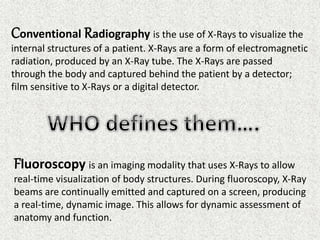
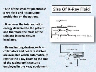
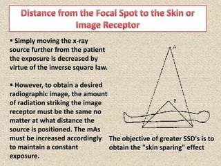
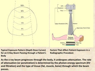
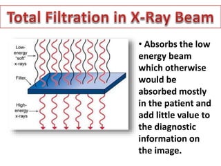
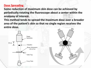
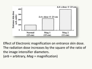
This document summarizes techniques to reduce radiation dose in diagnostic radiology, including fluoroscopy. It discusses using the smallest x-ray field, increasing distance between the patient and x-ray source, using filters, grids, and intensifying screens. It also covers automatic processing, avoiding unnecessary repeat images, and techniques to reduce dose in fluoroscopy like intermittent exposure, removal of grids, and last image hold. The document emphasizes training operators and using radiation only when necessary to obtain diagnostic information.
this power-point slide presentation includes lots of information like how MRI coil works. what is shimming, magnet, fringe, and design of mri coil and also magnet. this will help a lot for radiologist and technician radiographers.. thanks.
Recent advancements in modern x ray tubeSantosh Ojha
All the advancements in X-ray tubes till date are done to increase the Tube heat storage capacity thus increasing the lifetime of x -ray tubes. This slide explains about these recent advancements in x-ray tubes.
Recent advancements in modern x ray tubeSantosh Ojha
All the advancements in X-ray tubes till date are done to increase the Tube heat storage capacity thus increasing the lifetime of x -ray tubes. This slide explains about these recent advancements in x-ray tubes.
Those who administer ionizing radiation must become familiar with the magnitude of exposure encountered in medicine, dentistry and every day life; the possible risks associated with such exposure; and the methods used to affect exposure.
Practitioners should remain informed about safety updates to further improve diagnostic quality of radiographs and decrease radiation exposure.
Nuclear medicine is a branch of medical imaging that uses small amounts of radioactive material to diagnose and determine the severity of or treat a variety of diseases, including many types of cancers, heart disease, gastrointestinal, endocrine, neurological disorders and other abnormalities within the body.
Fluoroscopy ,Radiation safety and contrast agents including adverse effect an...Dr Ravi Shankar Sharma
IT includes everything related to fluoroscopy, radiation exposure, it,s effects, contrast agents , and it,s newer variants including gadolinium, anaphylaxis reactions and it,s management, images for epidural,intrathecal,subdural, intrarterial and intravenous contrast picture.
Similar to Dose reduction in Conventional Radiography and Fluoroscopy (20)
Title: Sense of Taste
Presenter: Dr. Faiza, Assistant Professor of Physiology
Qualifications:
MBBS (Best Graduate, AIMC Lahore)
FCPS Physiology
ICMT, CHPE, DHPE (STMU)
MPH (GC University, Faisalabad)
MBA (Virtual University of Pakistan)
Learning Objectives:
Describe the structure and function of taste buds.
Describe the relationship between the taste threshold and taste index of common substances.
Explain the chemical basis and signal transduction of taste perception for each type of primary taste sensation.
Recognize different abnormalities of taste perception and their causes.
Key Topics:
Significance of Taste Sensation:
Differentiation between pleasant and harmful food
Influence on behavior
Selection of food based on metabolic needs
Receptors of Taste:
Taste buds on the tongue
Influence of sense of smell, texture of food, and pain stimulation (e.g., by pepper)
Primary and Secondary Taste Sensations:
Primary taste sensations: Sweet, Sour, Salty, Bitter, Umami
Chemical basis and signal transduction mechanisms for each taste
Taste Threshold and Index:
Taste threshold values for Sweet (sucrose), Salty (NaCl), Sour (HCl), and Bitter (Quinine)
Taste index relationship: Inversely proportional to taste threshold
Taste Blindness:
Inability to taste certain substances, particularly thiourea compounds
Example: Phenylthiocarbamide
Structure and Function of Taste Buds:
Composition: Epithelial cells, Sustentacular/Supporting cells, Taste cells, Basal cells
Features: Taste pores, Taste hairs/microvilli, and Taste nerve fibers
Location of Taste Buds:
Found in papillae of the tongue (Fungiform, Circumvallate, Foliate)
Also present on the palate, tonsillar pillars, epiglottis, and proximal esophagus
Mechanism of Taste Stimulation:
Interaction of taste substances with receptors on microvilli
Signal transduction pathways for Umami, Sweet, Bitter, Sour, and Salty tastes
Taste Sensitivity and Adaptation:
Decrease in sensitivity with age
Rapid adaptation of taste sensation
Role of Saliva in Taste:
Dissolution of tastants to reach receptors
Washing away the stimulus
Taste Preferences and Aversions:
Mechanisms behind taste preference and aversion
Influence of receptors and neural pathways
Impact of Sensory Nerve Damage:
Degeneration of taste buds if the sensory nerve fiber is cut
Abnormalities of Taste Detection:
Conditions: Ageusia, Hypogeusia, Dysgeusia (parageusia)
Causes: Nerve damage, neurological disorders, infections, poor oral hygiene, adverse drug effects, deficiencies, aging, tobacco use, altered neurotransmitter levels
Neurotransmitters and Taste Threshold:
Effects of serotonin (5-HT) and norepinephrine (NE) on taste sensitivity
Supertasters:
25% of the population with heightened sensitivity to taste, especially bitterness
Increased number of fungiform papillae
Couples presenting to the infertility clinic- Do they really have infertility...Sujoy Dasgupta
Dr Sujoy Dasgupta presented the study on "Couples presenting to the infertility clinic- Do they really have infertility? – The unexplored stories of non-consummation" in the 13th Congress of the Asia Pacific Initiative on Reproduction (ASPIRE 2024) at Manila on 24 May, 2024.
- Video recording of this lecture in English language: https://youtu.be/lK81BzxMqdo
- Video recording of this lecture in Arabic language: https://youtu.be/Ve4P0COk9OI
- Link to download the book free: https://nephrotube.blogspot.com/p/nephrotube-nephrology-books.html
- Link to NephroTube website: www.NephroTube.com
- Link to NephroTube social media accounts: https://nephrotube.blogspot.com/p/join-nephrotube-on-social-media.html
Anti ulcer drugs and their Advance pharmacology ||
Anti-ulcer drugs are medications used to prevent and treat ulcers in the stomach and upper part of the small intestine (duodenal ulcers). These ulcers are often caused by an imbalance between stomach acid and the mucosal lining, which protects the stomach lining.
||Scope: Overview of various classes of anti-ulcer drugs, their mechanisms of action, indications, side effects, and clinical considerations.
These simplified slides by Dr. Sidra Arshad present an overview of the non-respiratory functions of the respiratory tract.
Learning objectives:
1. Enlist the non-respiratory functions of the respiratory tract
2. Briefly explain how these functions are carried out
3. Discuss the significance of dead space
4. Differentiate between minute ventilation and alveolar ventilation
5. Describe the cough and sneeze reflexes
Study Resources:
1. Chapter 39, Guyton and Hall Textbook of Medical Physiology, 14th edition
2. Chapter 34, Ganong’s Review of Medical Physiology, 26th edition
3. Chapter 17, Human Physiology by Lauralee Sherwood, 9th edition
4. Non-respiratory functions of the lungs https://academic.oup.com/bjaed/article/13/3/98/278874
Ethanol (CH3CH2OH), or beverage alcohol, is a two-carbon alcohol
that is rapidly distributed in the body and brain. Ethanol alters many
neurochemical systems and has rewarding and addictive properties. It
is the oldest recreational drug and likely contributes to more morbidity,
mortality, and public health costs than all illicit drugs combined. The
5th edition of the Diagnostic and Statistical Manual of Mental Disorders
(DSM-5) integrates alcohol abuse and alcohol dependence into a single
disorder called alcohol use disorder (AUD), with mild, moderate,
and severe subclassifications (American Psychiatric Association, 2013).
In the DSM-5, all types of substance abuse and dependence have been
combined into a single substance use disorder (SUD) on a continuum
from mild to severe. A diagnosis of AUD requires that at least two of
the 11 DSM-5 behaviors be present within a 12-month period (mild
AUD: 2–3 criteria; moderate AUD: 4–5 criteria; severe AUD: 6–11 criteria).
The four main behavioral effects of AUD are impaired control over
drinking, negative social consequences, risky use, and altered physiological
effects (tolerance, withdrawal). This chapter presents an overview
of the prevalence and harmful consequences of AUD in the U.S.,
the systemic nature of the disease, neurocircuitry and stages of AUD,
comorbidities, fetal alcohol spectrum disorders, genetic risk factors, and
pharmacotherapies for AUD.
Prix Galien International 2024 Forum ProgramLevi Shapiro
June 20, 2024, Prix Galien International and Jerusalem Ethics Forum in ROME. Detailed agenda including panels:
- ADVANCES IN CARDIOLOGY: A NEW PARADIGM IS COMING
- WOMEN’S HEALTH: FERTILITY PRESERVATION
- WHAT’S NEW IN THE TREATMENT OF INFECTIOUS,
ONCOLOGICAL AND INFLAMMATORY SKIN DISEASES?
- ARTIFICIAL INTELLIGENCE AND ETHICS
- GENE THERAPY
- BEYOND BORDERS: GLOBAL INITIATIVES FOR DEMOCRATIZING LIFE SCIENCE TECHNOLOGIES AND PROMOTING ACCESS TO HEALTHCARE
- ETHICAL CHALLENGES IN LIFE SCIENCES
- Prix Galien International Awards Ceremony
MANAGEMENT OF ATRIOVENTRICULAR CONDUCTION BLOCK.pdfJim Jacob Roy
Cardiac conduction defects can occur due to various causes.
Atrioventricular conduction blocks ( AV blocks ) are classified into 3 types.
This document describes the acute management of AV block.
Ozempic: Preoperative Management of Patients on GLP-1 Receptor Agonists Saeid Safari
Preoperative Management of Patients on GLP-1 Receptor Agonists like Ozempic and Semiglutide
ASA GUIDELINE
NYSORA Guideline
2 Case Reports of Gastric Ultrasound
Tom Selleck Health: A Comprehensive Look at the Iconic Actor’s Wellness Journeygreendigital
Tom Selleck, an enduring figure in Hollywood. has captivated audiences for decades with his rugged charm, iconic moustache. and memorable roles in television and film. From his breakout role as Thomas Magnum in Magnum P.I. to his current portrayal of Frank Reagan in Blue Bloods. Selleck's career has spanned over 50 years. But beyond his professional achievements. fans have often been curious about Tom Selleck Health. especially as he has aged in the public eye.
Follow us on: Pinterest
Introduction
Many have been interested in Tom Selleck health. not only because of his enduring presence on screen but also because of the challenges. and lifestyle choices he has faced and made over the years. This article delves into the various aspects of Tom Selleck health. exploring his fitness regimen, diet, mental health. and the challenges he has encountered as he ages. We'll look at how he maintains his well-being. the health issues he has faced, and his approach to ageing .
Early Life and Career
Childhood and Athletic Beginnings
Tom Selleck was born on January 29, 1945, in Detroit, Michigan, and grew up in Sherman Oaks, California. From an early age, he was involved in sports, particularly basketball. which played a significant role in his physical development. His athletic pursuits continued into college. where he attended the University of Southern California (USC) on a basketball scholarship. This early involvement in sports laid a strong foundation for his physical health and disciplined lifestyle.
Transition to Acting
Selleck's transition from an athlete to an actor came with its physical demands. His first significant role in "Magnum P.I." required him to perform various stunts and maintain a fit appearance. This role, which he played from 1980 to 1988. necessitated a rigorous fitness routine to meet the show's demands. setting the stage for his long-term commitment to health and wellness.
Fitness Regimen
Workout Routine
Tom Selleck health and fitness regimen has evolved. adapting to his changing roles and age. During his "Magnum, P.I." days. Selleck's workouts were intense and focused on building and maintaining muscle mass. His routine included weightlifting, cardiovascular exercises. and specific training for the stunts he performed on the show.
Selleck adjusted his fitness routine as he aged to suit his body's needs. Today, his workouts focus on maintaining flexibility, strength, and cardiovascular health. He incorporates low-impact exercises such as swimming, walking, and light weightlifting. This balanced approach helps him stay fit without putting undue strain on his joints and muscles.
Importance of Flexibility and Mobility
In recent years, Selleck has emphasized the importance of flexibility and mobility in his fitness regimen. Understanding the natural decline in muscle mass and joint flexibility with age. he includes stretching and yoga in his routine. These practices help prevent injuries, improve posture, and maintain mobilit
Flu Vaccine Alert in Bangalore Karnatakaaddon Scans
As flu season approaches, health officials in Bangalore, Karnataka, are urging residents to get their flu vaccinations. The seasonal flu, while common, can lead to severe health complications, particularly for vulnerable populations such as young children, the elderly, and those with underlying health conditions.
Dr. Vidisha Kumari, a leading epidemiologist in Bangalore, emphasizes the importance of getting vaccinated. "The flu vaccine is our best defense against the influenza virus. It not only protects individuals but also helps prevent the spread of the virus in our communities," he says.
This year, the flu season is expected to coincide with a potential increase in other respiratory illnesses. The Karnataka Health Department has launched an awareness campaign highlighting the significance of flu vaccinations. They have set up multiple vaccination centers across Bangalore, making it convenient for residents to receive their shots.
To encourage widespread vaccination, the government is also collaborating with local schools, workplaces, and community centers to facilitate vaccination drives. Special attention is being given to ensuring that the vaccine is accessible to all, including marginalized communities who may have limited access to healthcare.
Residents are reminded that the flu vaccine is safe and effective. Common side effects are mild and may include soreness at the injection site, mild fever, or muscle aches. These side effects are generally short-lived and far less severe than the flu itself.
Healthcare providers are also stressing the importance of continuing COVID-19 precautions. Wearing masks, practicing good hand hygiene, and maintaining social distancing are still crucial, especially in crowded places.
Protect yourself and your loved ones by getting vaccinated. Together, we can help keep Bangalore healthy and safe this flu season. For more information on vaccination centers and schedules, residents can visit the Karnataka Health Department’s official website or follow their social media pages.
Stay informed, stay safe, and get your flu shot today!
Title: Sense of Smell
Presenter: Dr. Faiza, Assistant Professor of Physiology
Qualifications:
MBBS (Best Graduate, AIMC Lahore)
FCPS Physiology
ICMT, CHPE, DHPE (STMU)
MPH (GC University, Faisalabad)
MBA (Virtual University of Pakistan)
Learning Objectives:
Describe the primary categories of smells and the concept of odor blindness.
Explain the structure and location of the olfactory membrane and mucosa, including the types and roles of cells involved in olfaction.
Describe the pathway and mechanisms of olfactory signal transmission from the olfactory receptors to the brain.
Illustrate the biochemical cascade triggered by odorant binding to olfactory receptors, including the role of G-proteins and second messengers in generating an action potential.
Identify different types of olfactory disorders such as anosmia, hyposmia, hyperosmia, and dysosmia, including their potential causes.
Key Topics:
Olfactory Genes:
3% of the human genome accounts for olfactory genes.
400 genes for odorant receptors.
Olfactory Membrane:
Located in the superior part of the nasal cavity.
Medially: Folds downward along the superior septum.
Laterally: Folds over the superior turbinate and upper surface of the middle turbinate.
Total surface area: 5-10 square centimeters.
Olfactory Mucosa:
Olfactory Cells: Bipolar nerve cells derived from the CNS (100 million), with 4-25 olfactory cilia per cell.
Sustentacular Cells: Produce mucus and maintain ionic and molecular environment.
Basal Cells: Replace worn-out olfactory cells with an average lifespan of 1-2 months.
Bowman’s Gland: Secretes mucus.
Stimulation of Olfactory Cells:
Odorant dissolves in mucus and attaches to receptors on olfactory cilia.
Involves a cascade effect through G-proteins and second messengers, leading to depolarization and action potential generation in the olfactory nerve.
Quality of a Good Odorant:
Small (3-20 Carbon atoms), volatile, water-soluble, and lipid-soluble.
Facilitated by odorant-binding proteins in mucus.
Membrane Potential and Action Potential:
Resting membrane potential: -55mV.
Action potential frequency in the olfactory nerve increases with odorant strength.
Adaptation Towards the Sense of Smell:
Rapid adaptation within the first second, with further slow adaptation.
Psychological adaptation greater than receptor adaptation, involving feedback inhibition from the central nervous system.
Primary Sensations of Smell:
Camphoraceous, Musky, Floral, Pepperminty, Ethereal, Pungent, Putrid.
Odor Detection Threshold:
Examples: Hydrogen sulfide (0.0005 ppm), Methyl-mercaptan (0.002 ppm).
Some toxic substances are odorless at lethal concentrations.
Characteristics of Smell:
Odor blindness for single substances due to lack of appropriate receptor protein.
Behavioral and emotional influences of smell.
Transmission of Olfactory Signals:
From olfactory cells to glomeruli in the olfactory bulb, involving lateral inhibition.
Primitive, less old, and new olfactory systems with different path
The hemodynamic and autonomic determinants of elevated blood pressure in obes...
Dose reduction in Conventional Radiography and Fluoroscopy
1. Supervising Faculty:
Dr. Vishal Gupta,
Professor
Mentor:
Dr. Amit Mishra,
Senior Resident
Presented By:
Dr. Tarun Goyal,
PG 1
School Of Medical Sciences
and Research, Greater Noida,
2. Conventional Radiography is the use of X-Rays to visualize the
internal structures of a patient. X-Rays are a form of electromagnetic
radiation, produced by an X-Ray tube. The X-Rays are passed
through the body and captured behind the patient by a detector;
film sensitive to X-Rays or a digital detector.
Fluoroscopy is an imaging modality that uses X-Rays to allow
real-time visualization of body structures. During fluoroscopy, X-Ray
beams are continually emitted and captured on a screen, producing
a real-time, dynamic image. This allows for dynamic assessment of
anatomy and function.
3. History Talks About It….
• 1895: Discovery of X-Rays by Roentgen
• 1896: First biological effects in the form of skin
burns, hair loss were noticed
• 1911: Leukemia in 5 radiation workers
• 1915: British Roentgen Society introduced
proposal for radiation protection
• 1927: First observations on mutations by X-Rays in
Drosophila- fruit fly
4. UNIT EQUIVALENT
REM (Roentgen Equivalent
Man)
REM = RAD x Q Factor
Sievert (Sv) 1 Sv = 100 rem
Sv = Gy x Q
RAD (radiation absorbed
dose
1 RAD = 100
erg/gram
Gray (Gy) 1 Gy = 100 rad
Curie (Ci) 1 Ci = 37 billion dps = 37
billion Bq
Becquerel's (Bq) 1 Bq = 27 picoCi
Disintegrations per second
(dps)
1 dps = 1 Bq
5. OBJECTIVE OF REDUCTION OF DOSE……
ALARA
PRINCIPLE
(As Low As
Reasonably
Achievable)
WITH LEAST
POSSIBLE
DOSE OF
RADIATION
TRY TO GET A
DEATILED,
SHARP
IMAGE
6. Reduction of Patient Dose in
Diagnostic Radiology
• Avoidance of unnecessary dose
• Size of X-ray Field
• Distance from the Focal Spot to the Skin or Image Receptor
• Total Filtration in the X-ray Beam
• Grids
• Standby Radiation
• Control of Irradiating and Recording of Irradiating Time
• Intensifying Screen and Radiographic Films
• Radiographic Film Processing
• Reduction in Number of Repeat Irradiations
• Fluoroscopy
7. Basic principle of as low as reasonably achievable’,
economic and social factors being taken into account should
always apply.
No excuse for X-Ray examination to be carried out again
and again with unnecessarily high doses.
Factors leading to reduction of radiation include the
elimination of radiation not contributing to the formation
of the useful image and the correct choice of a sensitive
image receptor suitable for the diagnostic requirements of
a particular case.
8. It is necessary to:
1. Reduce the absorbed doses received by tissues in the
region of the body under examination to the minimum
compatible with obtaining the necessary information for
the particular patient
2. Limits as far as is practicable the irradiation of other parts
of the body.
3. Reduce the frequency of unnecessary repeat irradiation.
4. If another modality without involving radiation can be
used for assessment of disease with comparable accuracy
it should be preferred over X-Rays.
9. • Use of the smallest practicable
x-ray field and it’s accurate
positioning on the patient.
• It reduces the total radiation
energy delivered to the patient
and therefore the mass of the
skin and internal tissues
irradiated.
• Beam limiting devices such as
collimators and beam restrictors
are available which automatically
restrict the x-ray beam to the size
of the radiographic cassette
employed in the x-ray equipment.
10. Simply moving the x-ray
source further from the patient
the exposure is decreased by
virtue of the inverse square law.
However, to obtain a desired
radiographic image, the amount
of radiation striking the image
receptor must be the same no
matter at what distance the
source is positioned. The mAs
must be increased accordingly
to maintain a constant
exposure.
The objective of greater SSD's is to
obtain the "skin sparing" effect
11. Two related advantages of SSD are:
1. The "unsharpness" of the image is reduced due to a smaller
penumbra at the larger SSD
2. The magnification of the image at the image receptor is
reduced which is desirable for most diagnostic procedures
since the radiographer wants to see pathology of actual size
for proper comparison to other internal structures.
Radiography and fluoroscopy with mobile x-ray equipment,
the focal spot-to-skin distance should not be less than 30cm
whereas in stationary distance should not be less than 45cm.
longer focal spot-to-image receptor distances have clinical
advantages; Photofluorography and radiography of the chest
should be performed with a focal spot-to-image receptor
distance of at least 120cm.
12. Typical Exposure Pattern (Depth Dose Curves)
for an X-Ray Beam Passing through a Patient's
Body
Factors That Affect Patient Exposure in a
Radiographic Procedure
As the x-ray beam progresses through the body, it undergoes attenuation. The rate
of attenuation (or penetration) is determined by the photon-energy spectrum (KV
and filtration) and the type of tissue (fat, muscle, bone) through which the beam
passes.
13. • Absorbs the low
energy beam
which otherwise
would be
absorbed mostly
in the patient and
add little value to
the diagnostic
information on
the image.
14. Inherent Filters Added Filters / Compound
Filters.
1. Abortion of low energy photons
by the X-Ray tube components
itself
2. Glass Housing, Metal enclosure
and assembly oil are
responsible.
3. It is measured in ALUMINIUM
EQUIVQLENT which lies
between 0.5 to 1.00mm
4. Disadvantage is that it causes a
significant reduction in the
image contrast.
1. Any beam absorber which is
placed in the path of the X-Ray
beam, this absorber absorbs the
low or high energy photons.
2. Always use added filters
mostly in a group of Aluminum,
13 ( facing patient) + Copper, 29
(facing tube).
3. Patient radiation dose as well as
the image contrast is reduced.
4. Disadvantage is that it increases
the tube loading.
15. Grids are placed between the
patient and the x-ray film to
reduce the scattered radiation.
Consists of Lead Interspacing
aligned with geometry of X-Ray
beam particular for the tube.
The interspaces between lead
strips are made up of Al.
Grids were invented by Dr.
GUSTAVE BUCKY in 1913
The overall reduction of
absorbed dose in the skin of the
patient facing the x-ray tube,
from the combined use of
carbon fiber in patient supports,
anti-scatter grids and
radiographic cassettes, is in the
range of about 30% to more than
50%.
16. Ratio of the incident
radiation falling on the
grid to the transmitted
radiation passing through
the grid.
It indicates how much
we should increase or
decrease the factors
when doing X-Rays with
or without grid.
17. Radiation emitted from the x-ray
tube when the exposure switch or
timer is not activated shall not
exceed a rate of 0.03 mili
roentgens in one minute at 5
centimeters from any accessible
surface of the diagnostic source
assembly
Radiation discharged through
the X-Ray tube will not exceed
100 mR in 1 hour at 100 cm
from the x-ray source.
Applies to any capacitor
energy storage in
diagnostic X-Ray system
with full open Beam
Limiting Device.
18. • Operating switches should be constructed in a way that they
can be terminated manually whenever needed.
• In fluoroscopy, operator should be aware of the irradiation
time, hence it should be fitted with integrated timer which
terminates the irradiation after a pre-set time has elapsed
with an alarm.
• The recording of irradiation time in fluoroscopy is useful in
reminding operators that they should keep fluoroscopy time
to a minimum.
19. They contain high-efficiency phosphorescent materials,
such as rare earth, barium and tantalum, require less
radiation than conventional intensifying screens to
produce radiographs with similar image quality
USING INTENSIFYING SCREENS REDUCES THE DOSE
REQUIRED FOR AN EXAMINATION, WHICH CAN RESULT IN
SHORTER EXPOSURE TIMES AND HENCE LESS MOVEMENT
UNSHARPNESS.
20. Correct processing techniques are necessary to give
reproducible radiographs of optimum diagnostic value
with minimum dose to the patient.
Improper processing techniques can easily result in a
doubling of the dose required to produce a satisfactory
radiograph.
With automatic processing, QUALITY CONTROL is
particularly important.
21. Quality control should be carried out daily by use of film strips
exposed in sensitometer shortly before their processing. The
density and contrast of the film strips should then be quantitatively
evaluated.
It is desirable that radiographers see all their radiographs
immediately after processing so that they can recognize any faults
in technique, equipment or processing and can correct any errors.
AUTOMATIC FILM PROCESSING has got
advantages over the Manual one as it :
• provides compact size films
• faster
• more consistent
• time and temperature controlled
• produce dry radiograph immediately
22. X-Ray should be repeated until the new radiograph will
give added information which was not available on the
previous radiograph.
The major cause of retaken identified in most of these
studies was either errors in POSITIONING the patient or
radiographs that were TOO DARK or TOO LIGHT.
Use of a reference list of technical factors (i.e. kVp and
mAs based on patient size and shape) is strongly
recommended as an aid to proper irradiation.
23. The principal difficulty is the relative positions of the
x-ray tube and the radiographic film, particularly
when an anti-scatter grid was used, which leads to
unnecessary repeat of the X-Rays and thus
radiation.
Fluoroscopy should not be carried out with mobile x-
ray equipment unless an image intensifier is
employed.
24. Absorbed dose in breast tissue during mammography
should be kept as low as reasonably achievable without
sacrificing necessary diagnostic information
Mammography should be carried out with dedicated
mammography x-ray equipment and not with conventional
x-ray equipment intended for use at higher x-ray tube
voltages.
Under no circumstance should the total permanent filtration
be less than 0.03mm of molybdenum for screen-film
mammography.
25. Fluoroscopy should be carried out only if the required information
cannot be obtained by radiography alone.
The absorbed dose rate at the point of the entrance surface of the
patient should not exceed 50 mGy per minute and should be
typically much lower.
Direct fluoroscopy delivers higher doses to the patient than
fluoroscopy with image intensification and produces images of
lower quality, hence should be avoided.
With a properly operating image intensifier, the absorbed dose
rates can be reduced to about one-third of those in direct
fluoroscopy.
26. Dose Reduction Technique
Intermittent Fluoroscopy:
It is keeping the X-Rays on only a few seconds at a time, long enough to
view the current catheter position. It reduce total fluoroscopic times
considerably
Removal of Grid
Grid increase the dose to the patient and staff by a factor of two or more
although improves quality of the image .
Last Image Hold and Electronic Collimation
It allows the last image to be digitally “frozen” on the monitor after x-ray
exposure is terminated and thus is a dose-saving feature since it allows
physicians to contemplate the last image and plan the next move without
additional radiation exposure in an interventional procedure.
Electronic collimation, which overlays a collimator blade on the last image
hold so that one can adjust field dimensions without exposing the patient.
27. Dose Spreading
Some reduction of maximum skin dose can be achieved by
periodically rotating the fluoroscope about a center within the
anatomy of interest.
This method tends to spread the maximum dose over a broader
area of the patient’s skin so that no single region receives the
entire dose.
28. Adjustment of Beam Quality
Beam energy primarily depends on the peak kilovoltage
selected and the amount of filtration in the beam.
For a fixed receptor entrance exposure, the skin entrance
dose varies inversely with the kilovolt peak, more
precisely as (kVp)3.
Substantial reductions in skin dose are also achieved by
inserting appropriate metal filters (aluminum, copper, or
other materials) into the beam at the collimator.
29. Image Magnification
There are two basic ways to magnify the image in
fluoroscopy: Geometric and Electronic.
Geometric magnification takes advantage of the
diverging x-ray beam to project a smaller region in the
patient to a larger area on the image intensifier.
Most modern fluoroscopes can also magnify the image
electronically within the image intensifier. Usually, dose
increases with greater electronic magnification.
One rule of thumb is that the radiation dose to the
patient increases by the square of the ratio of the image
intensifier diameters
30. Effect of Electronic magnification on entrance skin dose.
The radiation dose increases by the square of the ratio of
the image intensifier diameters.
(arb = arbitrary, Mag = magnification)
31. Dose Level Settings
A typical configuration by one manufacturer is to provide
three settings: low, medium, and high—with the dose being
half or twice the medium level at the low and high settings,
respectively.
Mostly, the medium mode should be used.
The low dose setting tends to produce a very noisy image,
and the high dose setting should be used rarely when
viewing very low contrast information since the image noise
is diminished or in a very thick patient.
Also the mode is adjusted according to the built of the
patient as desired. (patient may be too lean or too thick
sometimes….)
32. Training of Fluoroscopic Operators
With the dramatic increase in fluoroscopy use in
medicine and advances in technology, it becomes
critical for fluoroscopy users to have specialized
training in proper use of radiation.
It is necessary to develop procedures for
managing safe use of radiation to ensure that
both patients and personnel are not exposed to
excessive radiation levels
33. X-Ray beam is emitted as a series of short
pulses rather than continuously.
Images may be acquired at 15 frames per
second rather than the usual 30 frames per
second. Pulsed fluoroscopy can also be
performed at even lower frame rates (e.g., 7.5
or 3 frames per second) at the expense of a
“choppy” display when imaging rapidly moving
regions like the heart.
34. One would expect a 50% dose reduction when
going from 30 to 15 frames per second, but,
because of increased milliamperage, the actual
dose savings are 25%–28%; this increase in
milliamperage is done to reduce the noise.
Pulsed fluoroscopy has a great advantage as
long as the radiation exposure is lower at lower
frame rates. If the tube current is set too high to
achieve better-quality images, the entire
advantage of pulsed operation is defeated and
there may be no actual dose savings.
35. Effect of pulsed fluoroscopy on entrance skin dose. For
example, by switching from continuous fluoroscopy (Cont
Fluoro) mode to 15 pulses per second, dose savings of nearly
22% are achieved