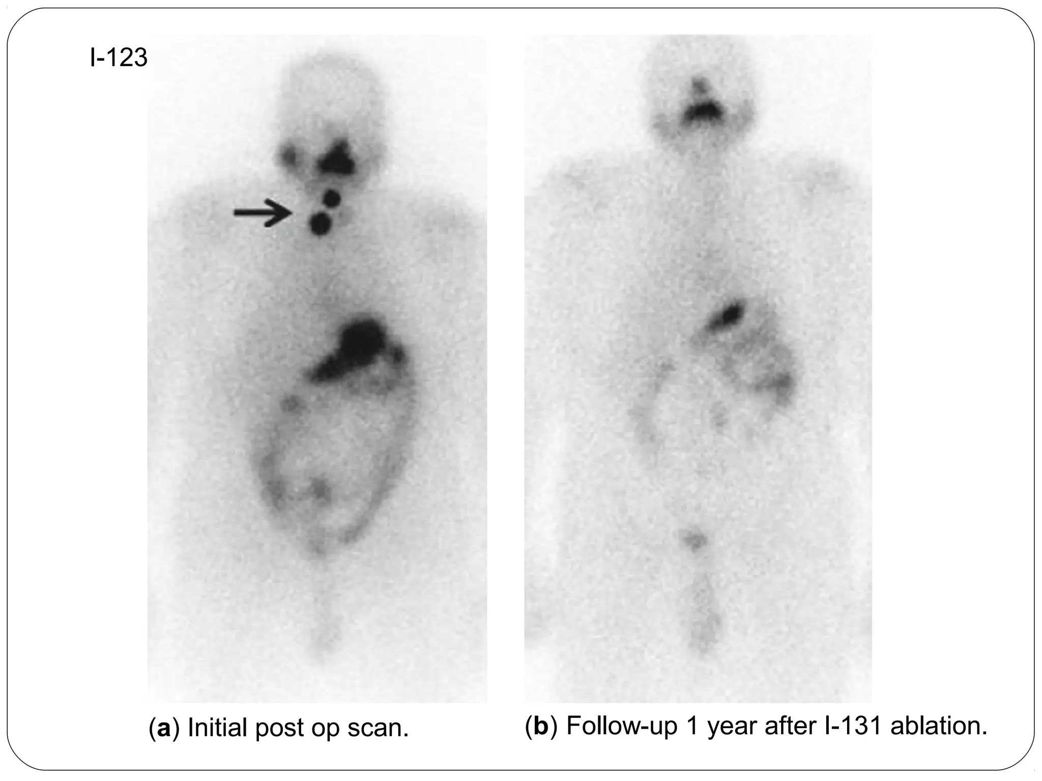
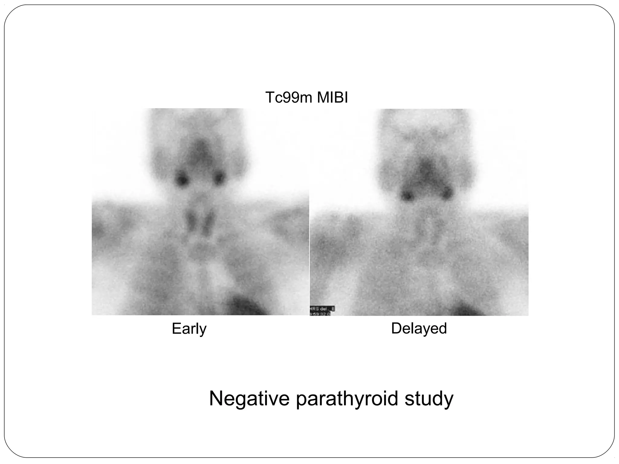
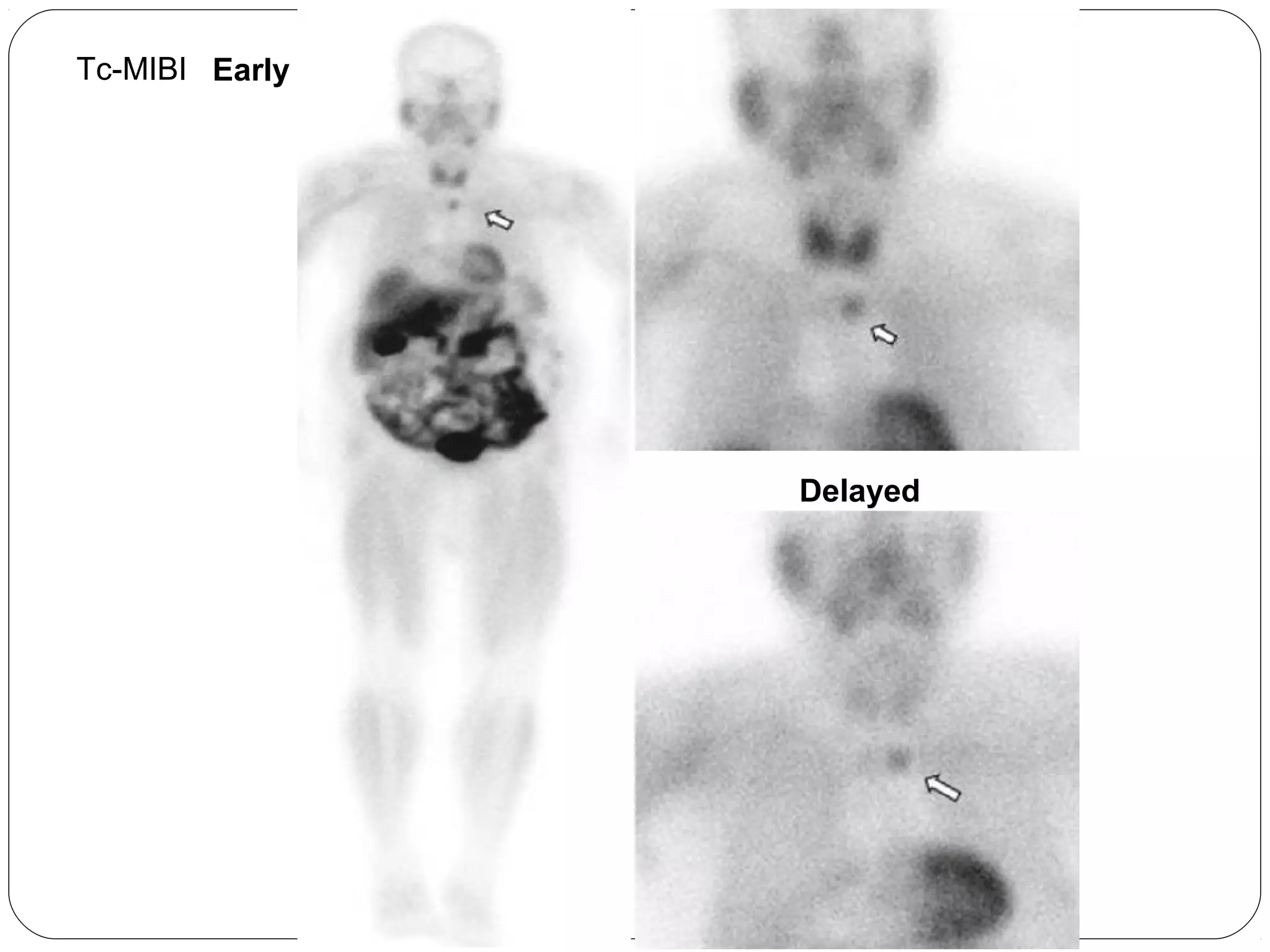
RADIONUCLIDE IMAGING OF 
THYROID AND PARATHYROID
THYROID SCAN 
A radionuclide thyroid scan is routinely used for 
the diagnosis of and guiding the management of 
many thyroid conditions.
RADIONUCLIDES 
I-123 is the most favorable because of its excellent 
image quality and relatively low radiation exposure. 
 I-131 is usually used for therapeutic purposes because 
of its beta emission and higher radiation dose.
Technetium-99m pertechnetate is used as a substitute 
for iodine because it is: 
Trapped by the thyroid 
Low radiation dose 
Readily available and less time consuming 
Inexpensive
INDICATIONS 
Thyrotoxicosis 
Thyroid goiter /nodules 
Ectopic thyroid 
Diagnosis and follow up of Thyroiditis.
 Detection and follow up of thyroid cancer recurrences or 
metastases. 
 Suspected Occult thyroid malignancy. 
 Evaluation of congenital thyroid abnormalities.
PREPERATION 
 Thyroid supplements and exogenous iodine 
source should be stopped. 
 Use of radiographic contrast media may 
depress the uptake of iodine by the thyroid for 
up to a month, so imaging studies should be 
scheduled accordingly.
Antithyroid drugs need not be stopped, as they do not interfere 
with the uptake of iodide, only with its organification.
TECHNIQUE 
I-123 labeled sodium iodide 
or 
Tc-99m sodium pertechnetate 
is given intravenously. 
With I-123 images are obtained 2-3hrs later, while with Tc-99m 
images are obtained 20-30 minutes post injection, using a high 
resolution or a pinhole collimator.
 Markers indicating the position of thyroid cartilage and sternal 
notch are helpful. 
 Additional markers may be used to indicate the site of palpable 
nodules.
 Usually anterior and oblique views are taken. 
 Lateral view of the neck should be added if ectopic thyroid is 
suspected.
THYROID UPTAKE MEASUREMENT 
Thyroid uptake indicates the level of functional activity of the 
gland by measuring the trapped proportion of ingested 
radioiodine at a certain time (2, 4 and/or 24 hrs) using a special 
probe.
Normal thyroid uptake is 10–35% in most laboratories at 4 
and 24 h
Normal thyroid uptake of the gland
24-year-old woman with raised T4, T3 and low TSH values. The 
24-hour RAIU was 84%.
Anterior Anterior with marker 
RAO LAO 
Grave’s disease
Thyroid images should be interpreted in association with clinical 
and lab data (TFTs) as well as the result of thyroid uptake 
especially in case of hyperthyroidism due to Grave’s disease since 
near normal images can be present in this condition.
Autonomous nodules
Tc99m Pertechnetate 
Anterior RAO LAO 
Multinodular gland
59-year-old woman with a very firm thyroid on examination. Lab 
data shows low T3 and T4 .The 24-hour RAIU was 7%.
ABSENT / REDUCED THYROID UPTAKE 
GENERALIZED 
o Sub acute thyroiditis 
o Ectopic thyroid 
o Ectopic hormone production 
o Thyroid supplements or exogenous iiooddiinnee ssoouurrccee
LOCALIZED 
o Colloid cyst 
o Non-functioning adenoma 
o Multinodular goiter 
o Carcinoma 
o Local thyroiditis
(A) (B) (C)
 “Hot” nodules (autonomously functioning thyroid nodules) are 
usually not malignant. 
 “Cold” nodules ( either hypo functioning or nonfunctioning) can 
be malignant in approximately 5-8% of cases.
ECTOPIC THYROID 
 Most commonly presents in childhood as a nodule or mass at the 
base of the tongue. 
 Retrosternal thyroids are usually extensions of a multinodular 
goiter in the neck.
 For imaging I-123 is preferable to Tc-99m because it shows 
greater uptake in the thyroid tissue compared with salivary glands 
and mediastinum.
A 20-year-old female presented with a submandibular neck swelling. Her 
biochemical profile was suggestive of hypothyroidism.
PERCHLORATE DISCHARGE TEST 
 Disorders characterised by a failure to organify the trapped 
iodide(of which the most common is Pendred's syndrome). 
 Serial images of thyroid are taken giving first I-123 and 2hrs later 
sodium or potassium perchlorate which rapidly discharges the 
unbound iodide.
The normal gland will retain that proportion of the iodide 
which has been bound, whereas with an organification 
defect most of the thyroid activity will be eliminated.
THYROID CANCER 
Well differentiated papillary and follicular tumors retain 
the ability to accumulate iodine although to a much 
lesser extent than normal thyroid tissue. 
 This property is used in the detection of local tumor 
recurrence as well as distant metastatic spread of thyroid 
cancer after surgery by the whole body iodine scan.
Post operative I-131 
scan 
Anterior
Post-op I-123 
scan 
Anterior 
Residual neck thyroid tissue
Anterior Posterior 
I-123 
whole body scan
I-123 
(a) Initial post op scan. (b) Follow-up 1 year after I-131 ablation.
Other imaging studies are used particularly when 
I-123 or 131 study is negative such as thallium 201 
and particularly in high-risk patients FDG-PET/CT 
study.
PARTATHYROID SCAN 
 Scintigraphy using Tc-99m MIBI(methoxy isobutyl-isonitrile) is 
currently the preferred nuclear medicine method for 
preoperative localization of hyperfunctioning parathyroid tissue. 
 it reduces operative time, cost, and operative failure rates.
 Normally Tc-99m MIBI is taken by thyroid gland and it clears 
over time. 
 In the presence of abnormal parathyroid glands the radiotracer is 
retained in these glands and are seen as foci of tracer 
accumulation.
Tc99m MIBI 
Early Delayed 
Negative parathyroid study
Tc99m MIBI Tc99m Pertechnetate
Early 
Early Delayed 
Tc-MIBI
THANK YOU

Radionuclide imaging thyroid & parathyroid

  • 1.
    RADIONUCLIDE IMAGING OF THYROID AND PARATHYROID
  • 2.
    THYROID SCAN Aradionuclide thyroid scan is routinely used for the diagnosis of and guiding the management of many thyroid conditions.
  • 3.
    RADIONUCLIDES I-123 isthe most favorable because of its excellent image quality and relatively low radiation exposure.  I-131 is usually used for therapeutic purposes because of its beta emission and higher radiation dose.
  • 4.
    Technetium-99m pertechnetate isused as a substitute for iodine because it is: Trapped by the thyroid Low radiation dose Readily available and less time consuming Inexpensive
  • 5.
    INDICATIONS Thyrotoxicosis Thyroidgoiter /nodules Ectopic thyroid Diagnosis and follow up of Thyroiditis.
  • 6.
     Detection andfollow up of thyroid cancer recurrences or metastases.  Suspected Occult thyroid malignancy.  Evaluation of congenital thyroid abnormalities.
  • 7.
    PREPERATION  Thyroidsupplements and exogenous iodine source should be stopped.  Use of radiographic contrast media may depress the uptake of iodine by the thyroid for up to a month, so imaging studies should be scheduled accordingly.
  • 8.
    Antithyroid drugs neednot be stopped, as they do not interfere with the uptake of iodide, only with its organification.
  • 9.
    TECHNIQUE I-123 labeledsodium iodide or Tc-99m sodium pertechnetate is given intravenously. With I-123 images are obtained 2-3hrs later, while with Tc-99m images are obtained 20-30 minutes post injection, using a high resolution or a pinhole collimator.
  • 11.
     Markers indicatingthe position of thyroid cartilage and sternal notch are helpful.  Additional markers may be used to indicate the site of palpable nodules.
  • 12.
     Usually anteriorand oblique views are taken.  Lateral view of the neck should be added if ectopic thyroid is suspected.
  • 13.
    THYROID UPTAKE MEASUREMENT Thyroid uptake indicates the level of functional activity of the gland by measuring the trapped proportion of ingested radioiodine at a certain time (2, 4 and/or 24 hrs) using a special probe.
  • 15.
    Normal thyroid uptakeis 10–35% in most laboratories at 4 and 24 h
  • 17.
  • 18.
    24-year-old woman withraised T4, T3 and low TSH values. The 24-hour RAIU was 84%.
  • 19.
    Anterior Anterior withmarker RAO LAO Grave’s disease
  • 20.
    Thyroid images shouldbe interpreted in association with clinical and lab data (TFTs) as well as the result of thyroid uptake especially in case of hyperthyroidism due to Grave’s disease since near normal images can be present in this condition.
  • 21.
  • 22.
    Tc99m Pertechnetate AnteriorRAO LAO Multinodular gland
  • 23.
    59-year-old woman witha very firm thyroid on examination. Lab data shows low T3 and T4 .The 24-hour RAIU was 7%.
  • 26.
    ABSENT / REDUCEDTHYROID UPTAKE GENERALIZED o Sub acute thyroiditis o Ectopic thyroid o Ectopic hormone production o Thyroid supplements or exogenous iiooddiinnee ssoouurrccee
  • 27.
    LOCALIZED o Colloidcyst o Non-functioning adenoma o Multinodular goiter o Carcinoma o Local thyroiditis
  • 28.
  • 29.
     “Hot” nodules(autonomously functioning thyroid nodules) are usually not malignant.  “Cold” nodules ( either hypo functioning or nonfunctioning) can be malignant in approximately 5-8% of cases.
  • 31.
    ECTOPIC THYROID Most commonly presents in childhood as a nodule or mass at the base of the tongue.  Retrosternal thyroids are usually extensions of a multinodular goiter in the neck.
  • 32.
     For imagingI-123 is preferable to Tc-99m because it shows greater uptake in the thyroid tissue compared with salivary glands and mediastinum.
  • 34.
    A 20-year-old femalepresented with a submandibular neck swelling. Her biochemical profile was suggestive of hypothyroidism.
  • 35.
    PERCHLORATE DISCHARGE TEST  Disorders characterised by a failure to organify the trapped iodide(of which the most common is Pendred's syndrome).  Serial images of thyroid are taken giving first I-123 and 2hrs later sodium or potassium perchlorate which rapidly discharges the unbound iodide.
  • 36.
    The normal glandwill retain that proportion of the iodide which has been bound, whereas with an organification defect most of the thyroid activity will be eliminated.
  • 37.
    THYROID CANCER Welldifferentiated papillary and follicular tumors retain the ability to accumulate iodine although to a much lesser extent than normal thyroid tissue.  This property is used in the detection of local tumor recurrence as well as distant metastatic spread of thyroid cancer after surgery by the whole body iodine scan.
  • 38.
    Post operative I-131 scan Anterior
  • 39.
    Post-op I-123 scan Anterior Residual neck thyroid tissue
  • 40.
    Anterior Posterior I-123 whole body scan
  • 41.
    I-123 (a) Initialpost op scan. (b) Follow-up 1 year after I-131 ablation.
  • 42.
    Other imaging studiesare used particularly when I-123 or 131 study is negative such as thallium 201 and particularly in high-risk patients FDG-PET/CT study.
  • 44.
    PARTATHYROID SCAN Scintigraphy using Tc-99m MIBI(methoxy isobutyl-isonitrile) is currently the preferred nuclear medicine method for preoperative localization of hyperfunctioning parathyroid tissue.  it reduces operative time, cost, and operative failure rates.
  • 45.
     Normally Tc-99mMIBI is taken by thyroid gland and it clears over time.  In the presence of abnormal parathyroid glands the radiotracer is retained in these glands and are seen as foci of tracer accumulation.
  • 46.
    Tc99m MIBI EarlyDelayed Negative parathyroid study
  • 47.
    Tc99m MIBI Tc99mPertechnetate
  • 48.
  • 49.

Editor's Notes

  • #15 Thyroid probe for radioiodine uptake study
  • #18 Thyroid scintigraphy. Normal thyroid uptake of the gland. 99 `TcO4
  • #19  Graves disease in a 24-year-old woman. Laboratory values were as follows: T4 = 16.7 μg/dL, T3 = 311 ng/dL, and TSH < 0.01 μIU/mL. The 24-hour RAIU was 84%. Anterior distant image obtained with Tc-99m pertechnetate shows an enlarged thyroid. The target-to-background activity is increased to such an extent that the submandibular salivary glands (arrowhead) are barely visualized. Note the appearance of the pyramidal lobe (large arrow). The round photopenic area (small arrow) in this and subsequent figures represents the 2-cm lead marker placed at the suprasternal notch.
  • #20 Typical pattern of Grave’s disease with uniform gland uptake and decreased background activity in the surrounding soft tissue
  • #22 Autonomous nodules. patients with solitary 'hot‘ nodules of overactive thyroid tissue and relative suppression of the remainders
  • #23 Multinodular gland
  • #24 Late-stage Hashimoto thyroiditis
  • #25 Scintigraphic pattern of thyroiditis where poor uptake and lack of delineation of thyroid gland borders are the typical features
  • #29 Solitary 'cold' nodules in three patients subjected to FNA. (A) Benign non-functioning adenoma (arrow); (B) carcinoma (arrows); (C) chronic thyroiditis affecting only the right lobe.
  • #31 Thyroid scan and CT image (sagittal section) showing dual ectopia - sublingual and suprahyoid - with absence of normal thyroid
  • #34 The patient with hemiagenesis presented with a neck swelling and a normal thyroid profile. The thyroid scan revealed normal tracer uptake in the right lobe of the thyroid and absent tracer uptake in the region of the left lobe of the thyroid. CT images confirmed the finding
  • #35 The case of triple ectopia was a 20-year-old female who presented with a submandibular neck swelling. Her biochemical profile was suggestive of subclinical hypothyroidism. The thyroid scan revealed three areas of abnormal tracer uptake in the region of the base of the tongue and the suprahyoid and the subhyoid locations. CT images showed hyperdense soft tissue in the region of base of tongue and hyperdense tissue with cystic degeneration in suprahyoid and subhyoid locations
  • #36 Imaging does not discriminate between iodide that is trapped in the thyroid and that which has been both trapped and organified.
  • #39 Postoperative I-131 whole-body study with no functioning thyroid tissue in the neck or the rest of the body. Note the physiologic uptake in the salivary glands, stomach, and urinary bladder
  • #40 I-123 24-h whole-body scan following surgical removal of thyroid gland for differentiated carcinoma. Residual neck thyroid tissue with or without residual tumor is evident (arrow)
  • #41 Anterior and posterior views of an I-123 whole body scan of a patient with thyroid cancer after surgical resection who is now presenting for I-131 therapy. The scan shows multiple foci of intense activity in the neck, mediastinum, thorax, abdomen, and right humerus. These findings suggest widespread disease with bone involvement
  • #42 Initial (a) and follow-up (b) I-123 24-h whole-body scans showing resolution of the neck activity (arrow) 1 year after I-131 postoperative ablation
  • #44 image of an F-18 FDG PET/CT study of a patient with differentiated thyroid cancer showing residual tissue in the nick (arrow head)
  • #47 Negative parathyroid study
  • #48 Parathyroid adenoma. 99 "'Tc-MIBI images at 10 min (A) and 3 h (B) showing a persistent focus of activity inferior to the right lobe of the thyroid; image (C) shows normal thyroid uptake
  • #49 Ectopic adenomas are usually sited in the superior mediastinum, often adjacent to the aortic arch