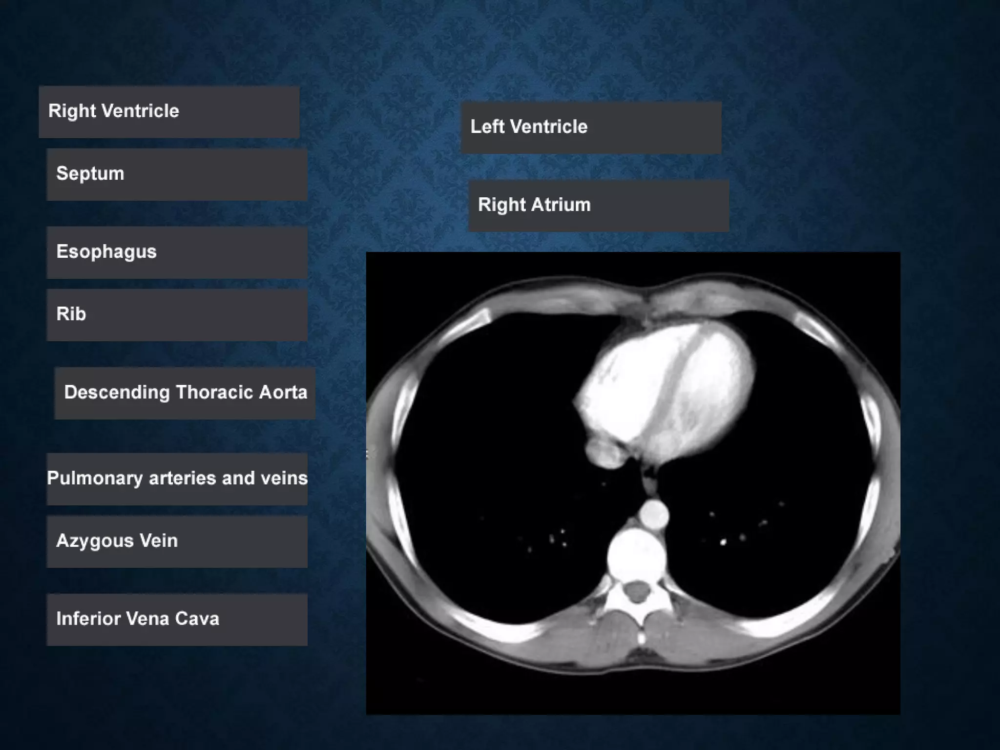
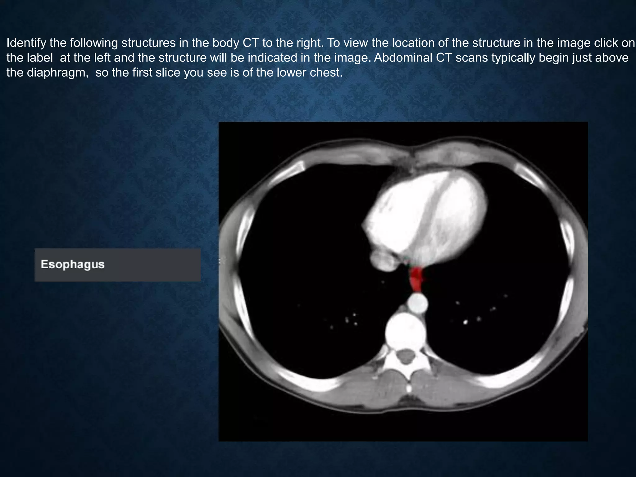
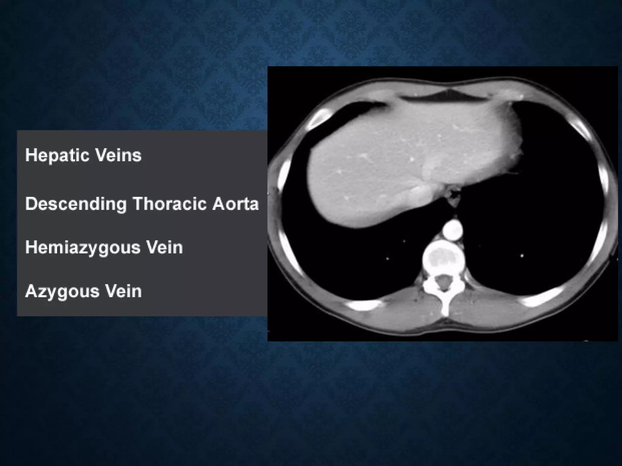
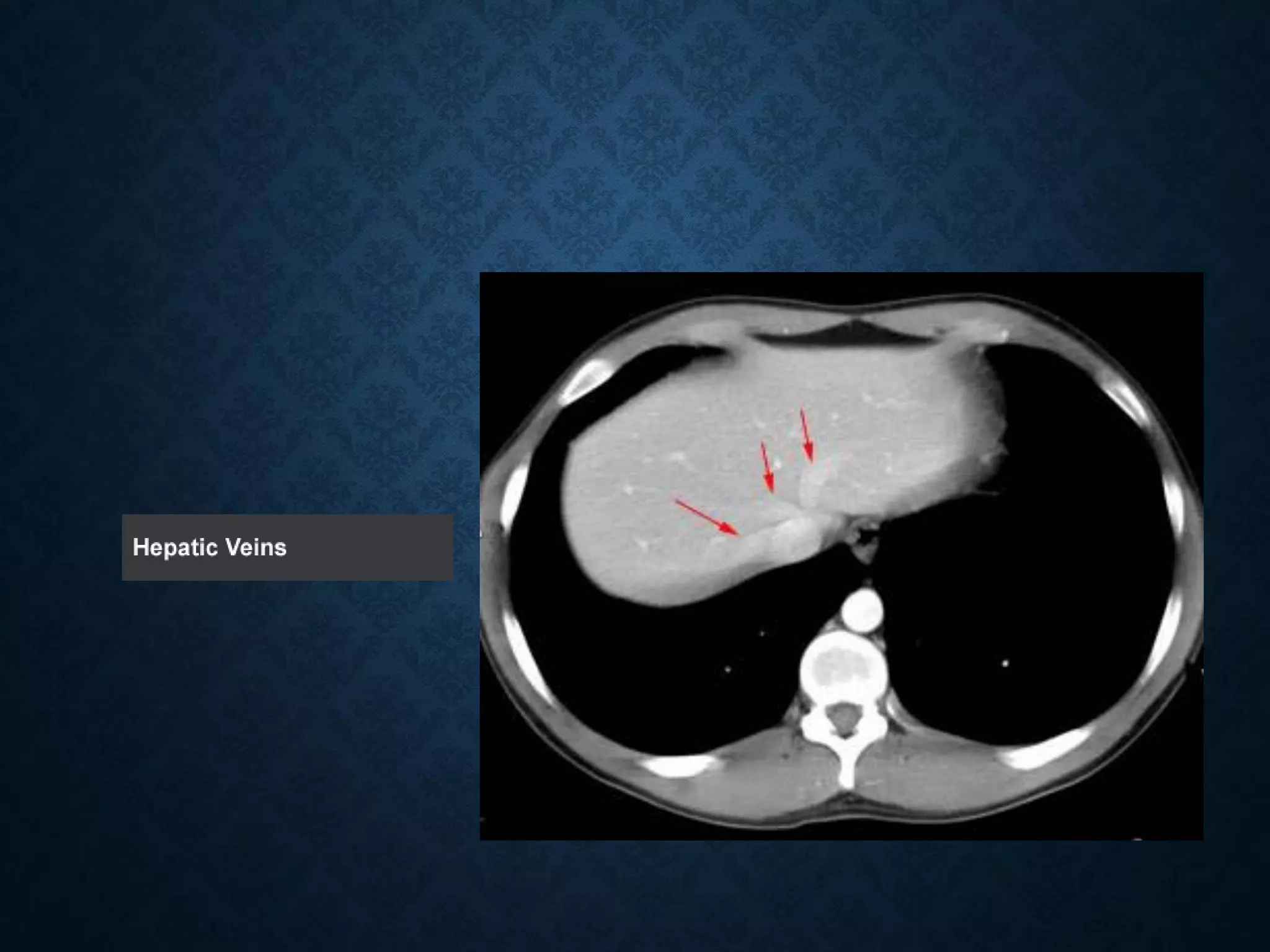
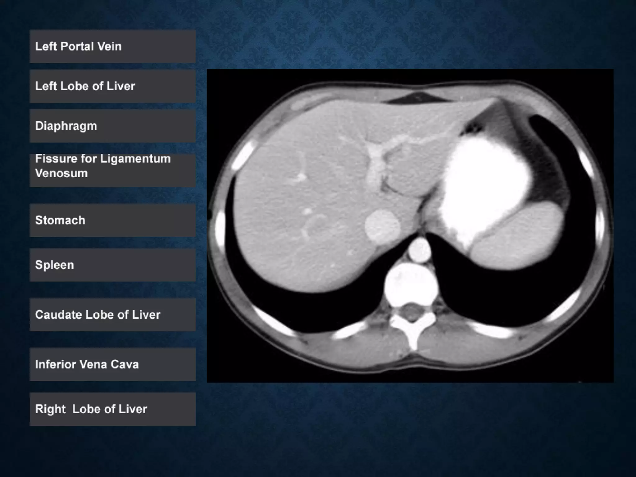
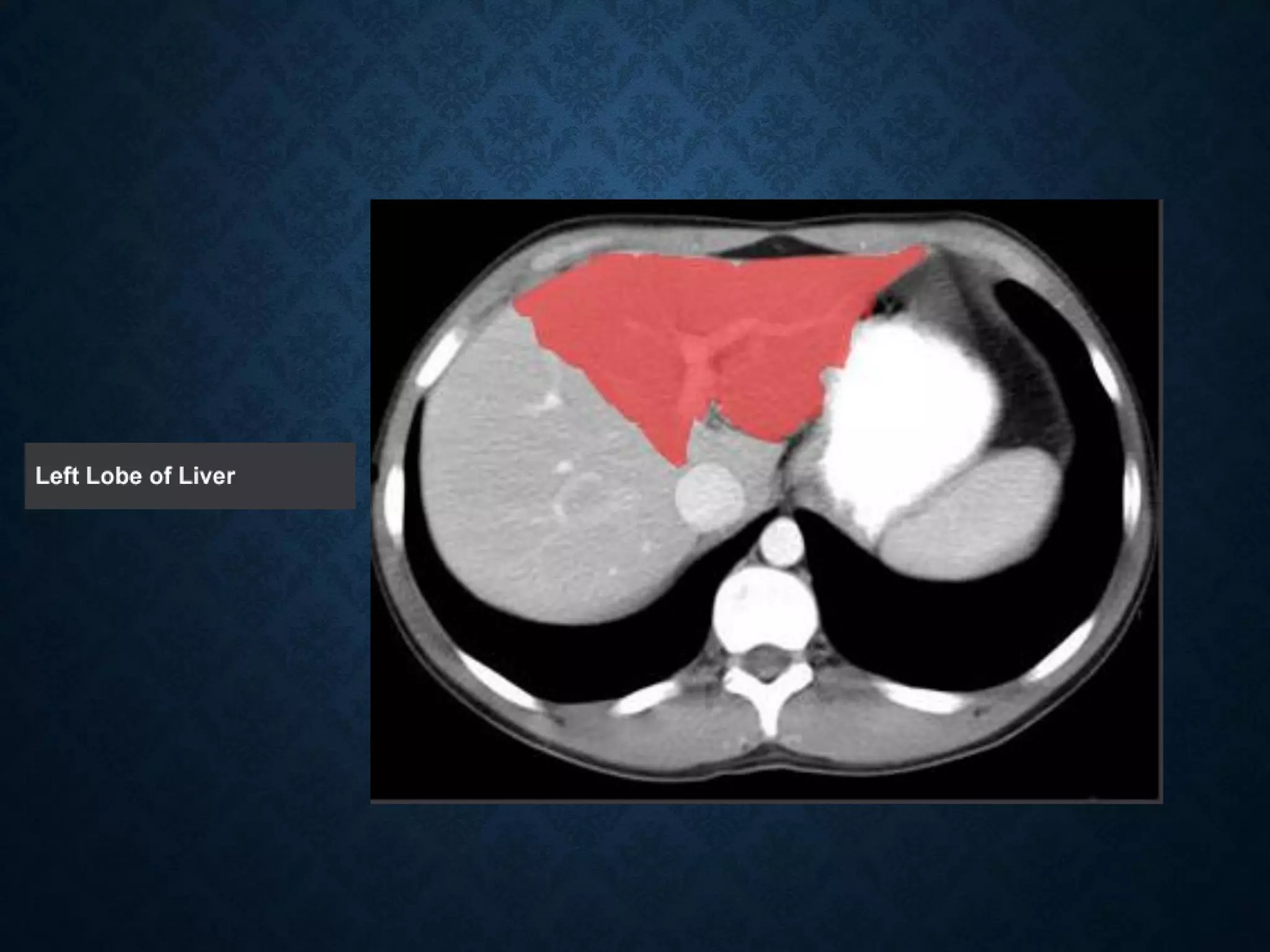
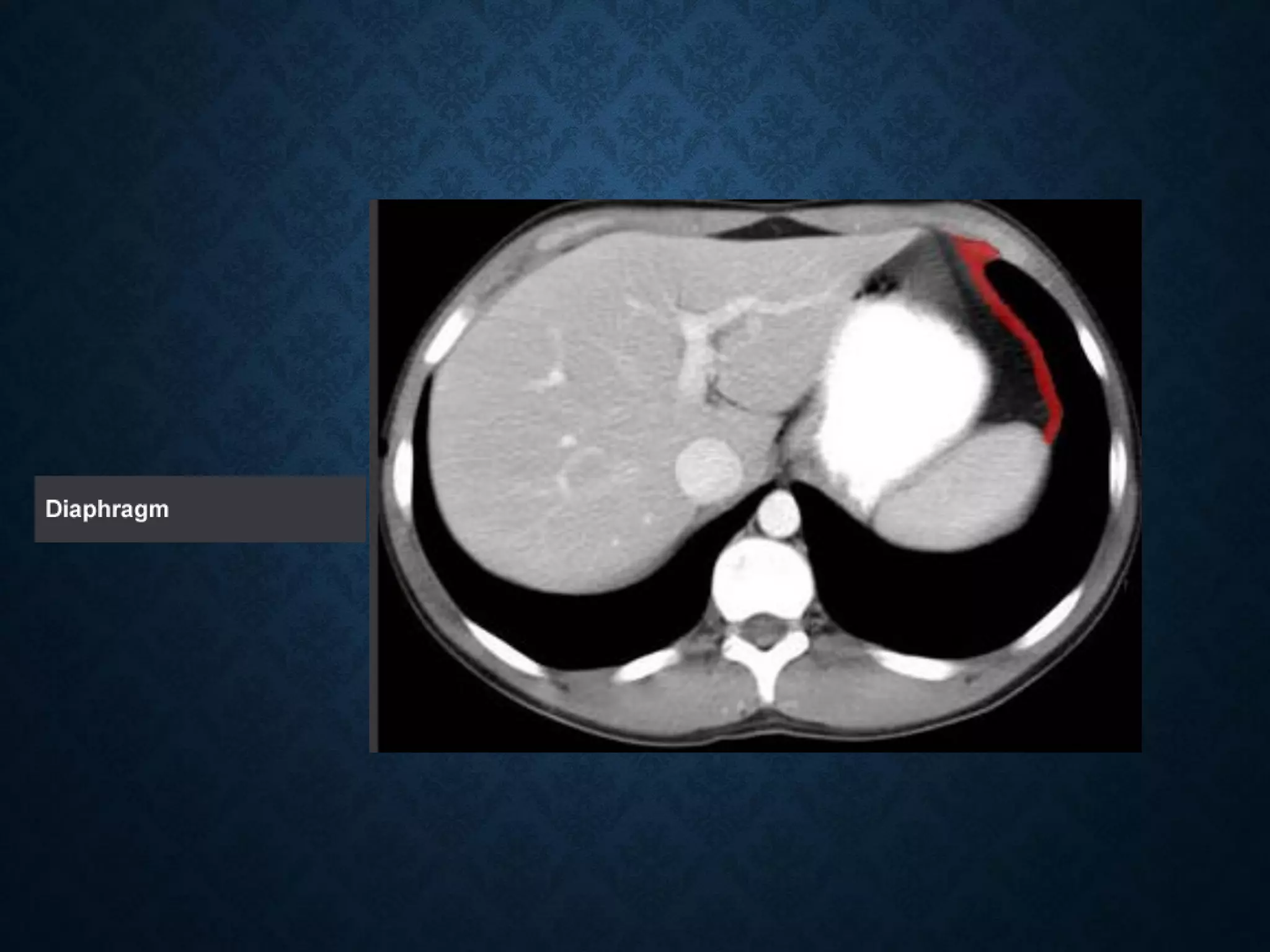
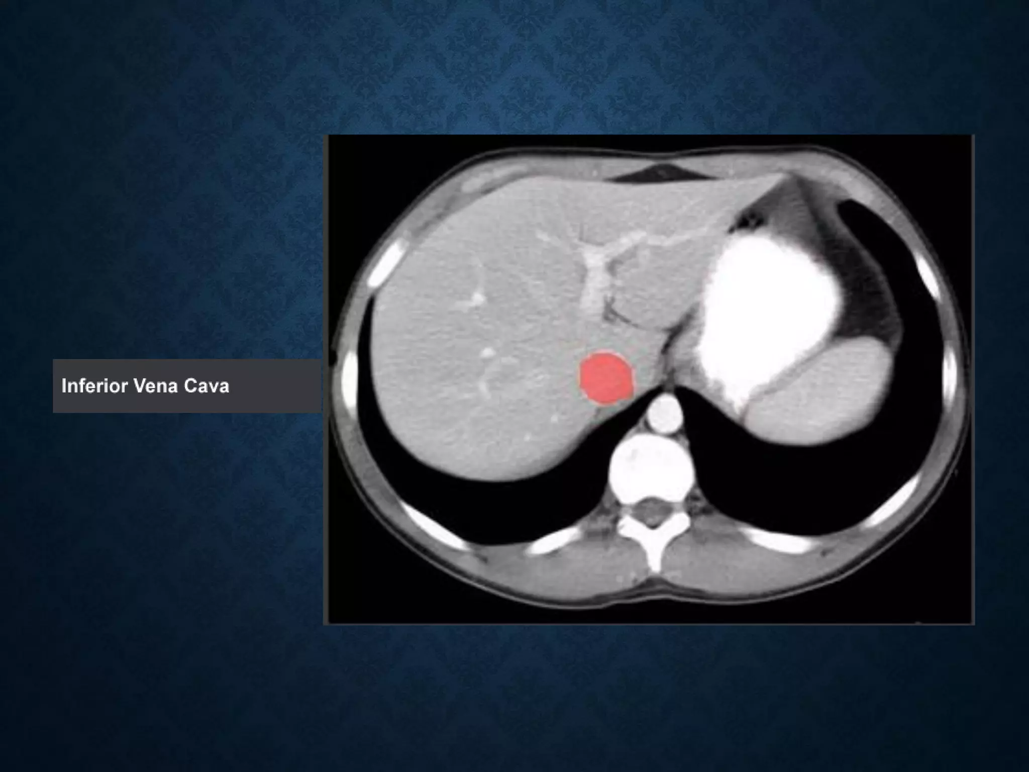
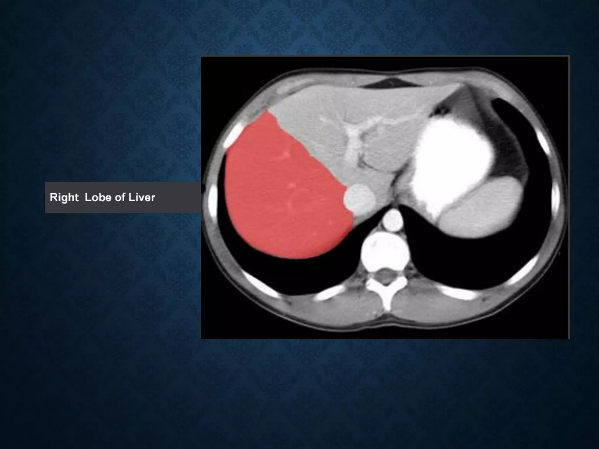
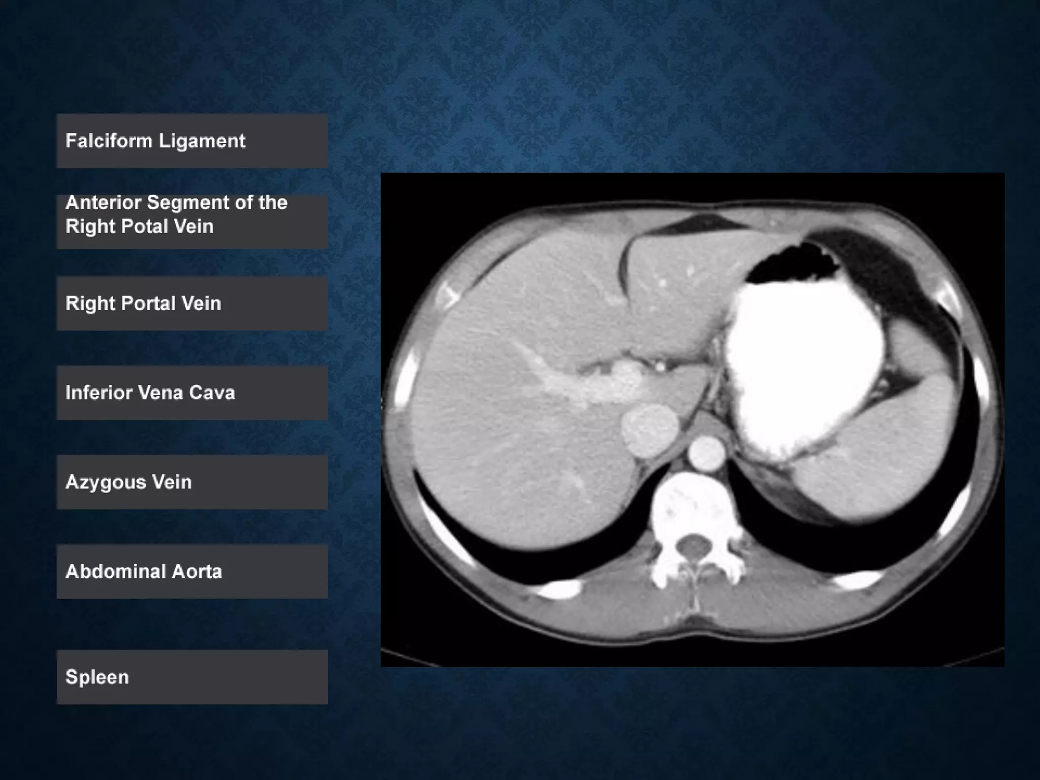
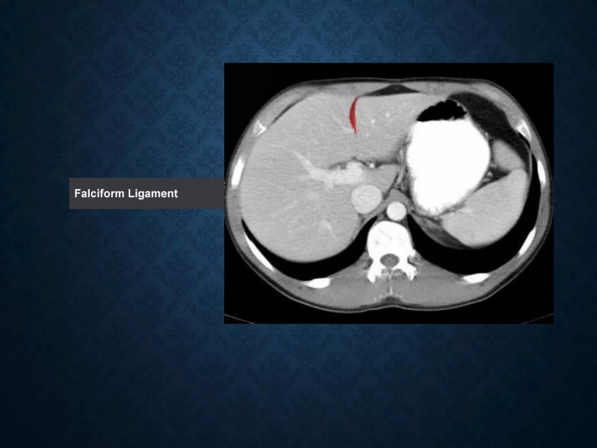
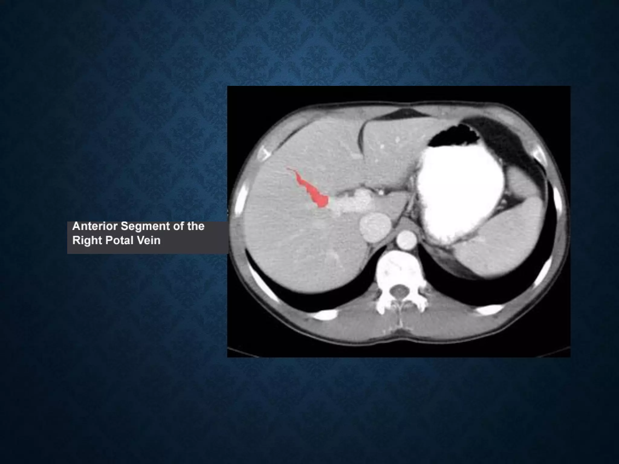
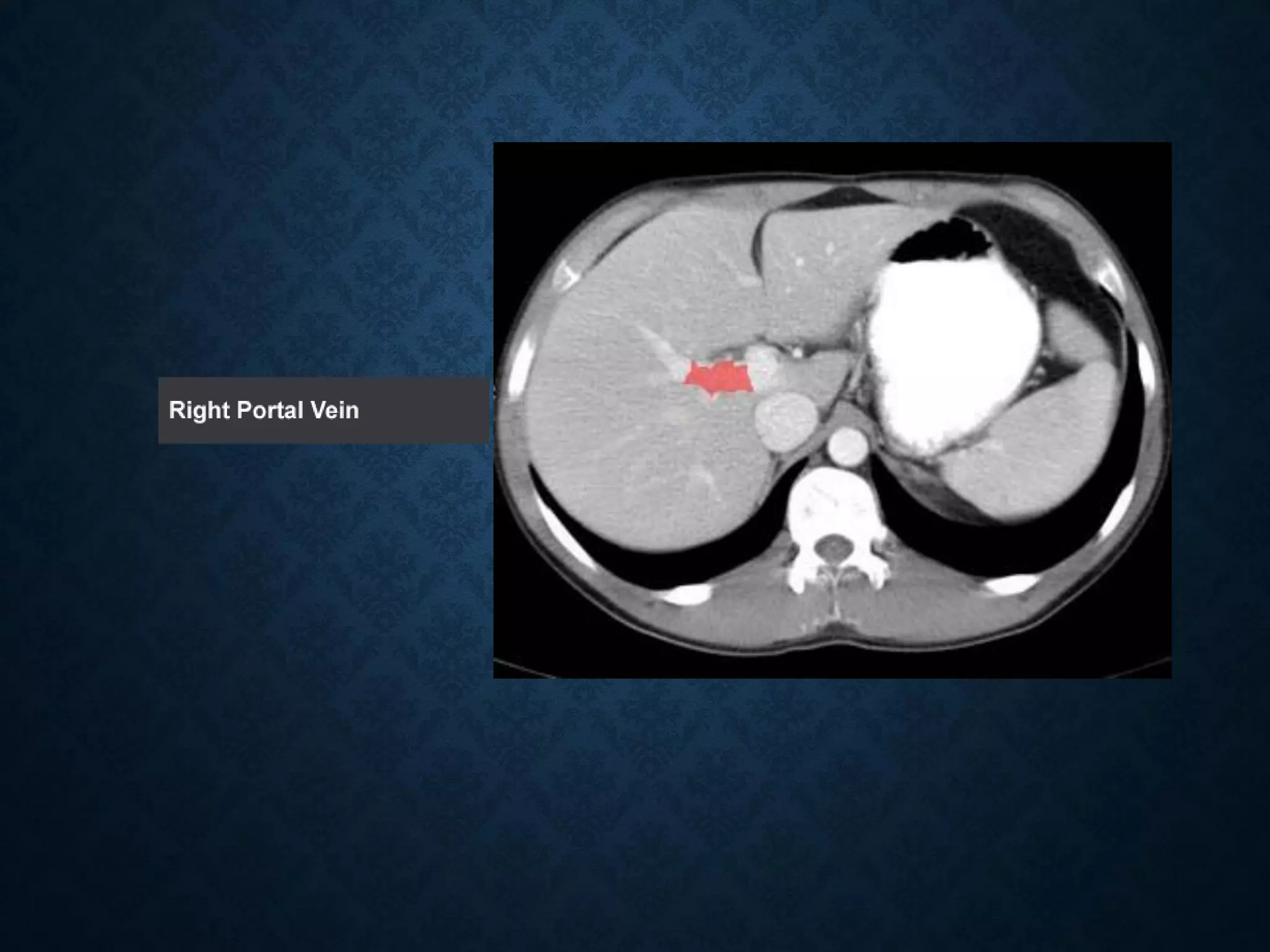
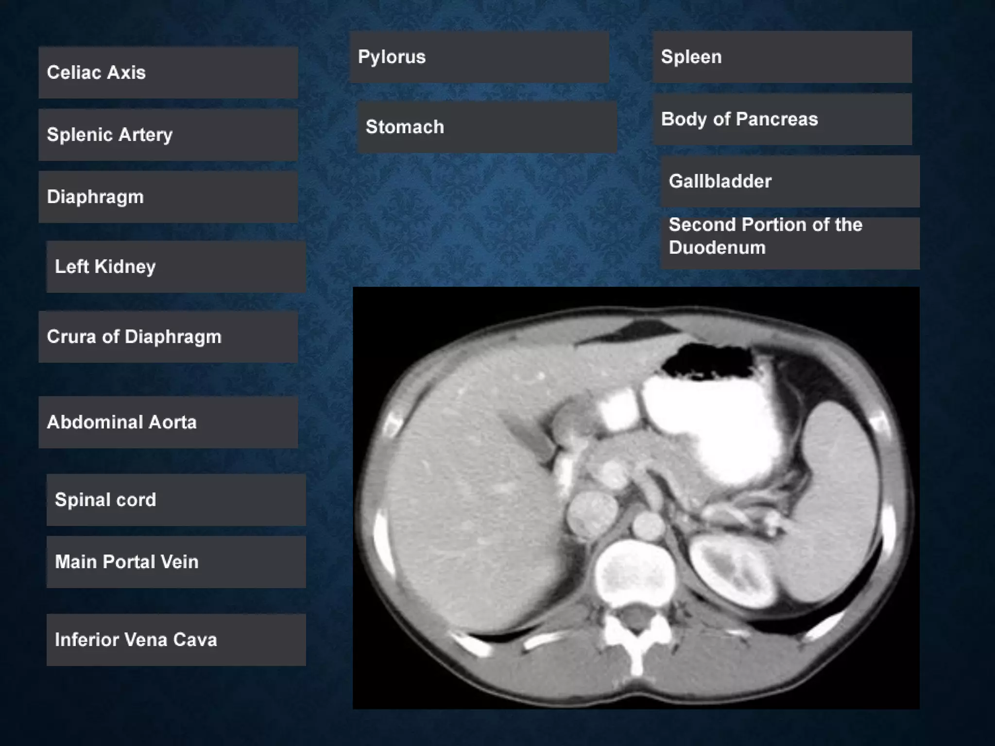
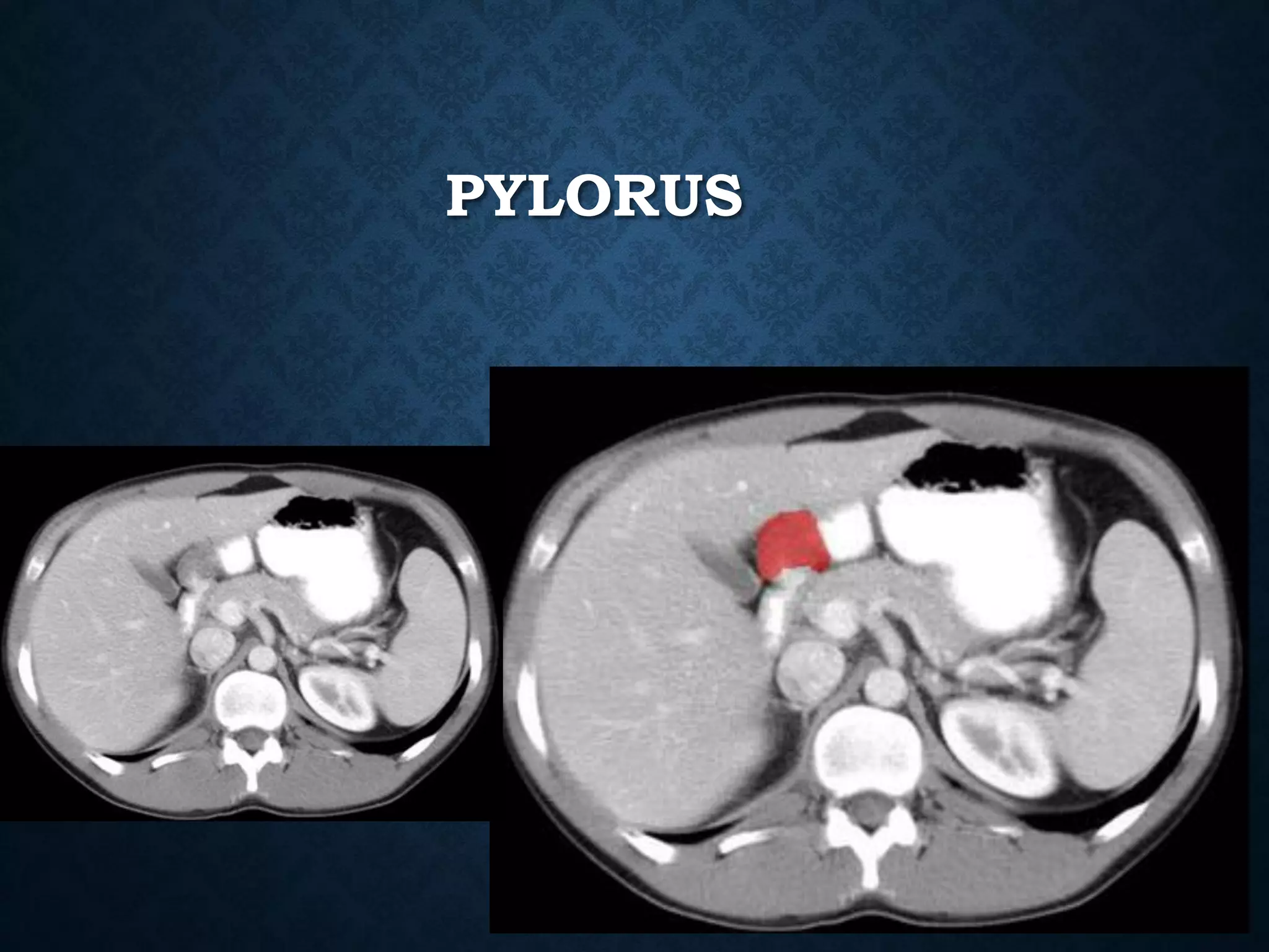
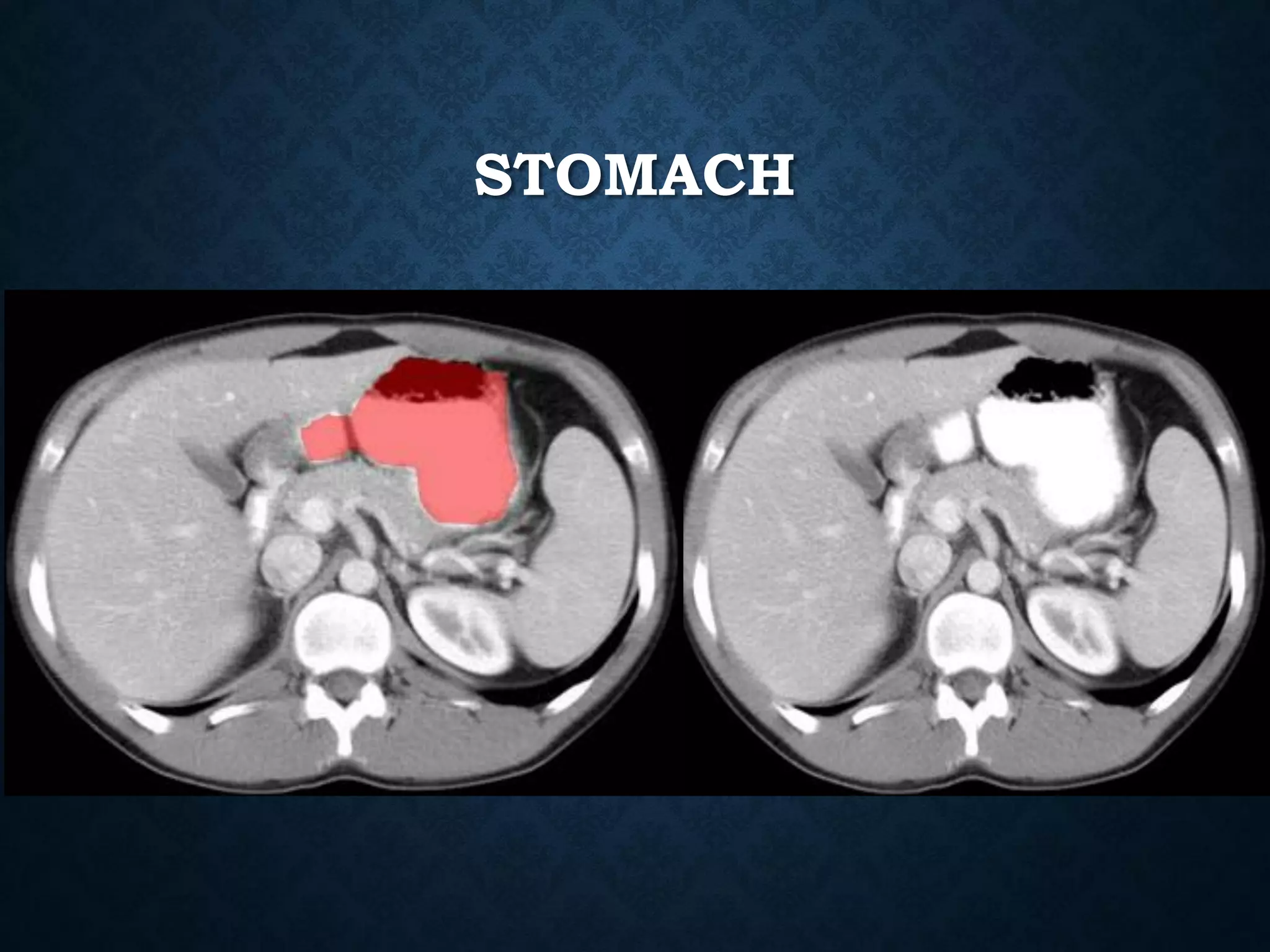
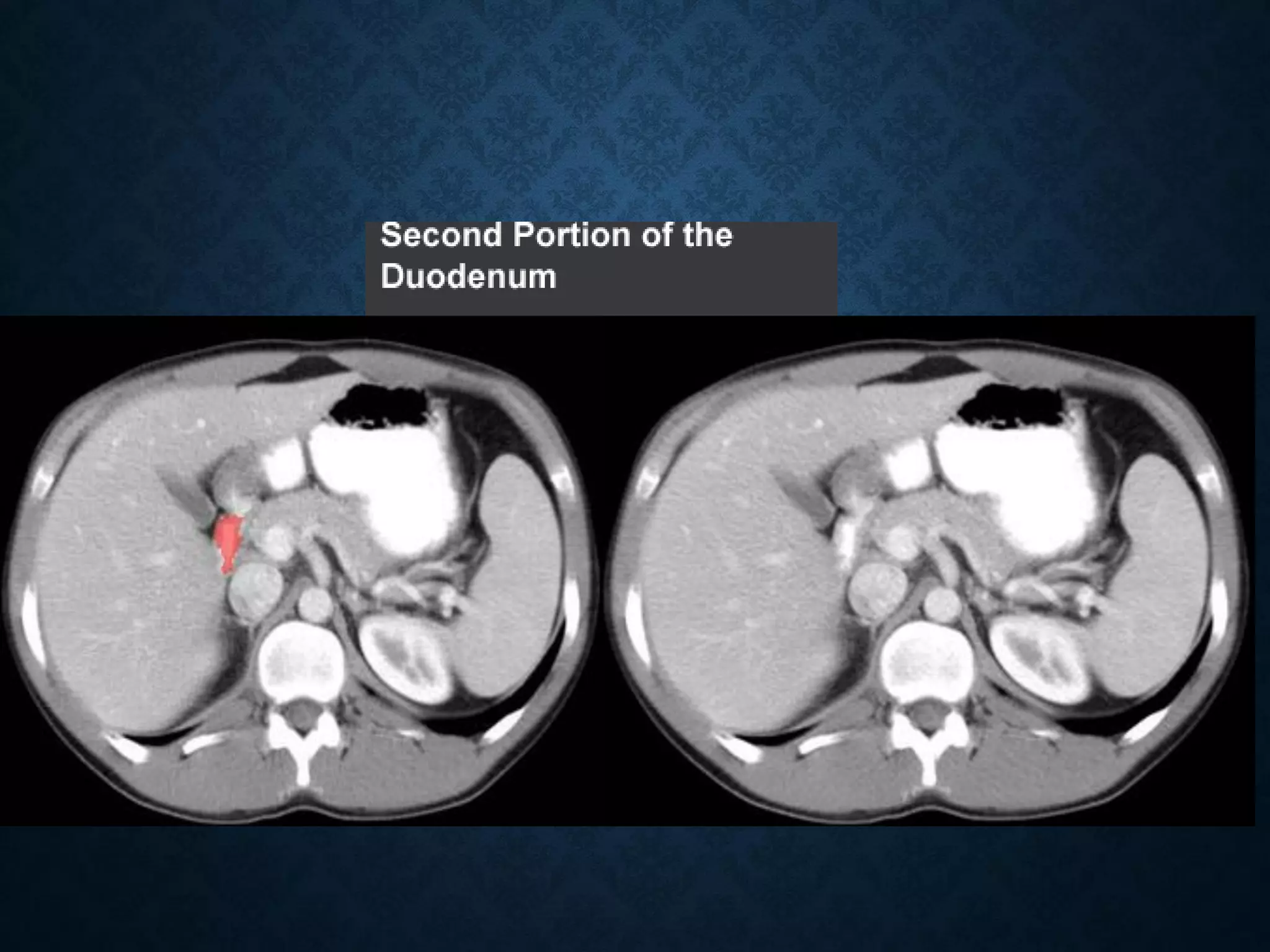
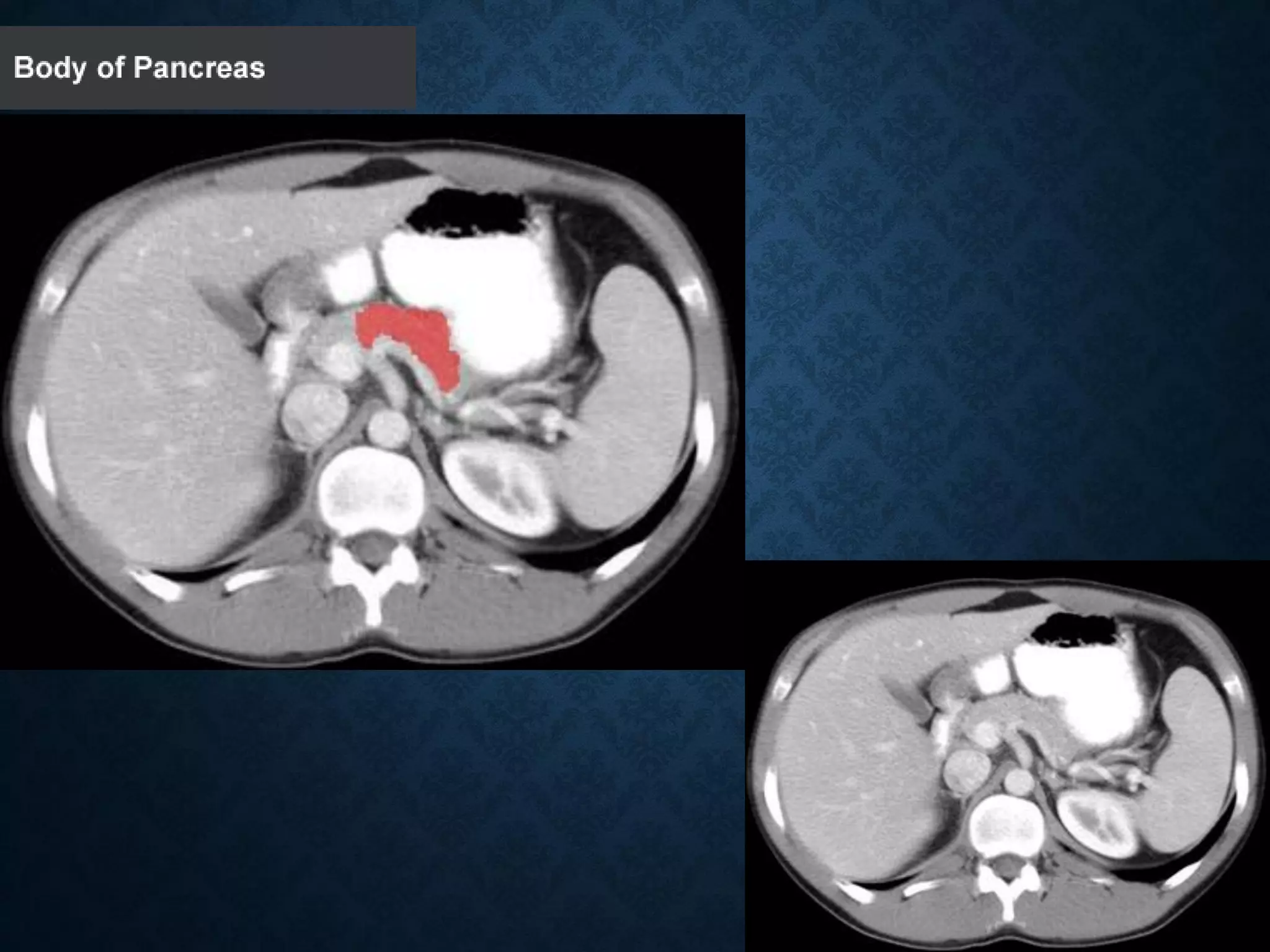
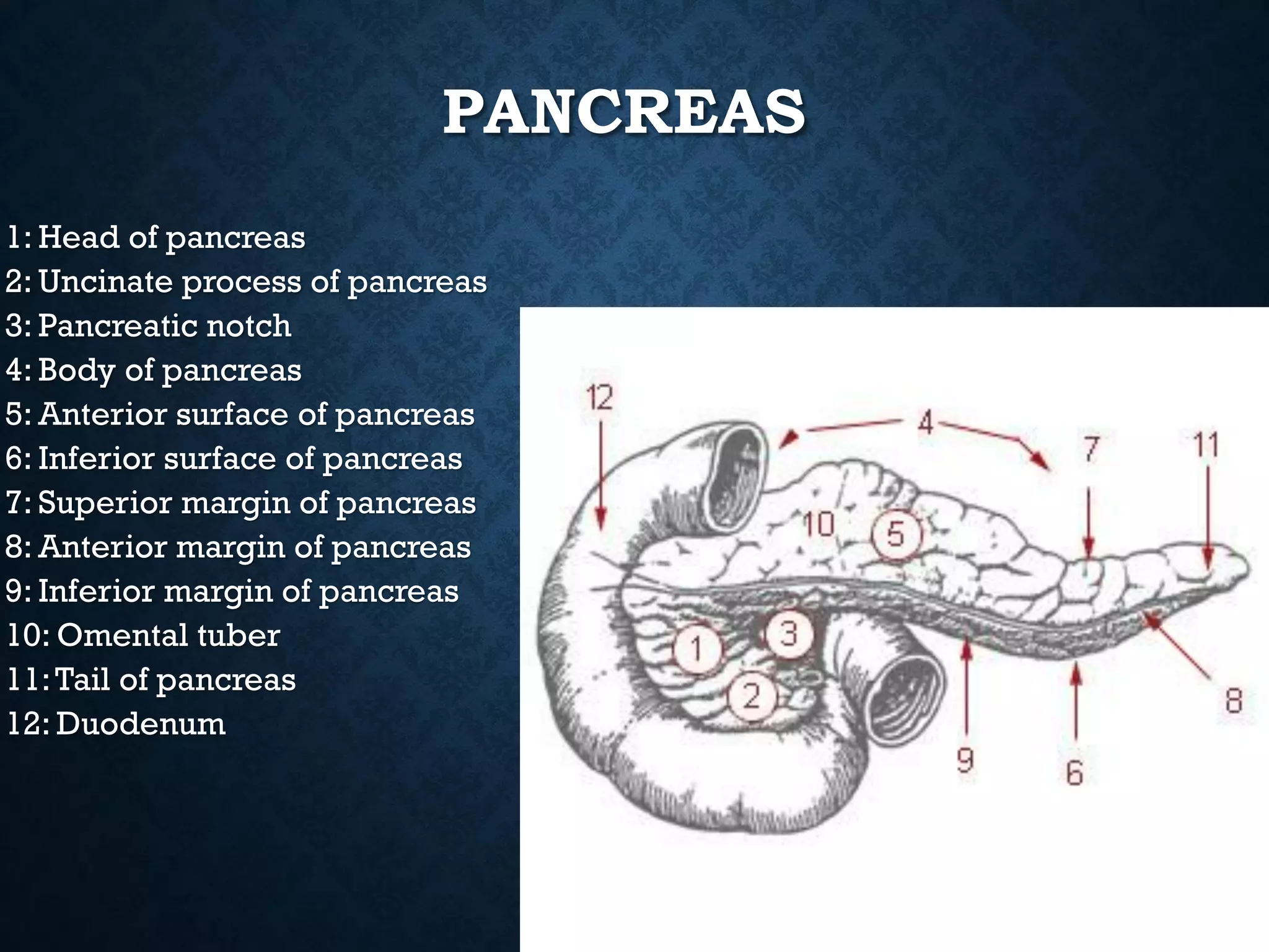
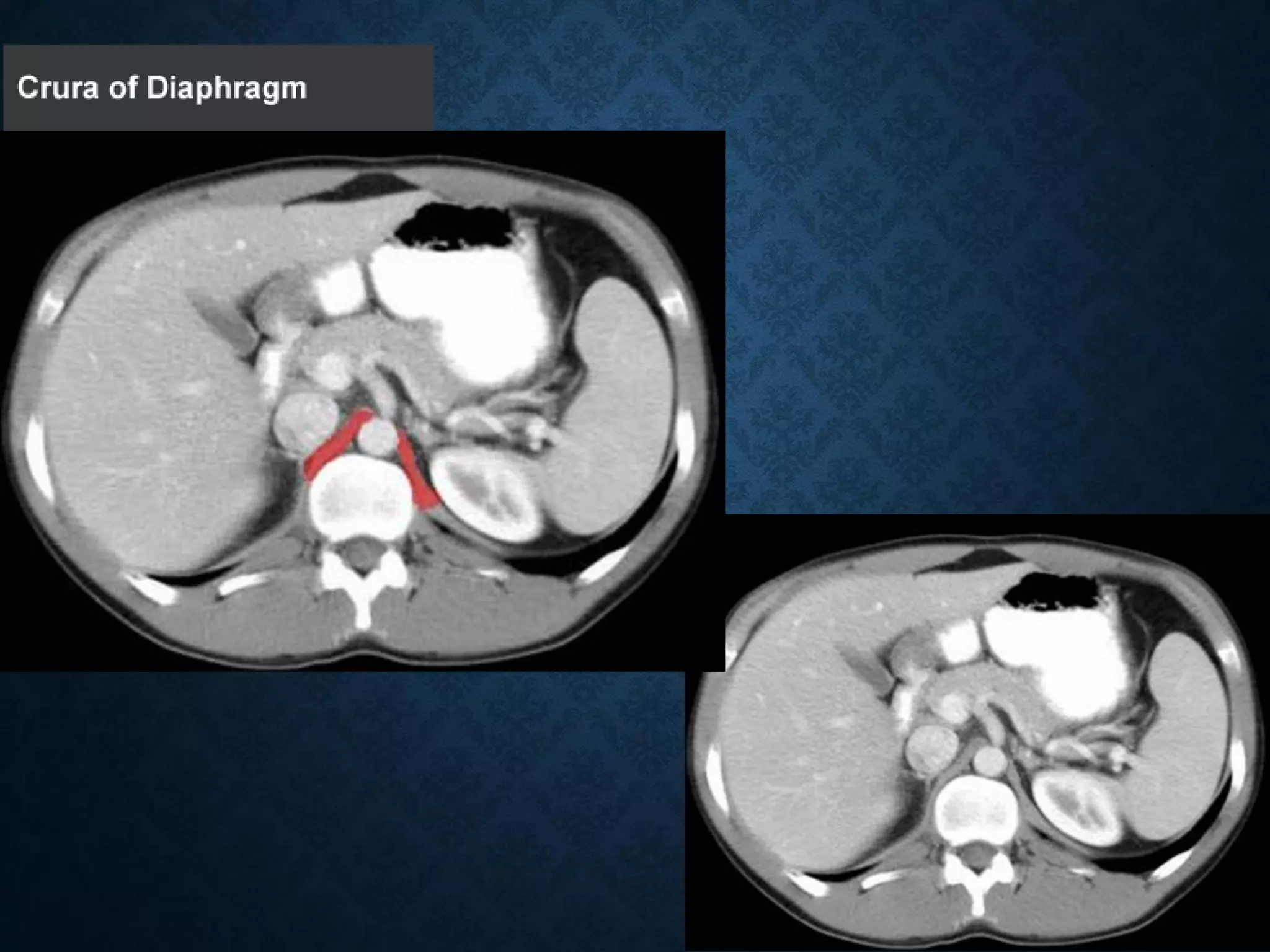
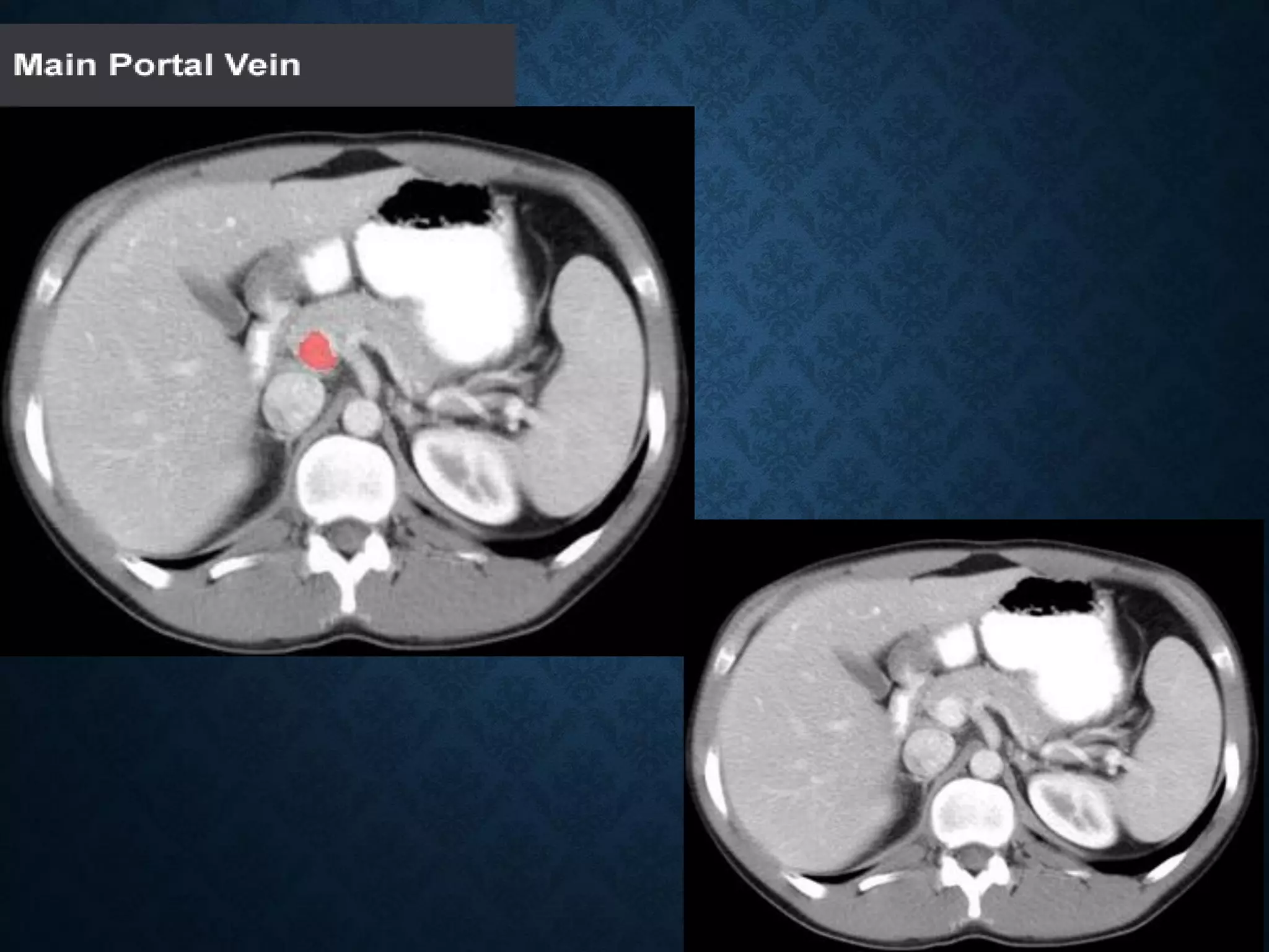
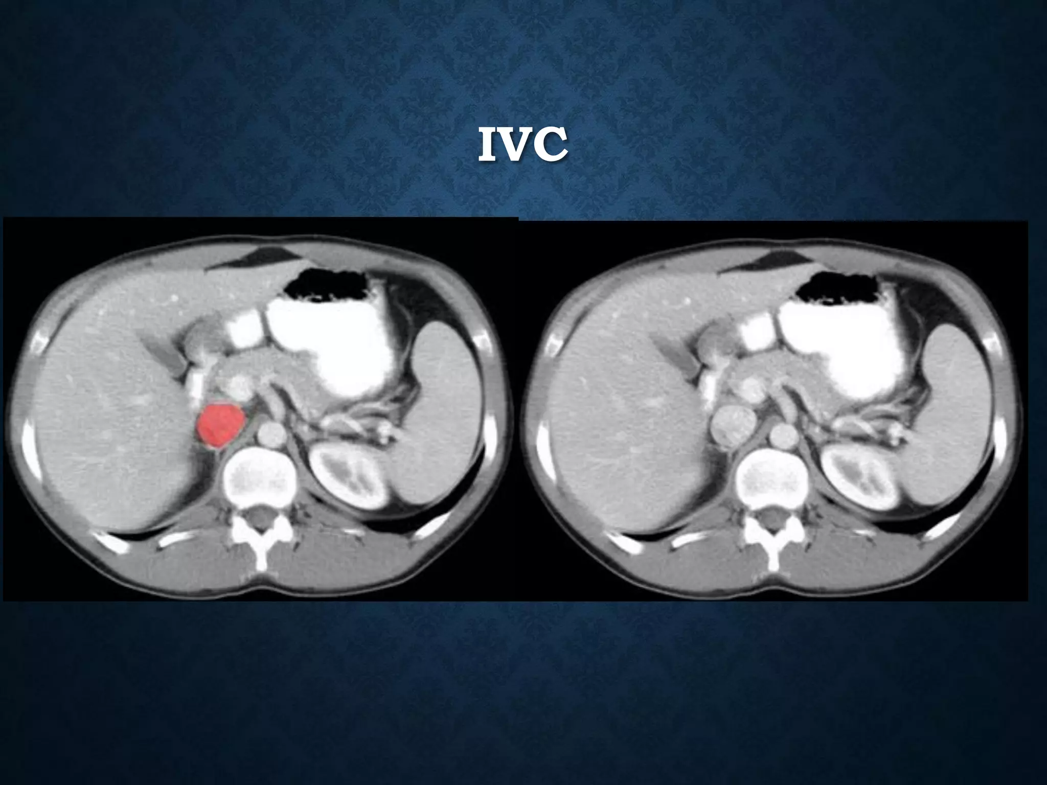
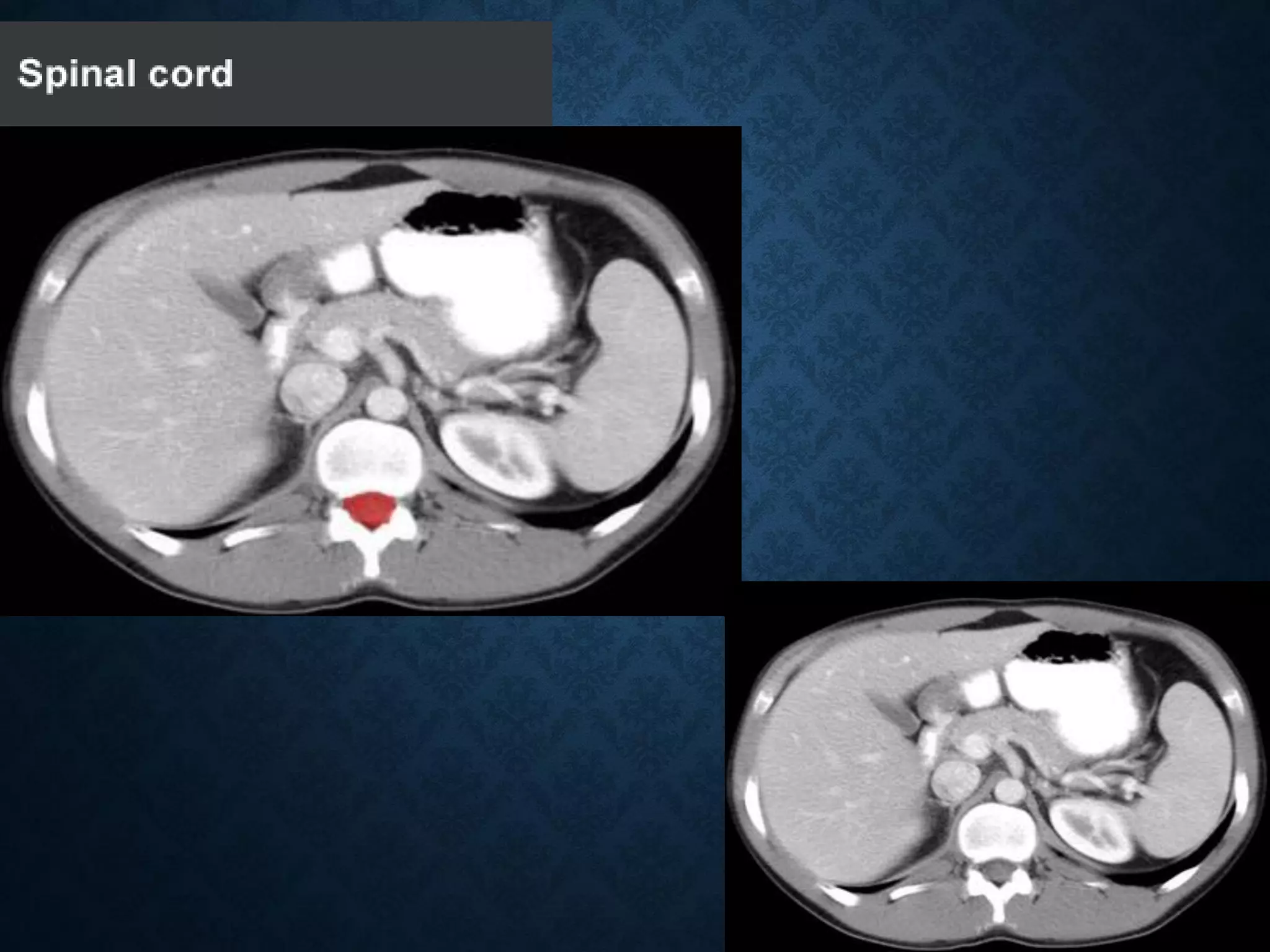
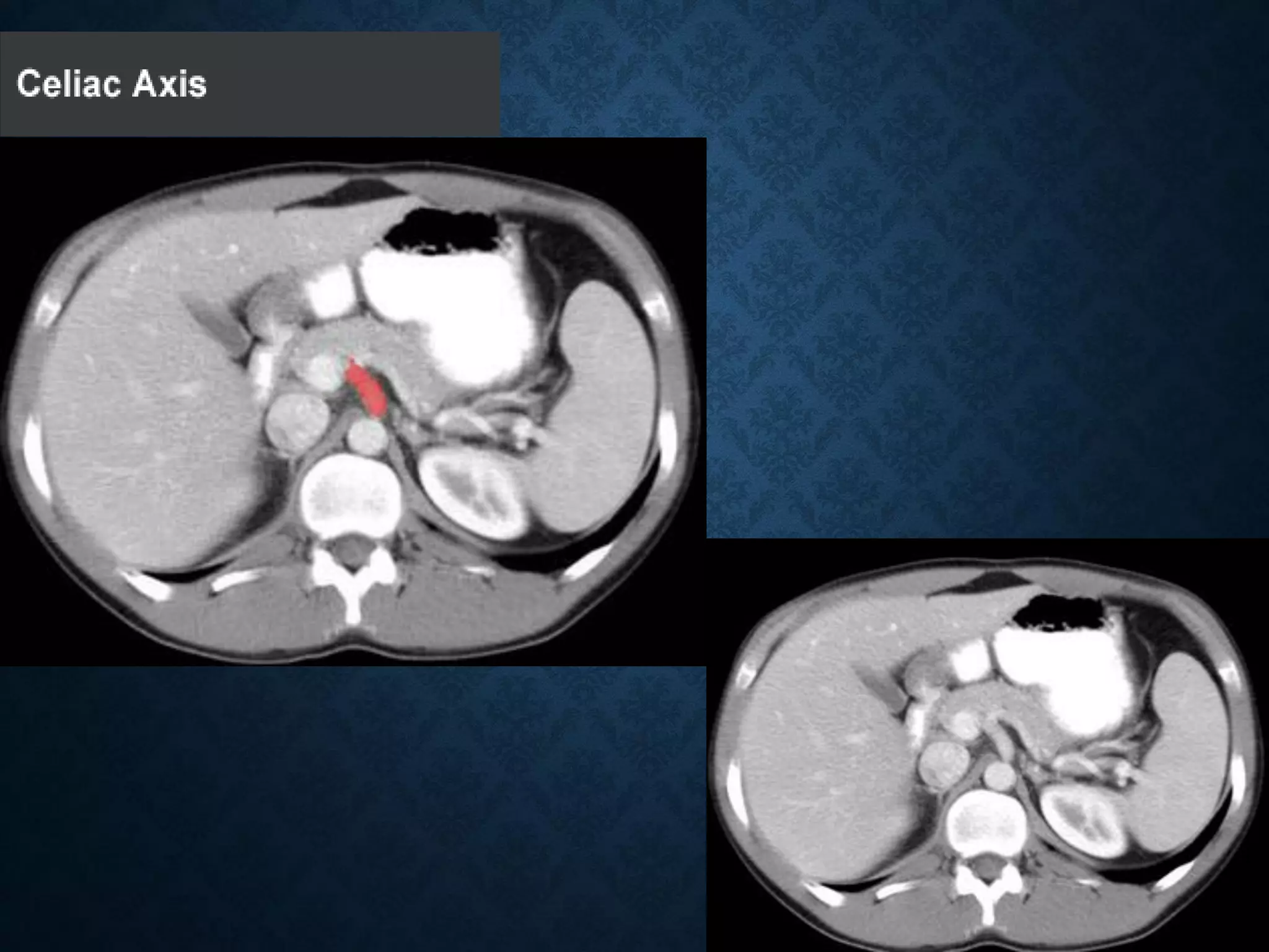
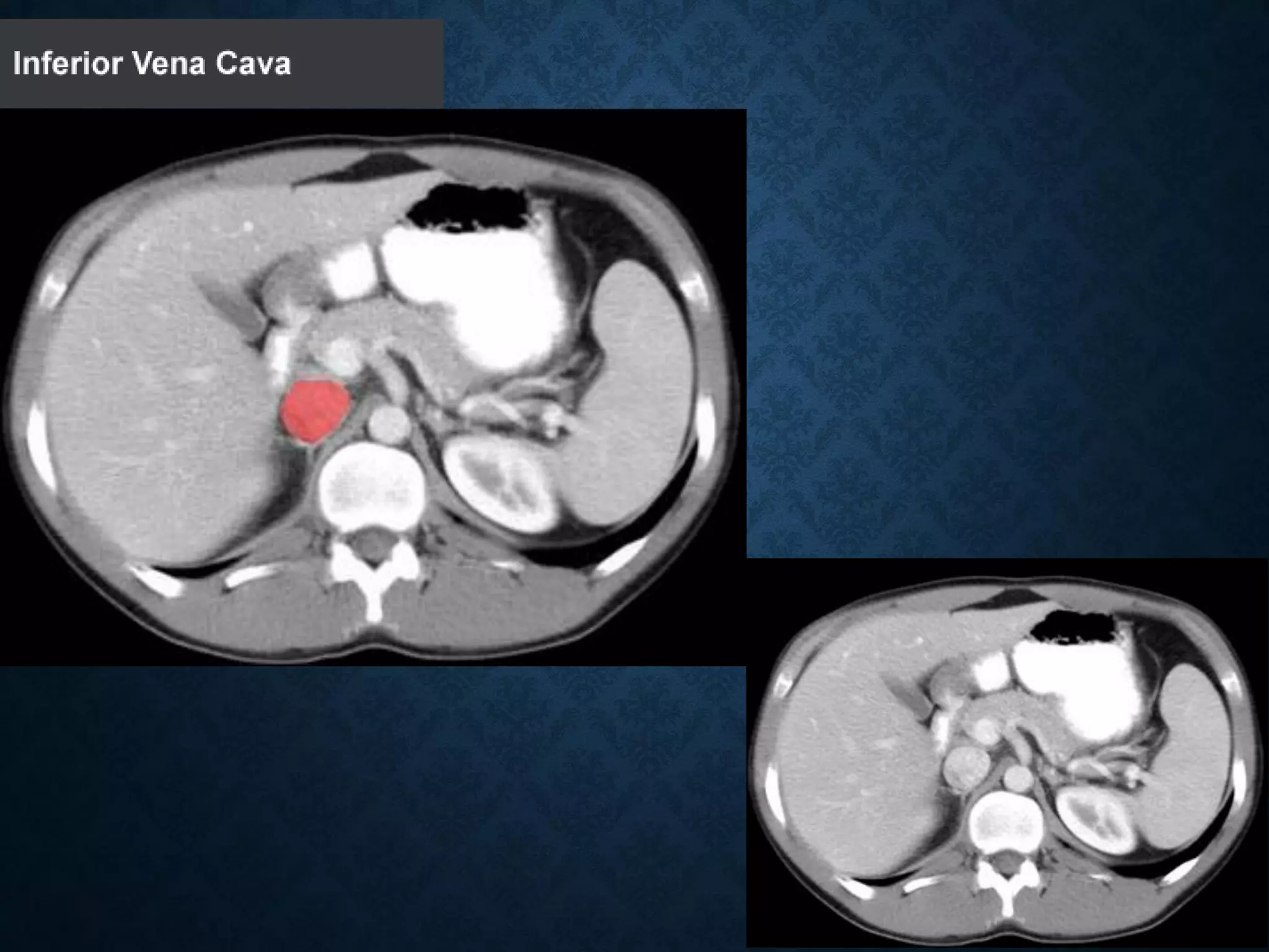
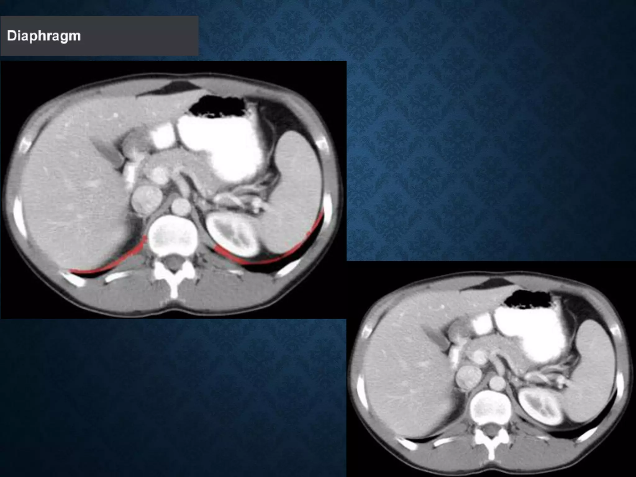
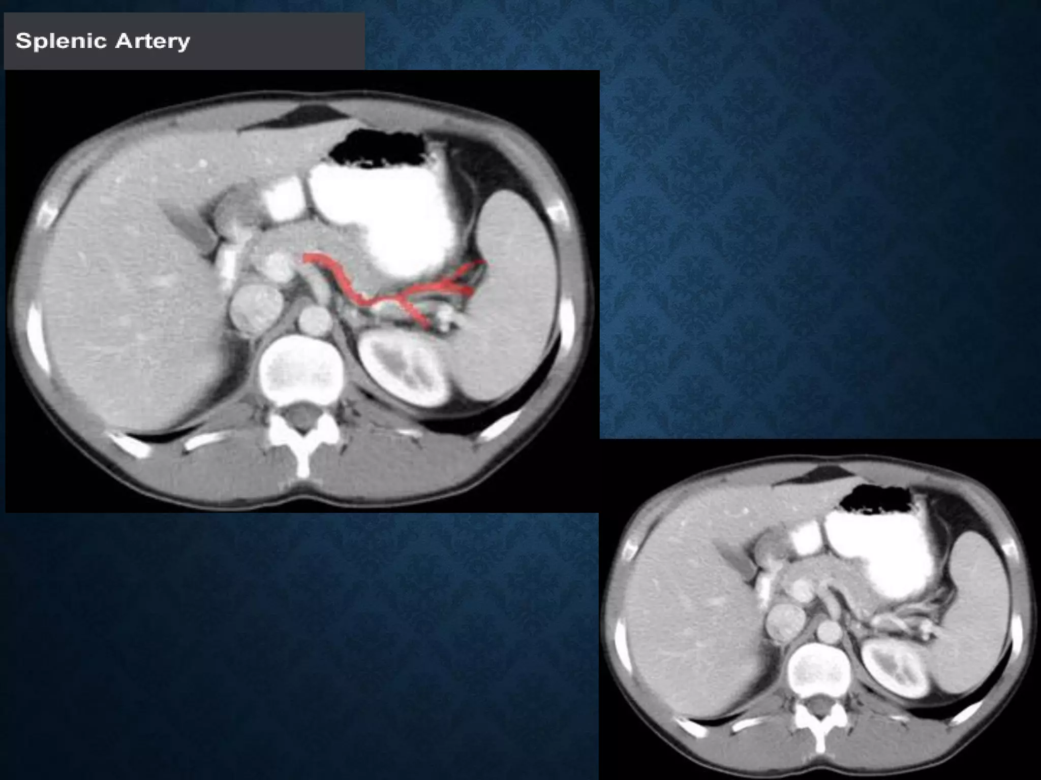
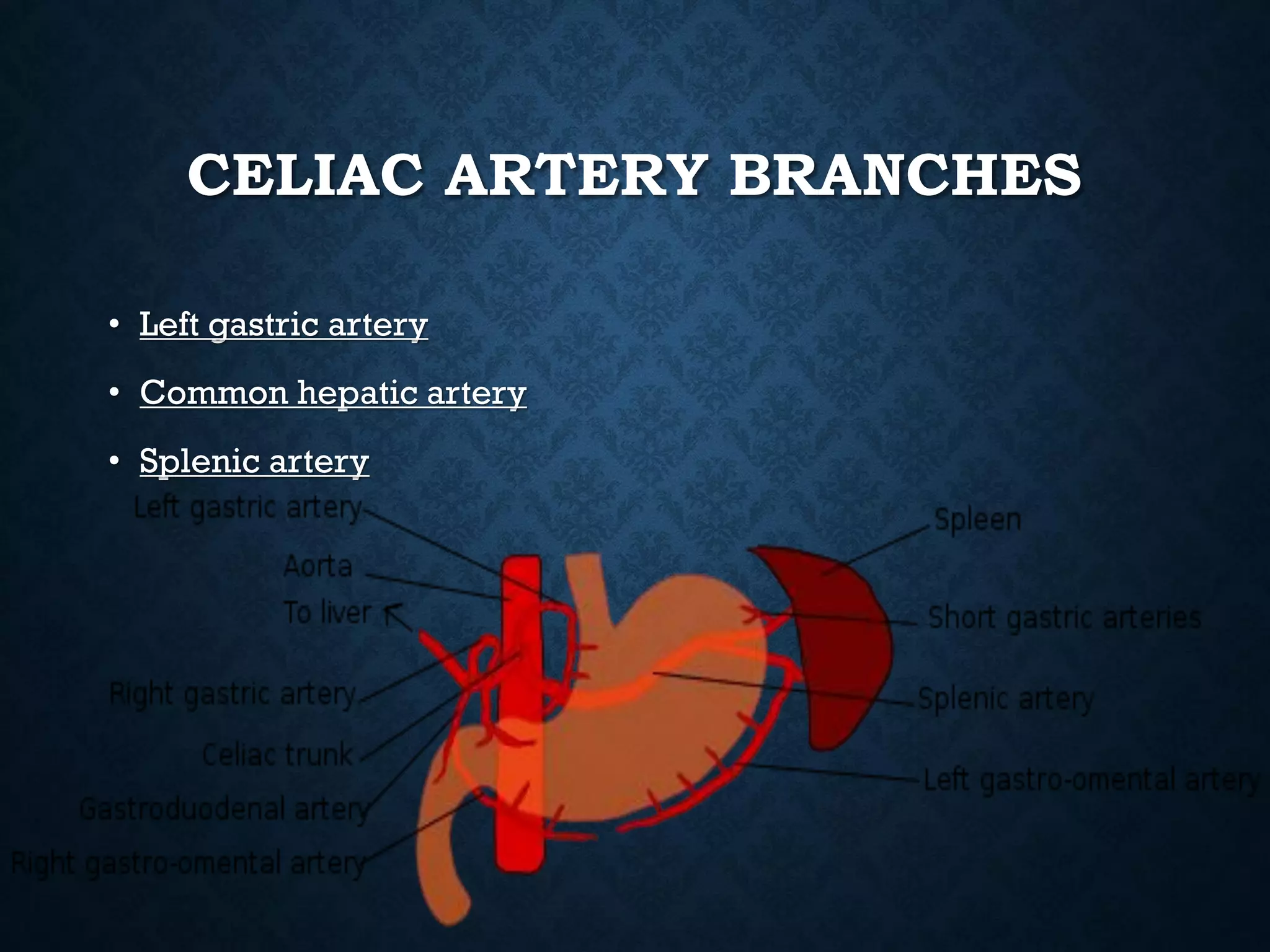
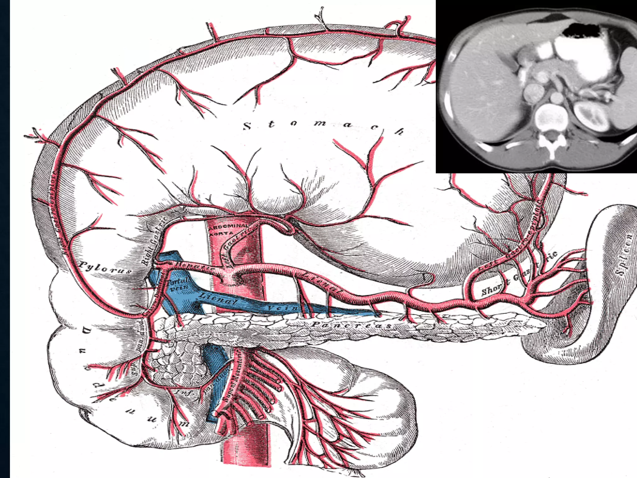
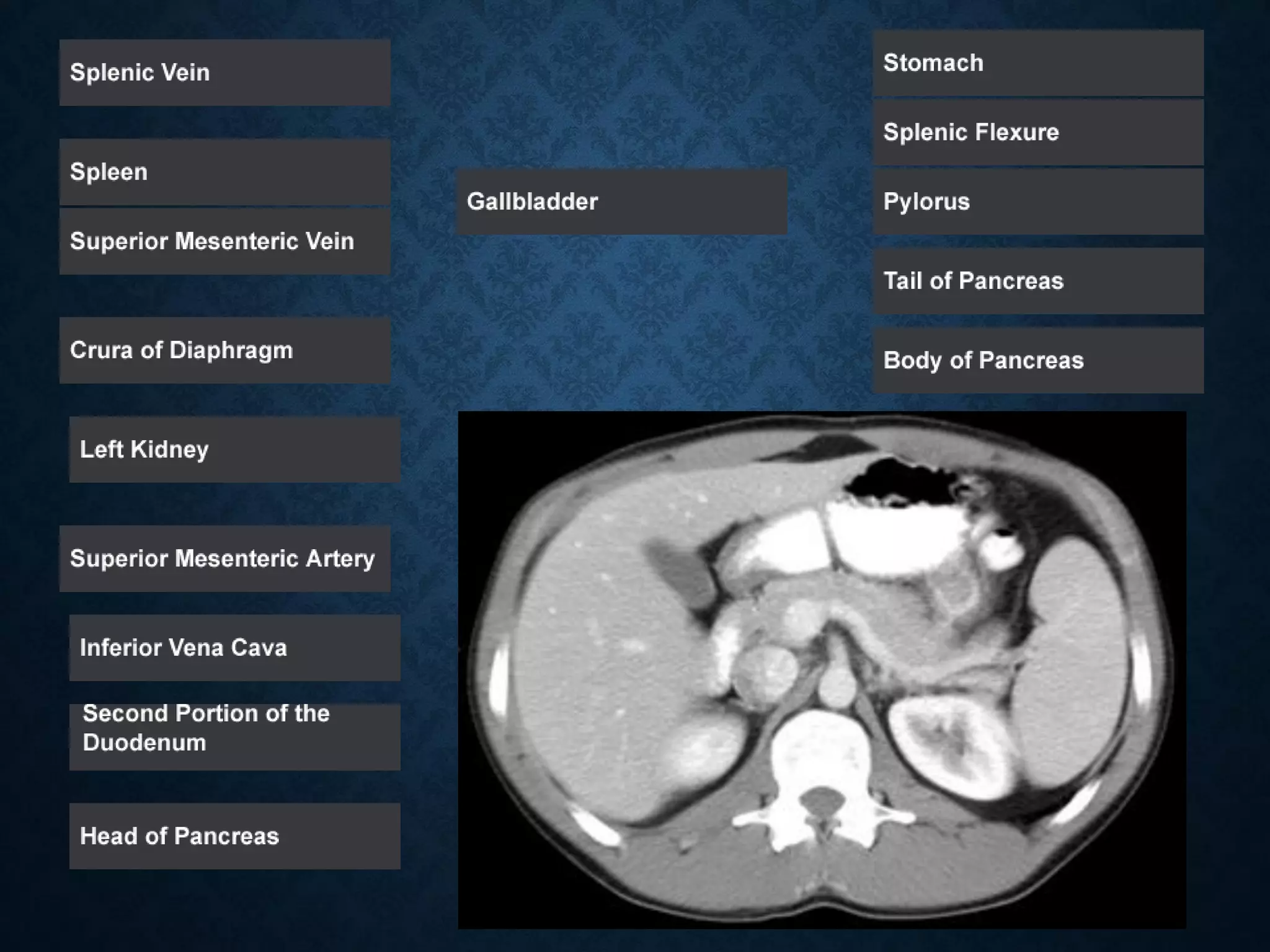
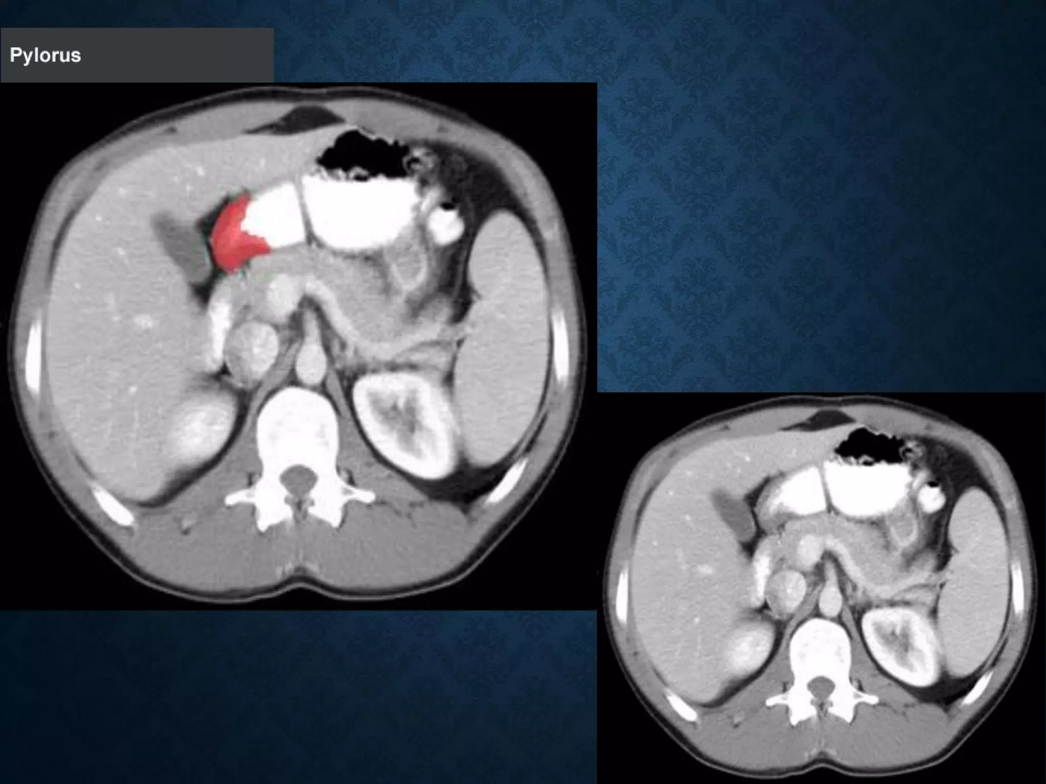
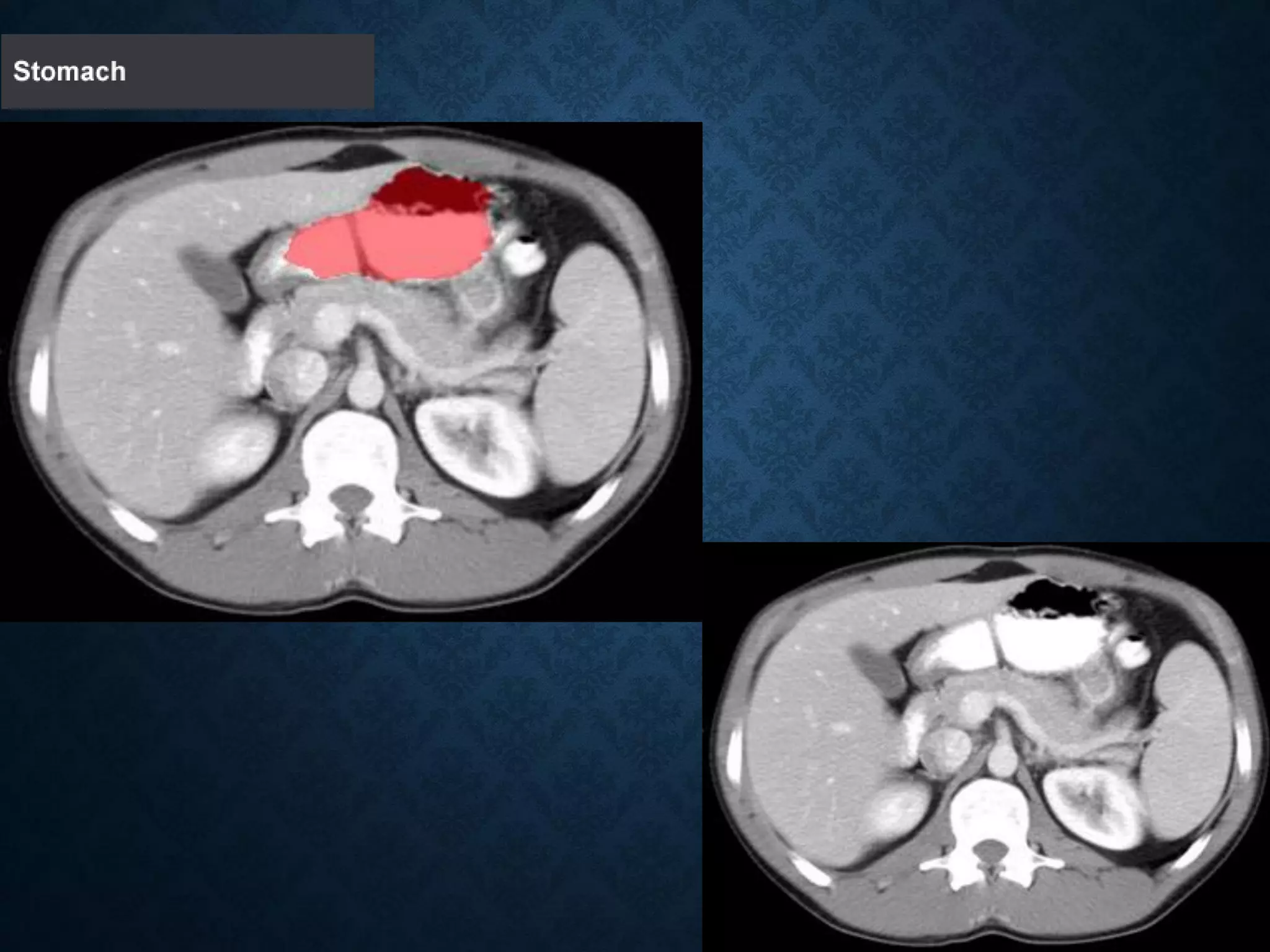
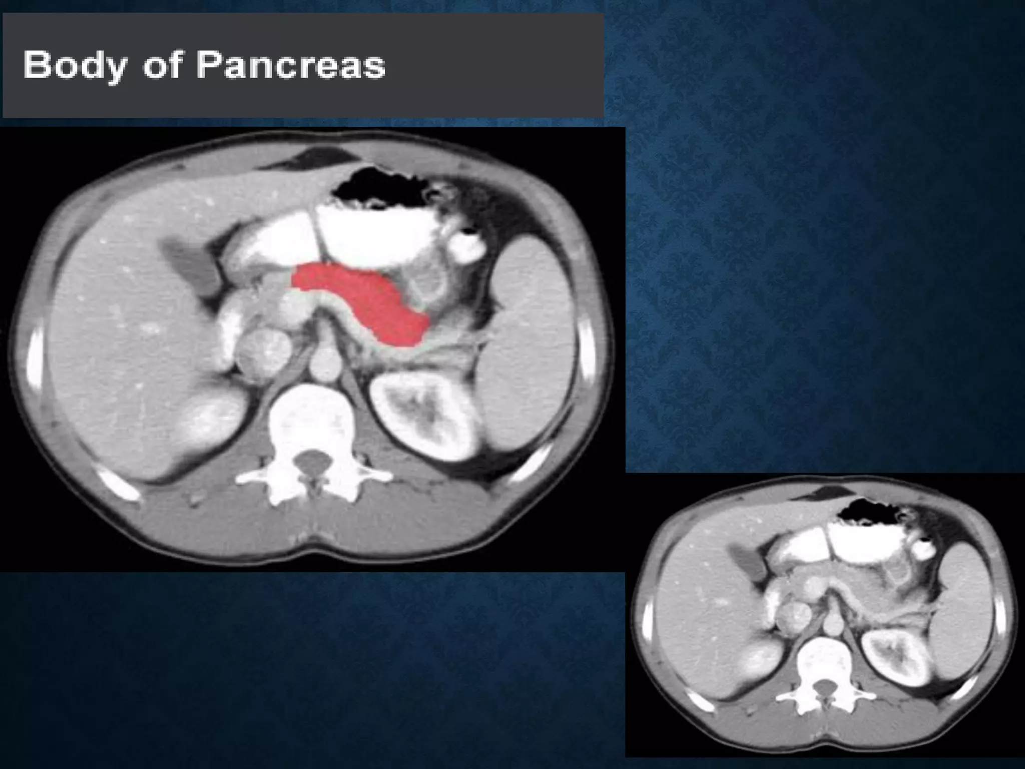
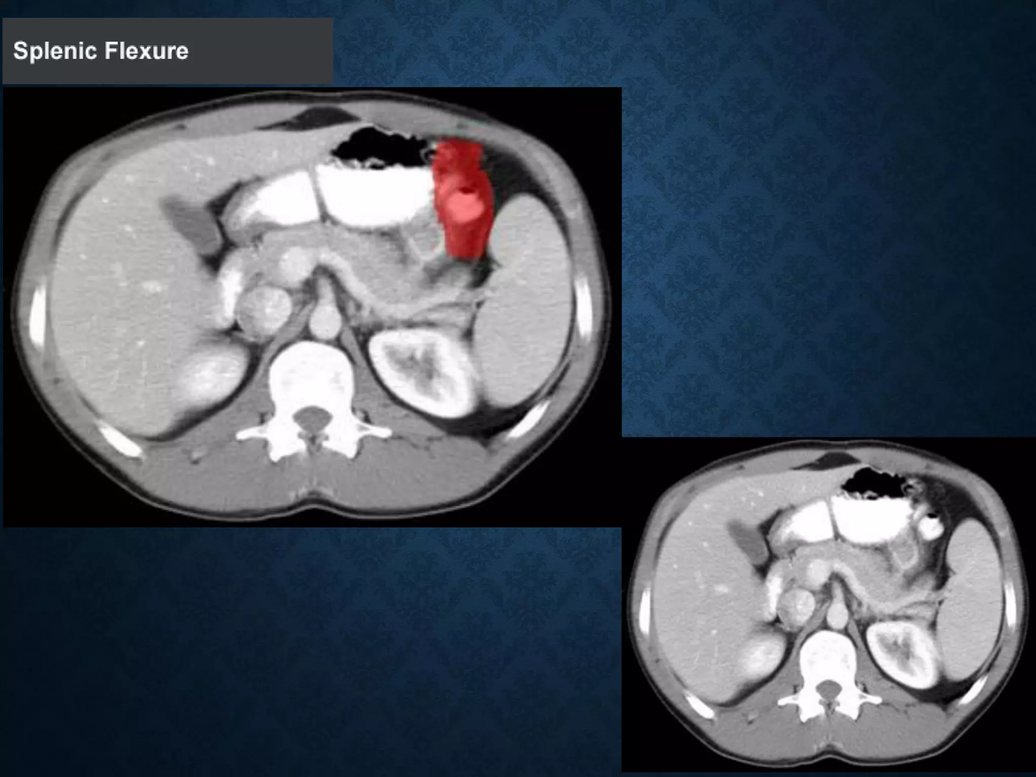
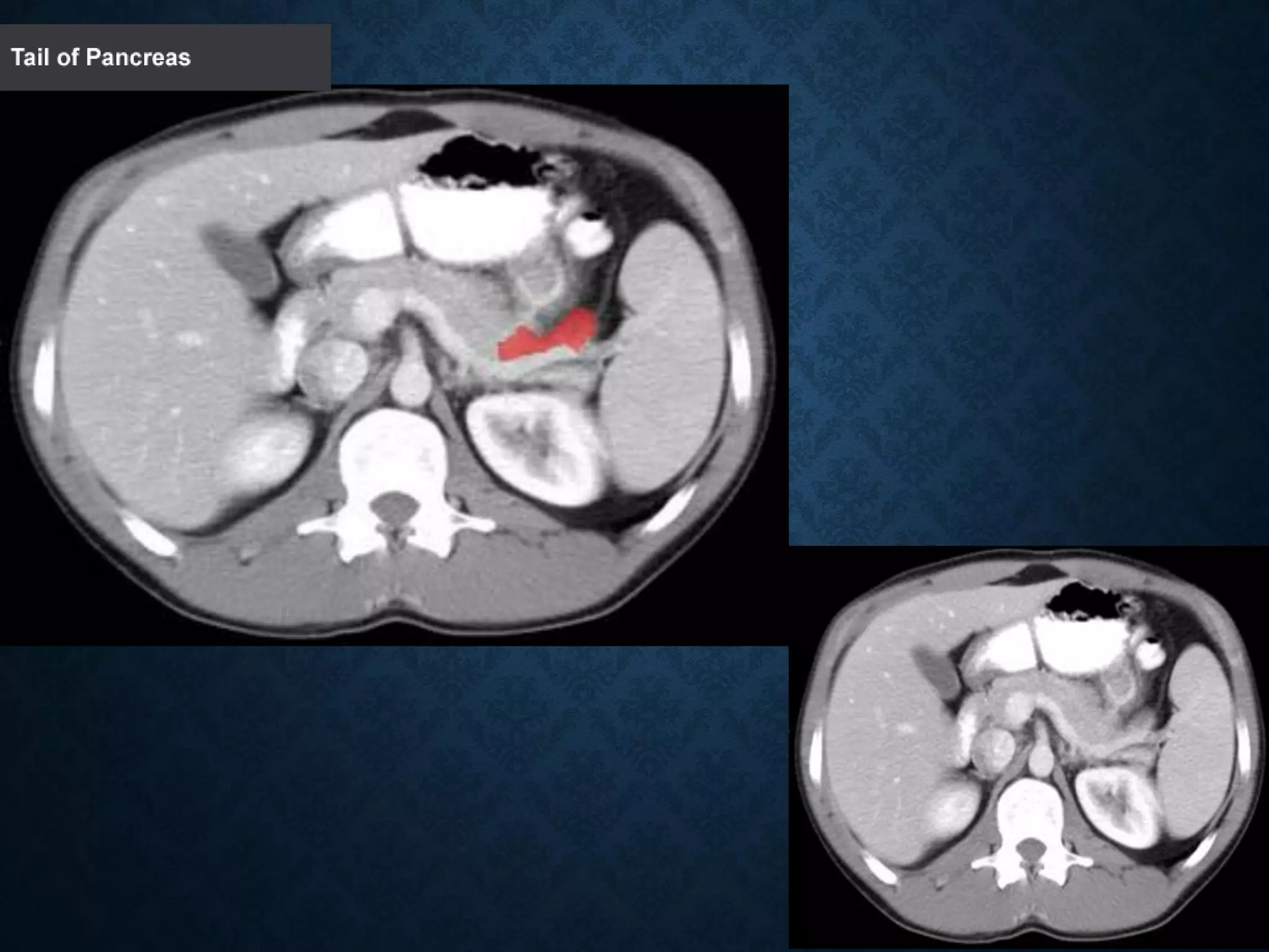
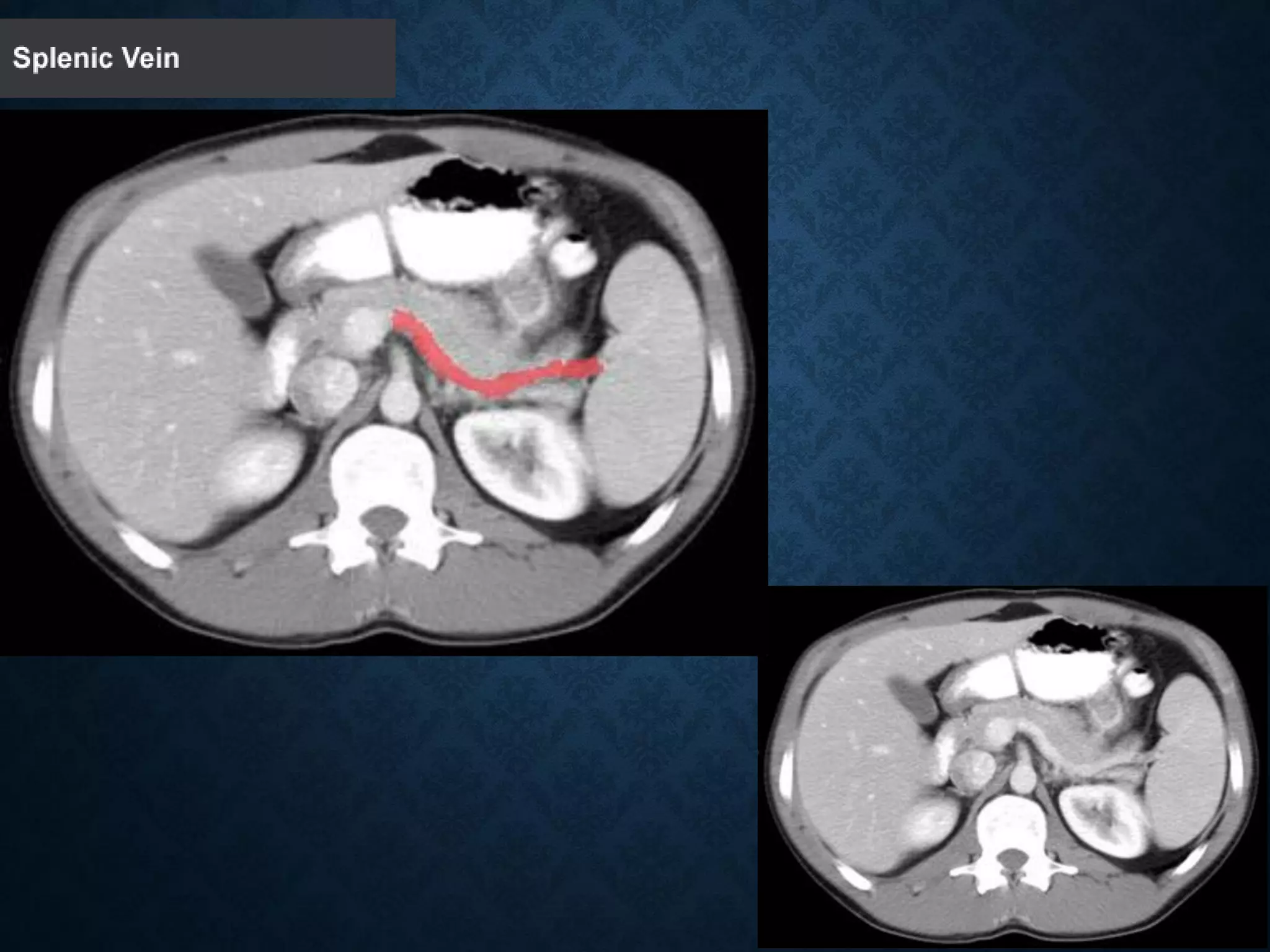
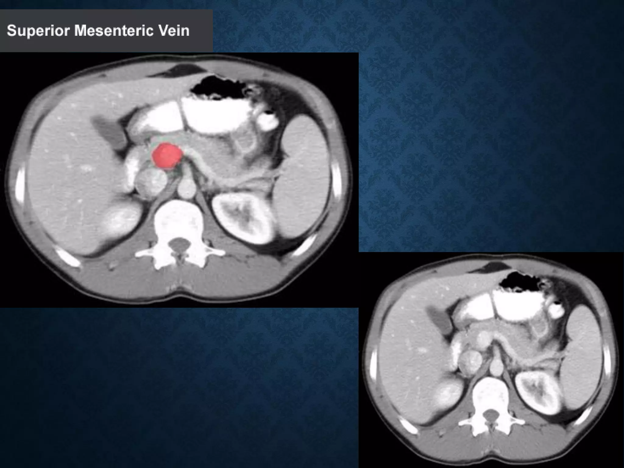
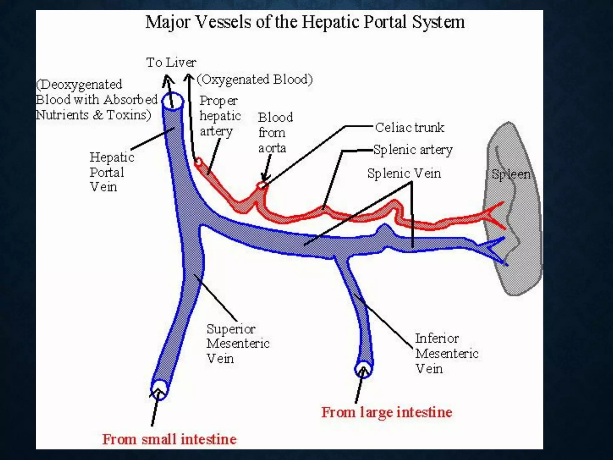
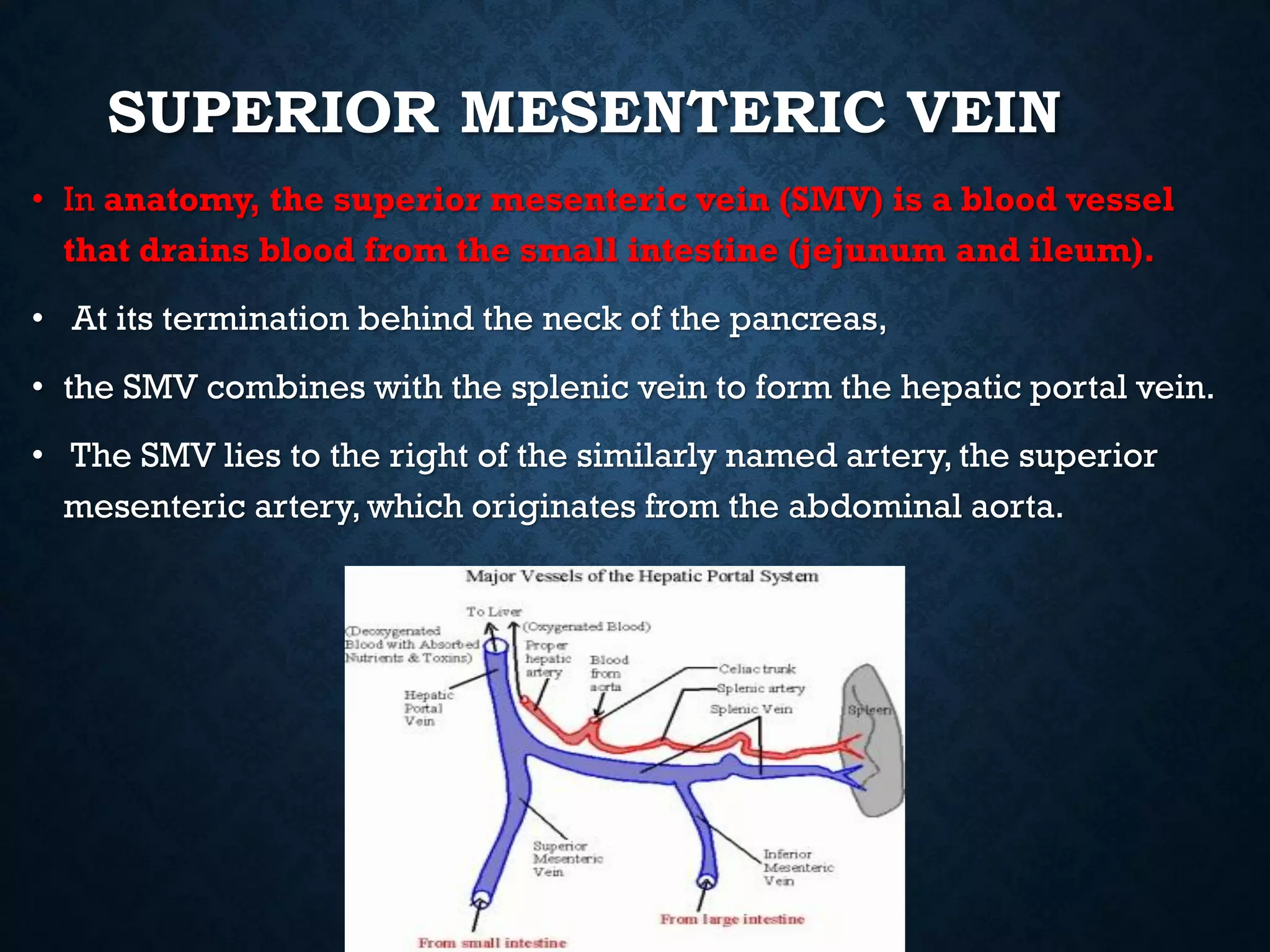
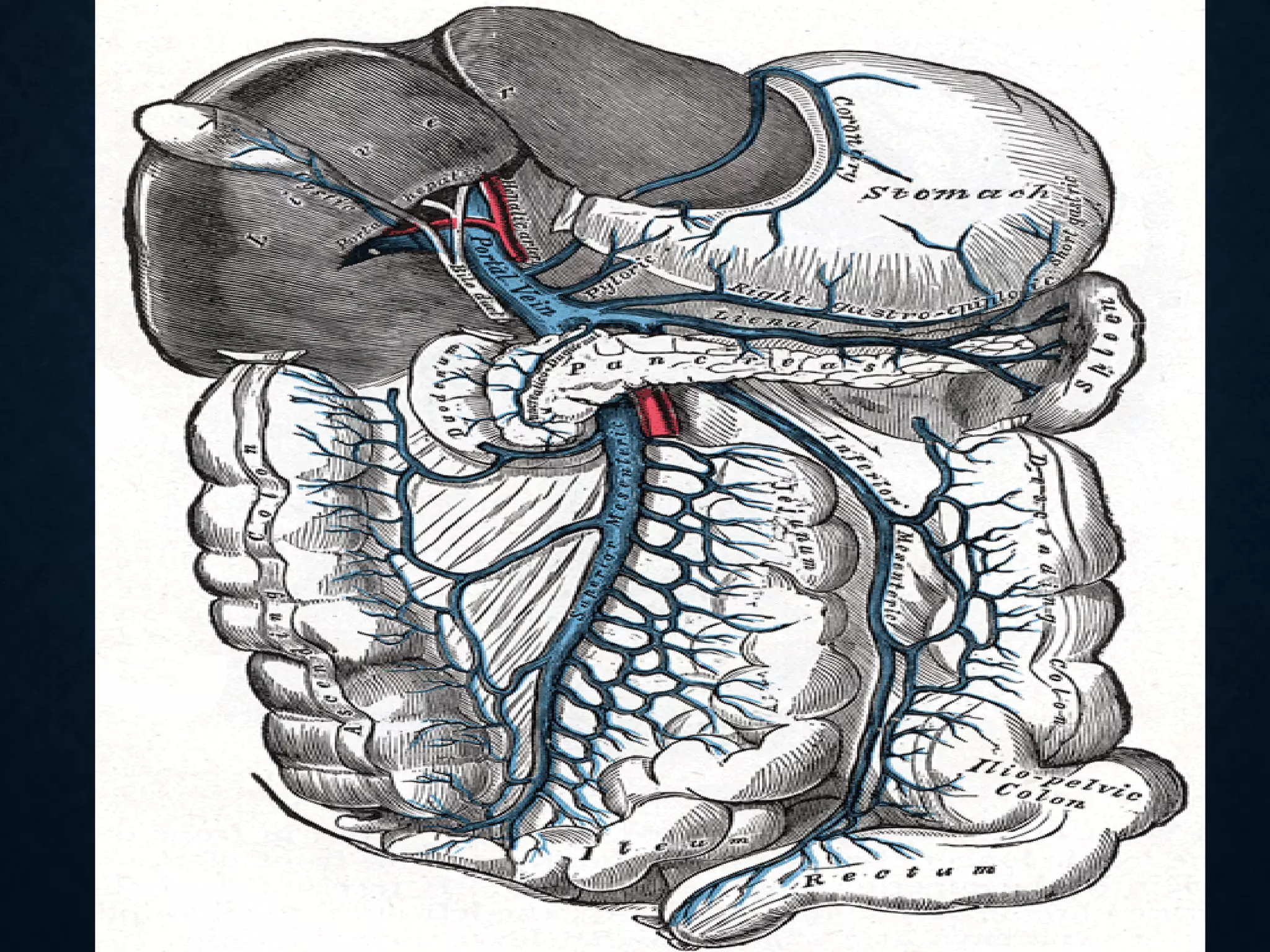
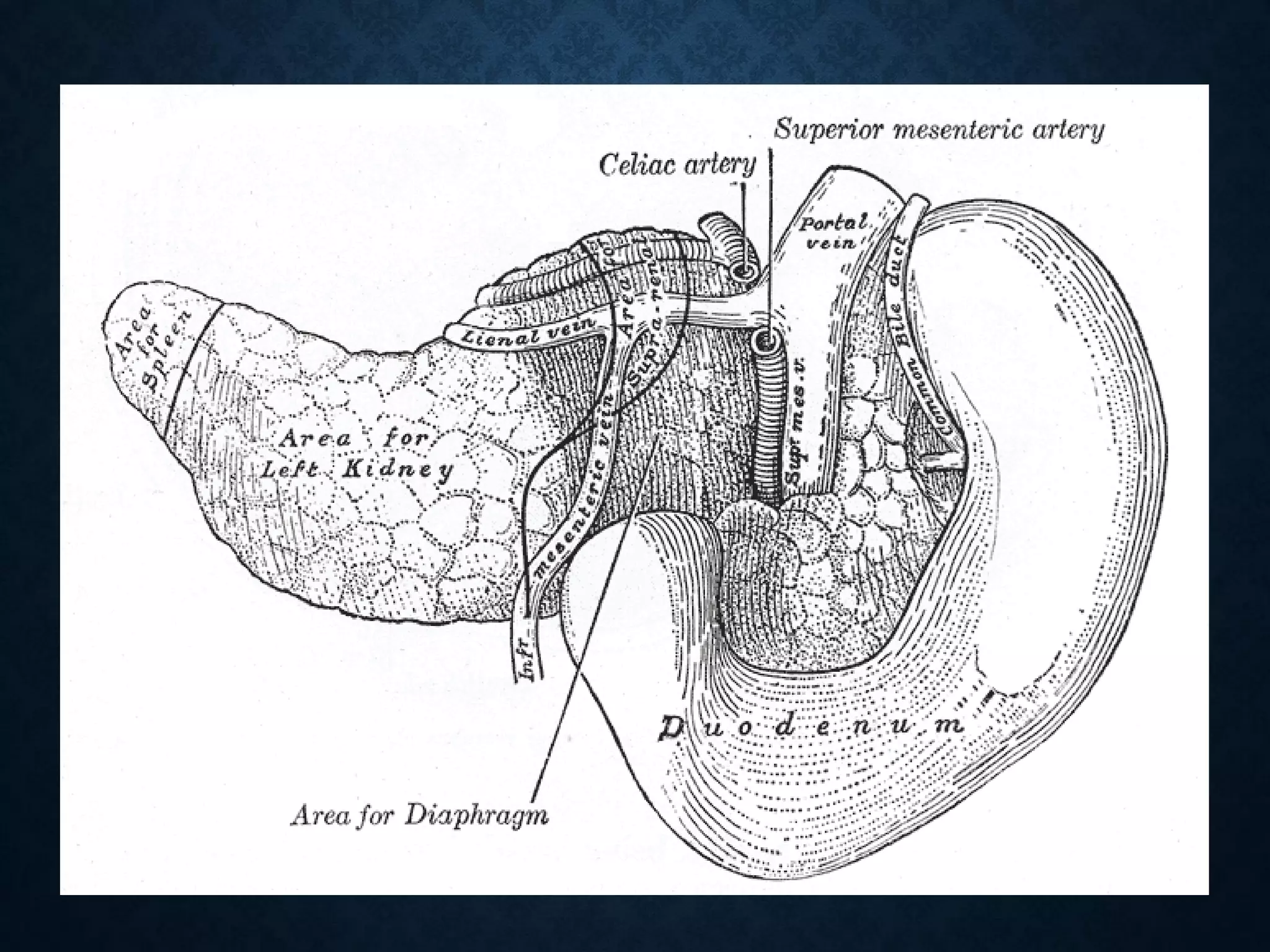
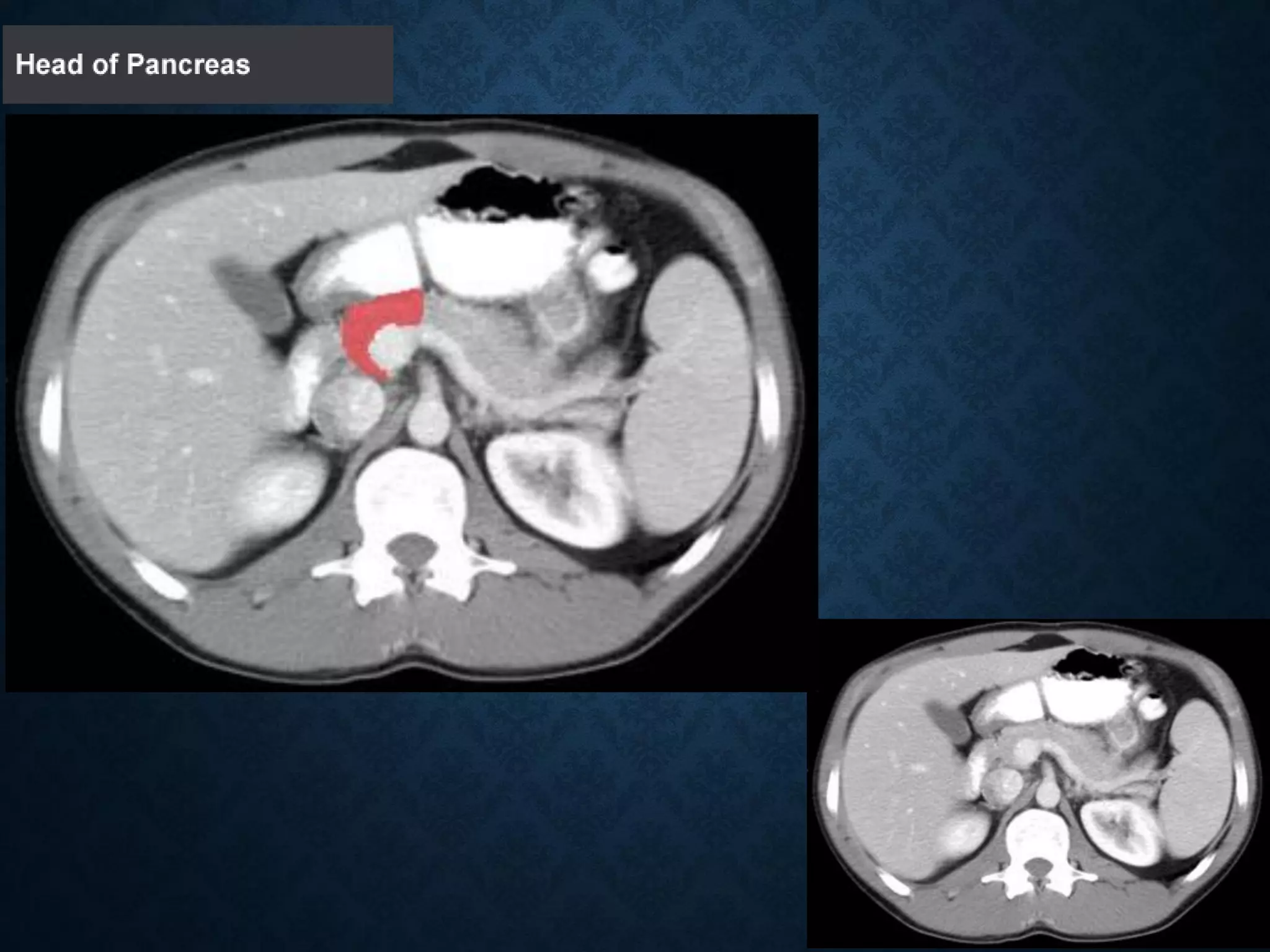
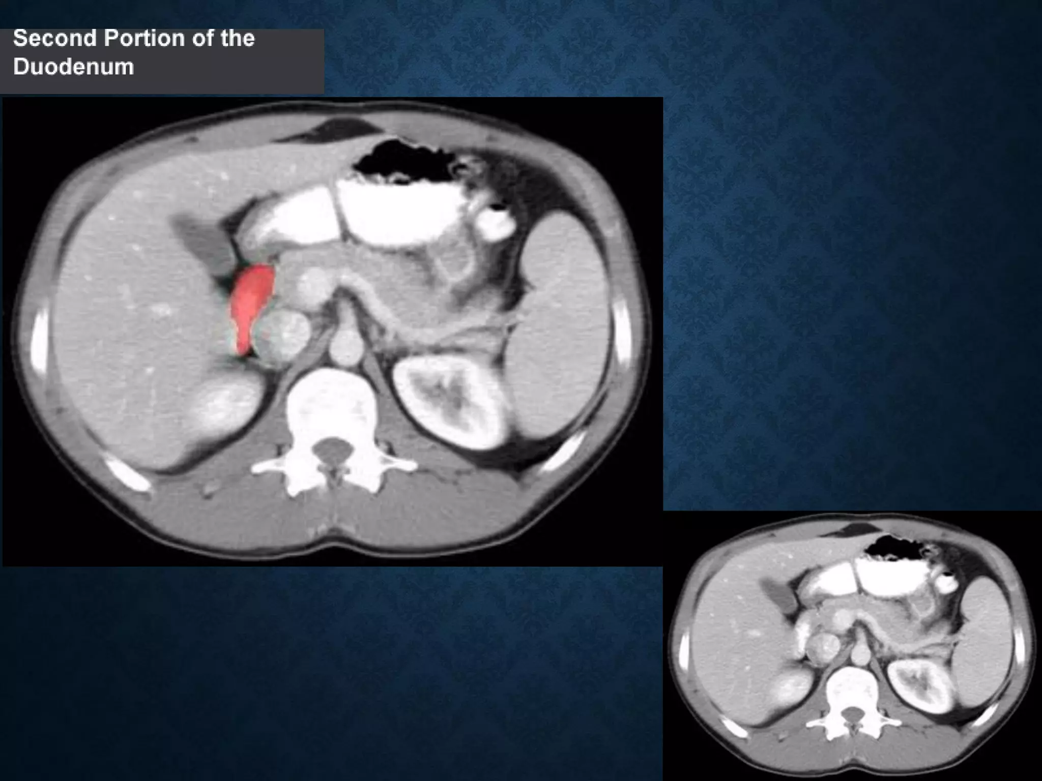
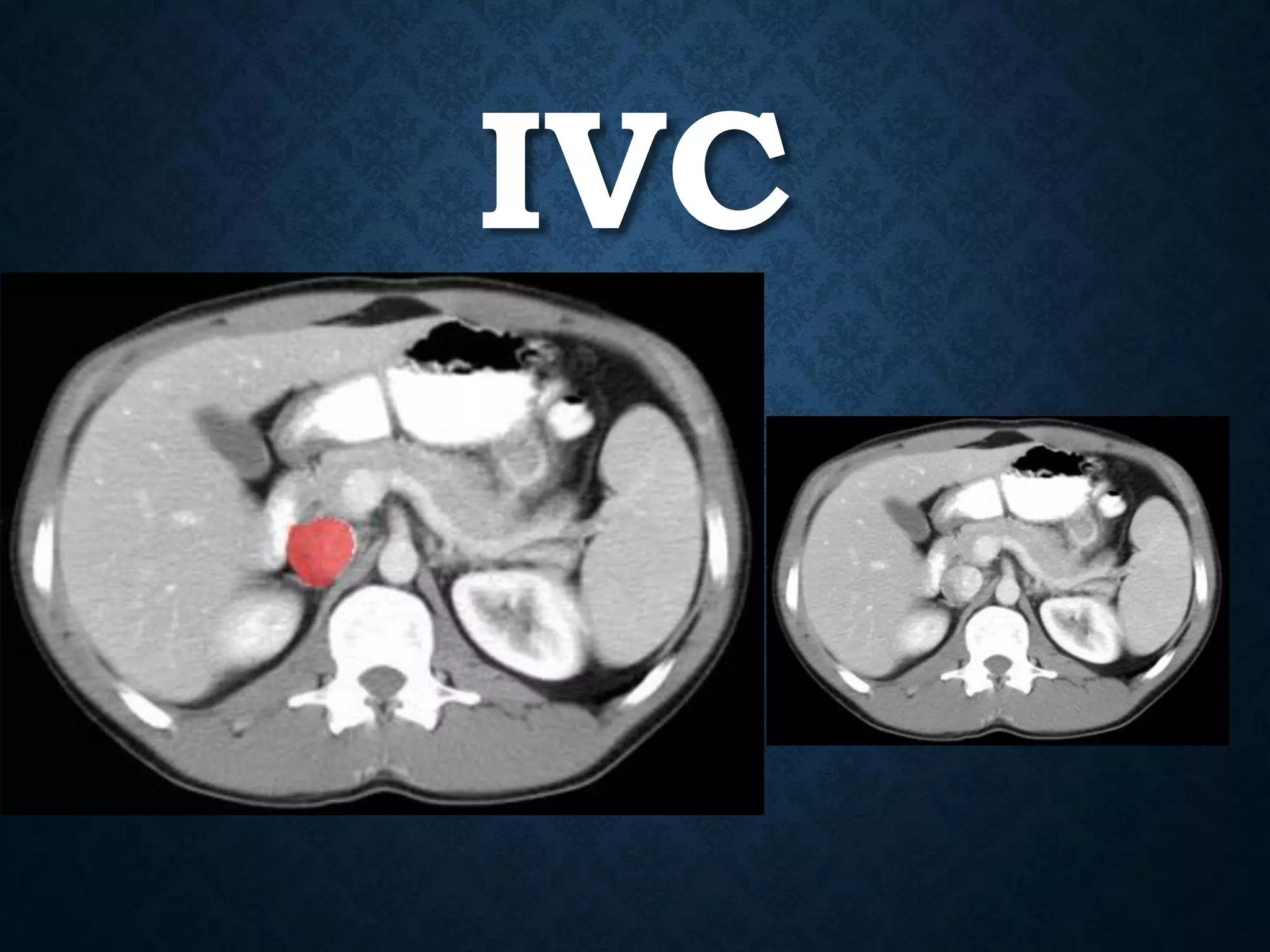
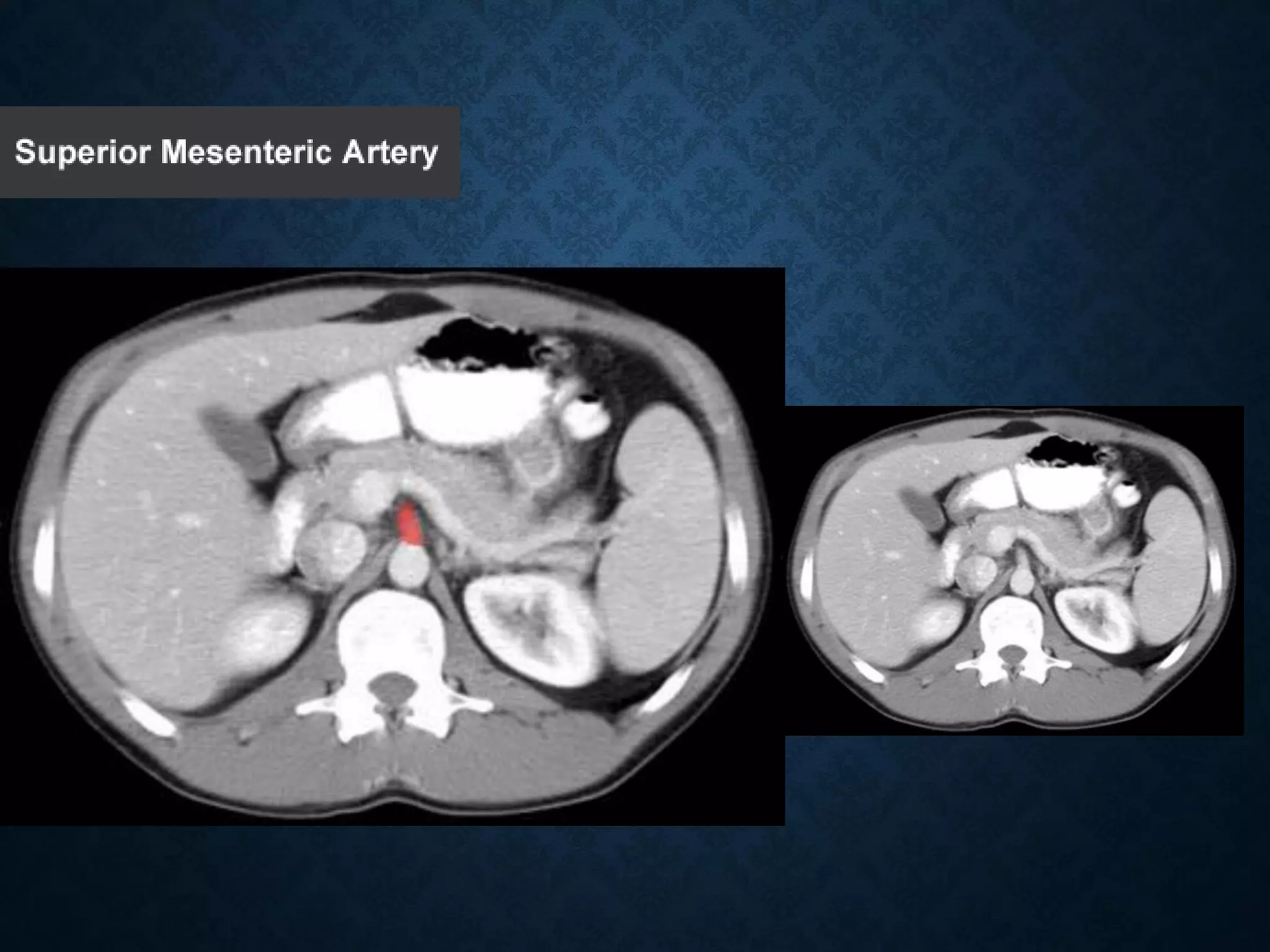
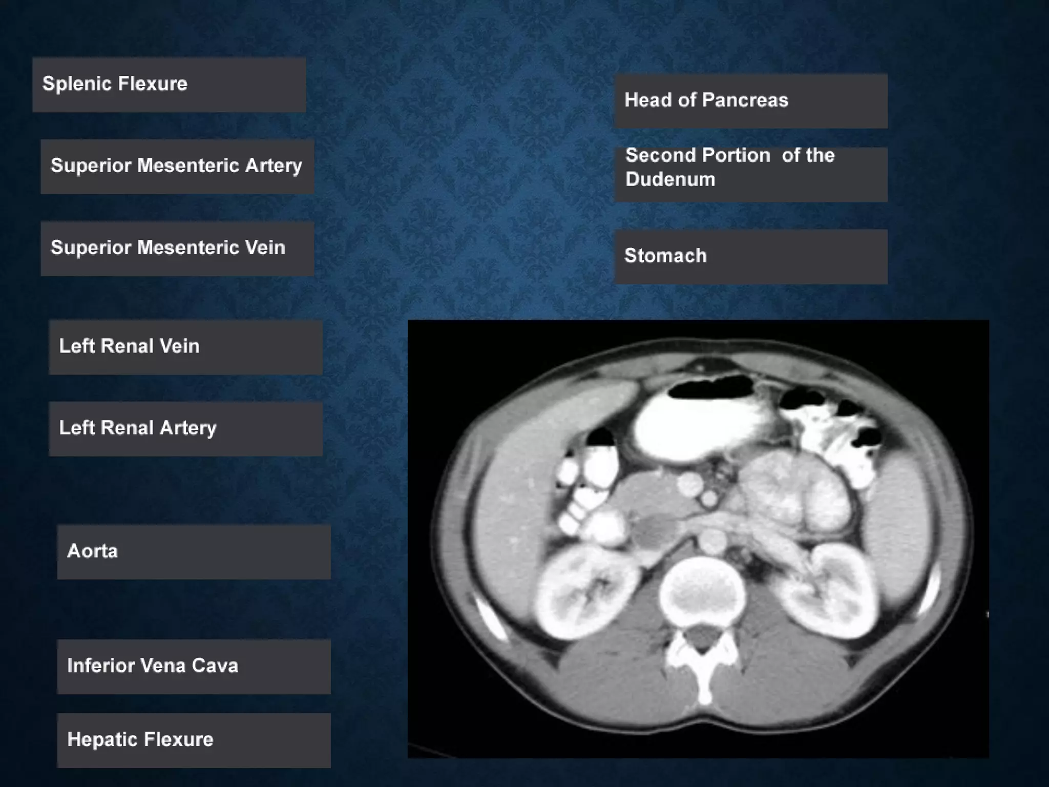
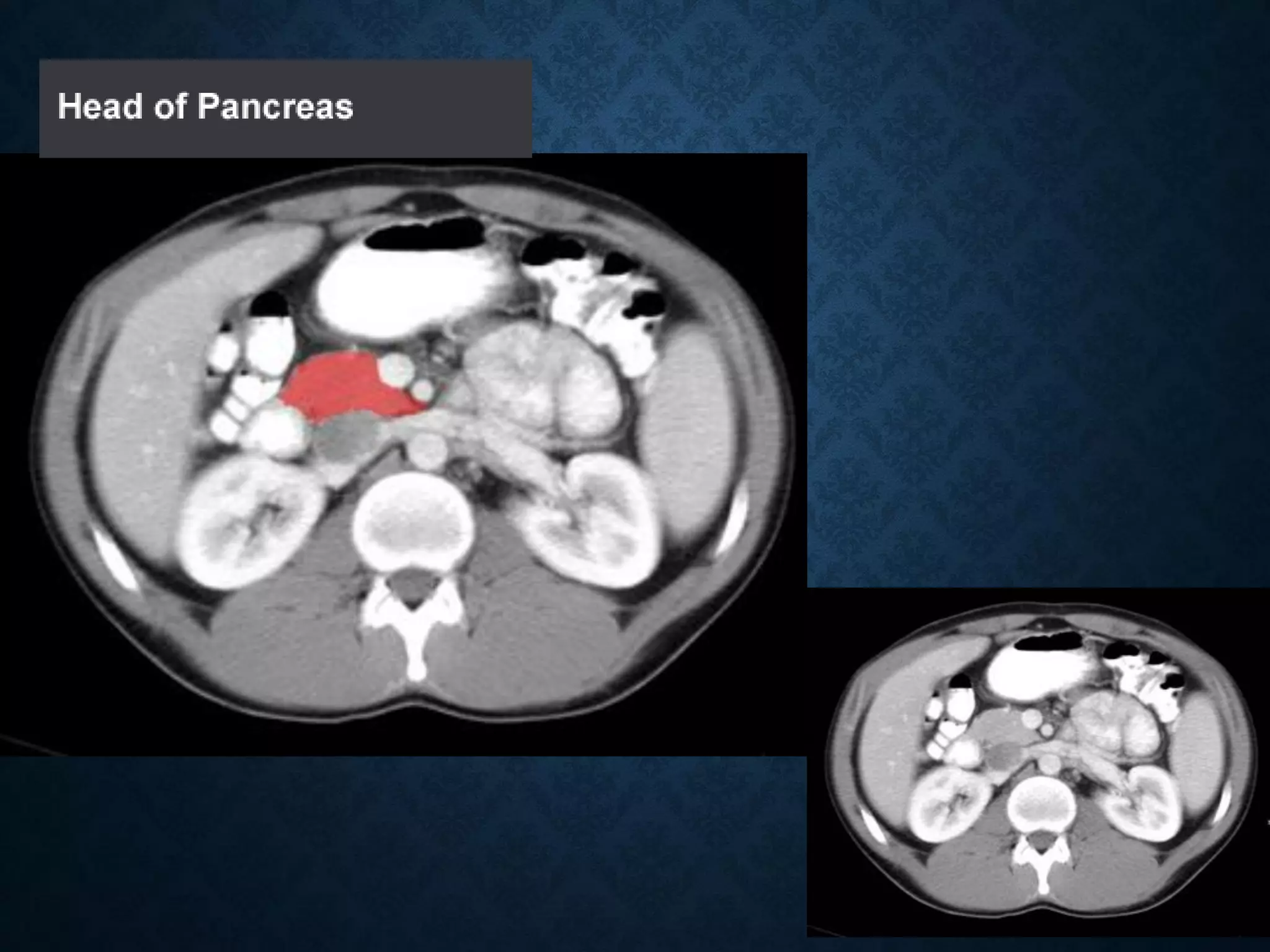
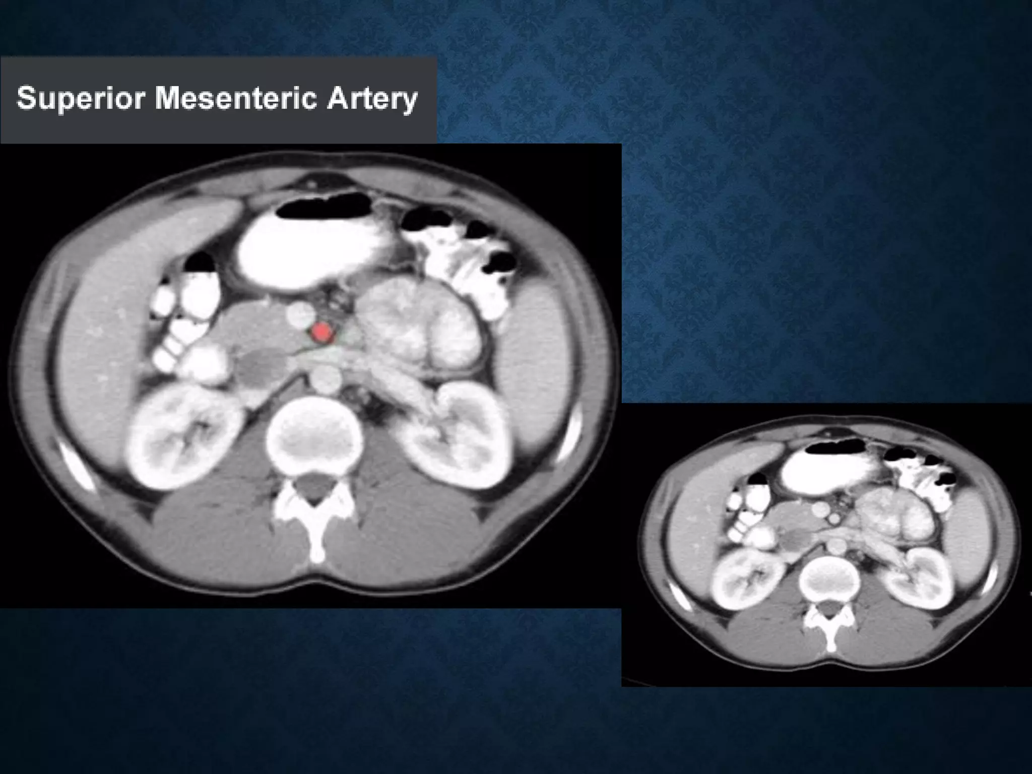
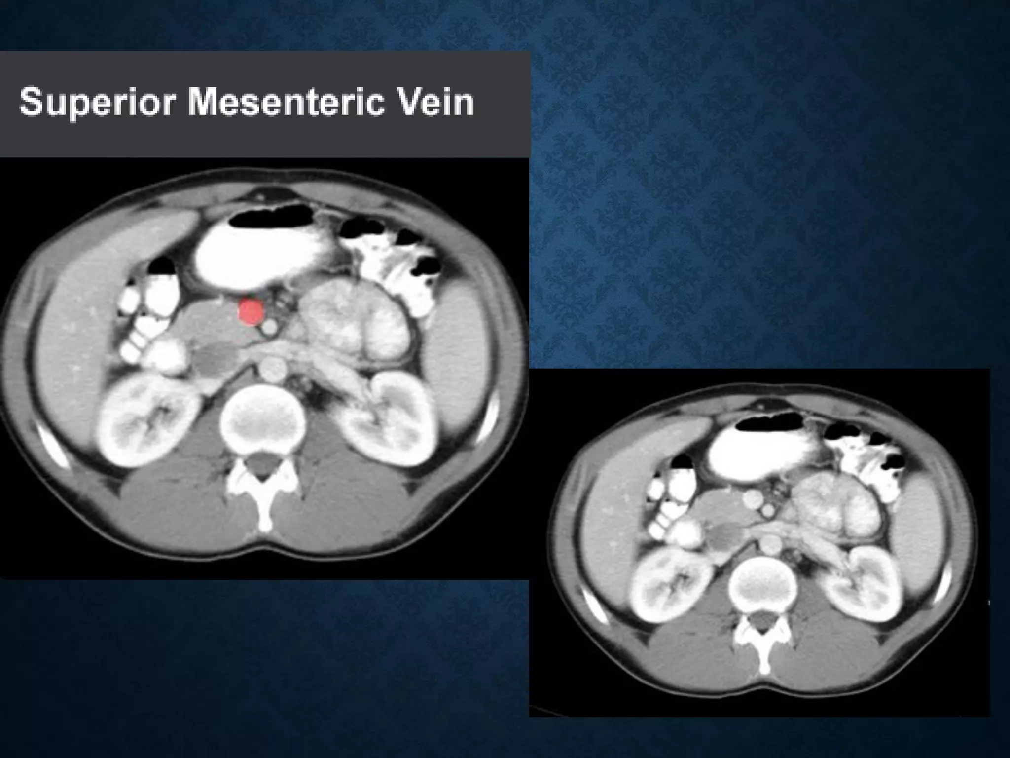
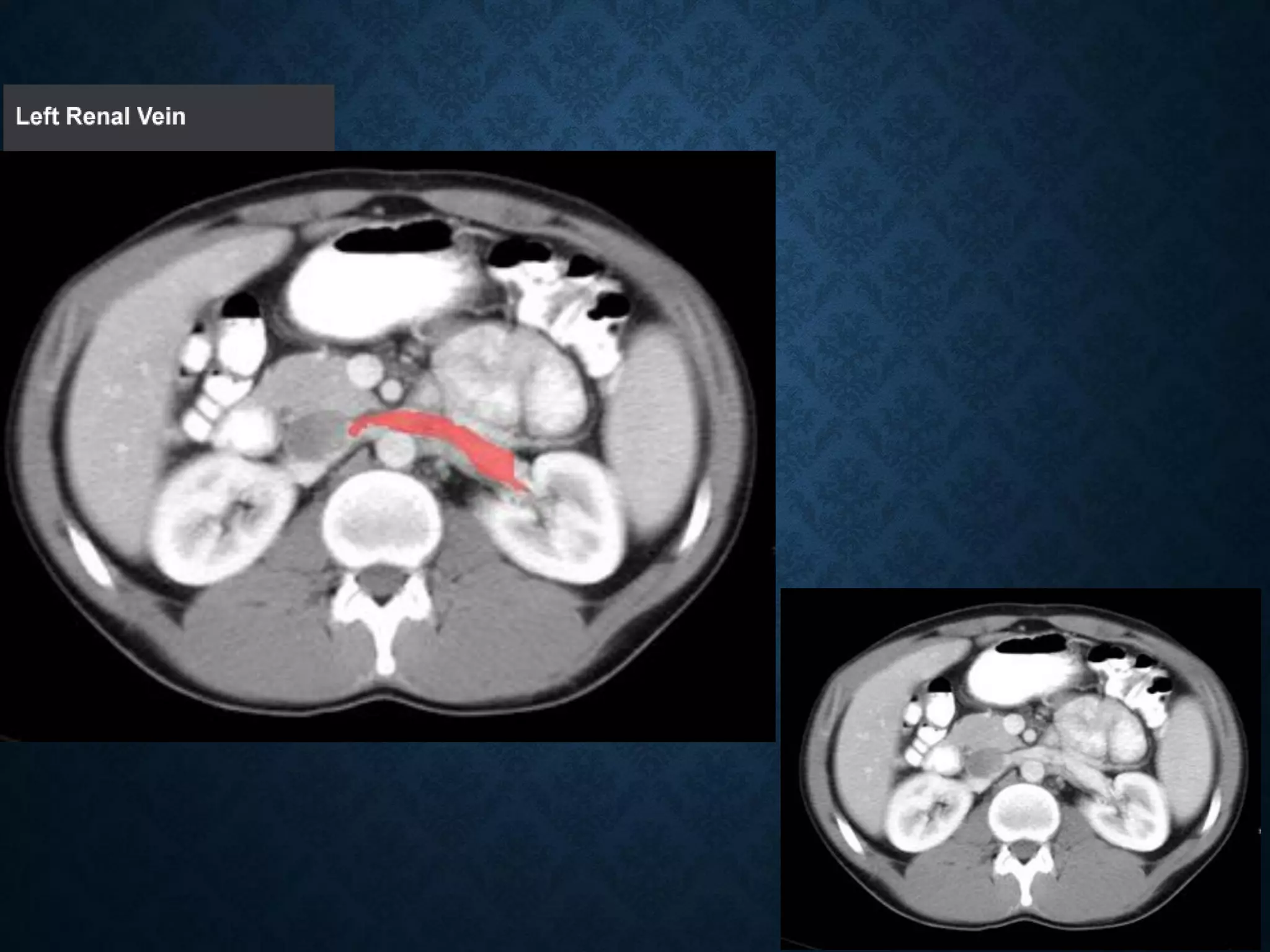
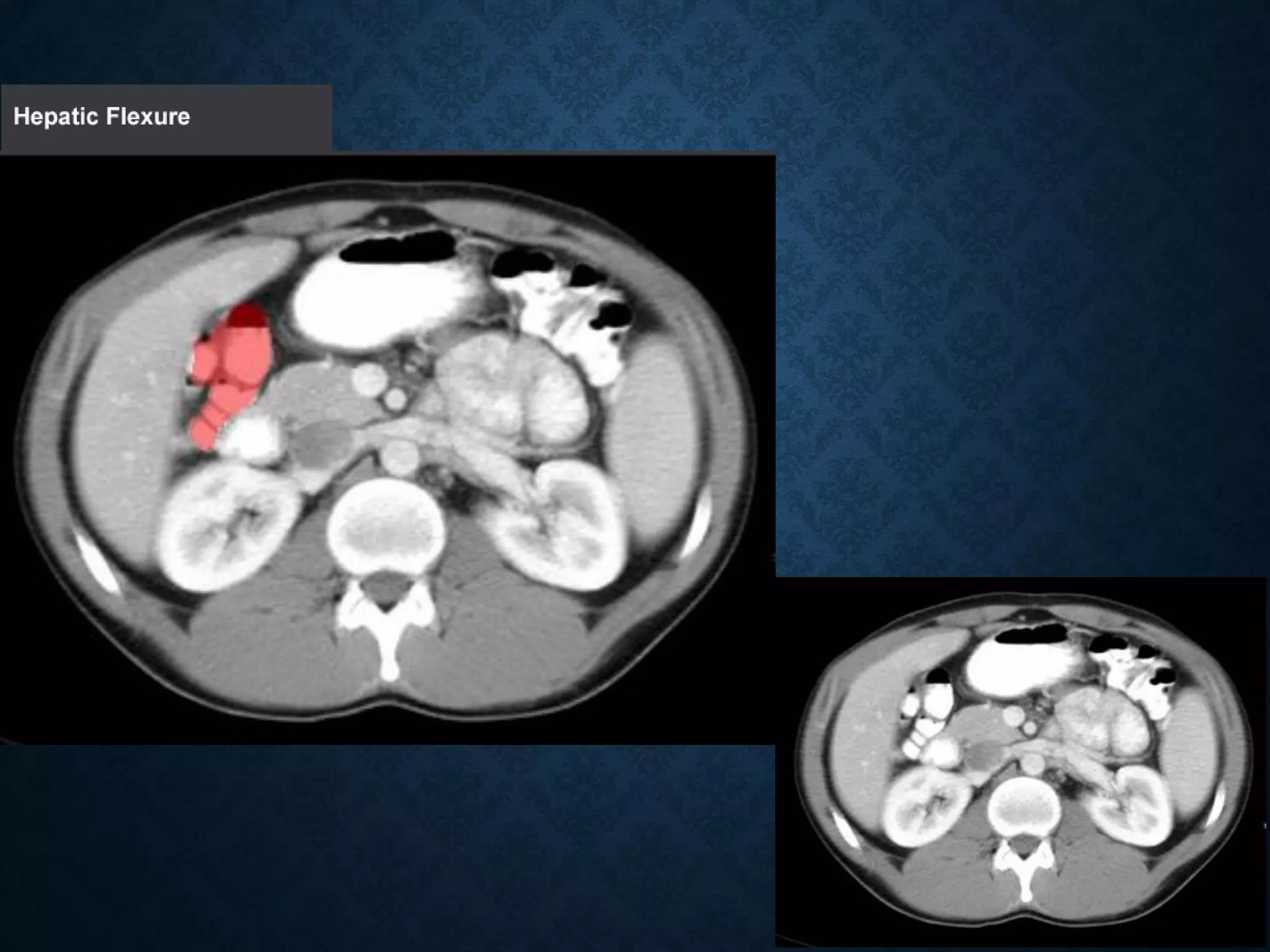
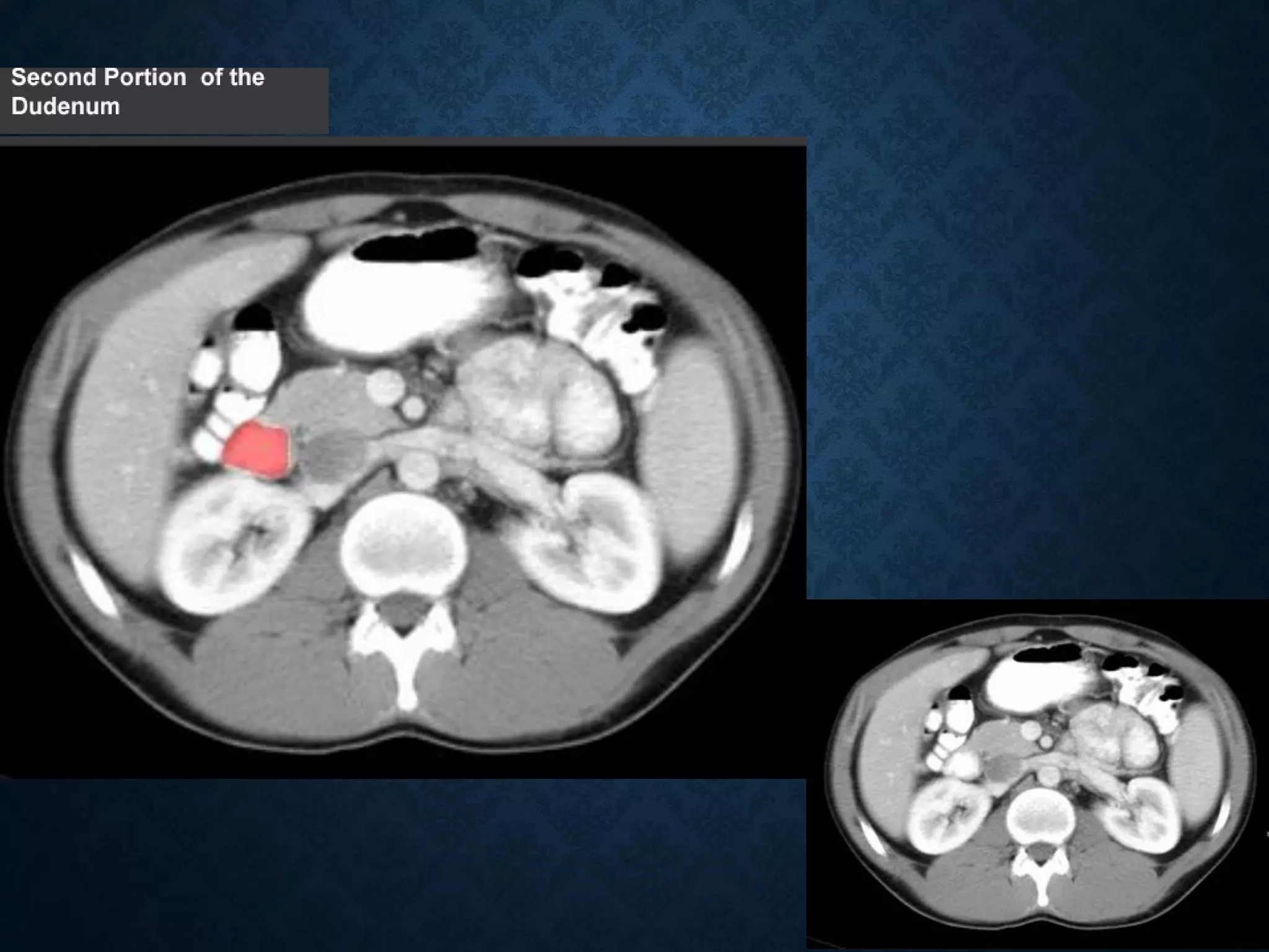
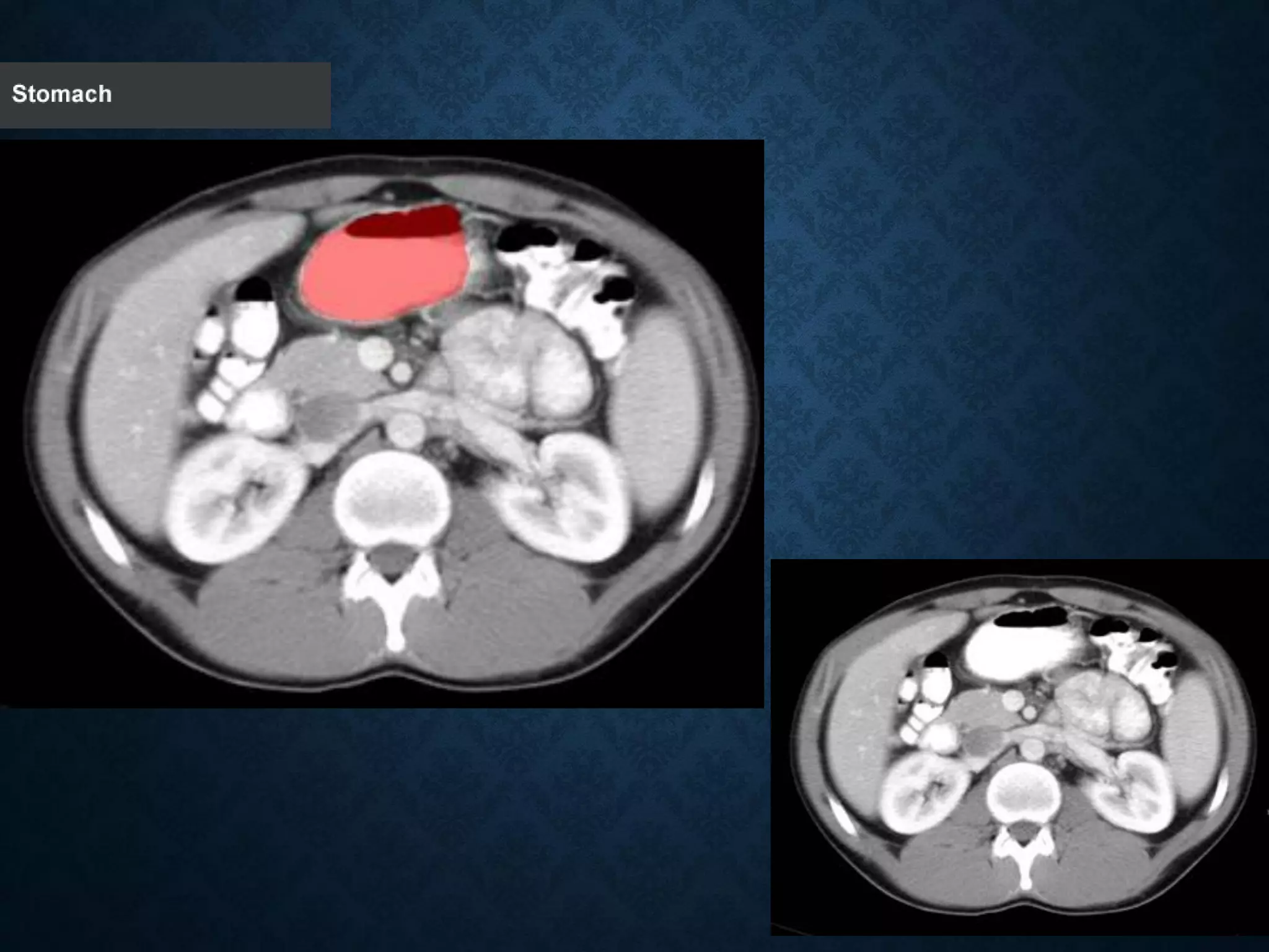
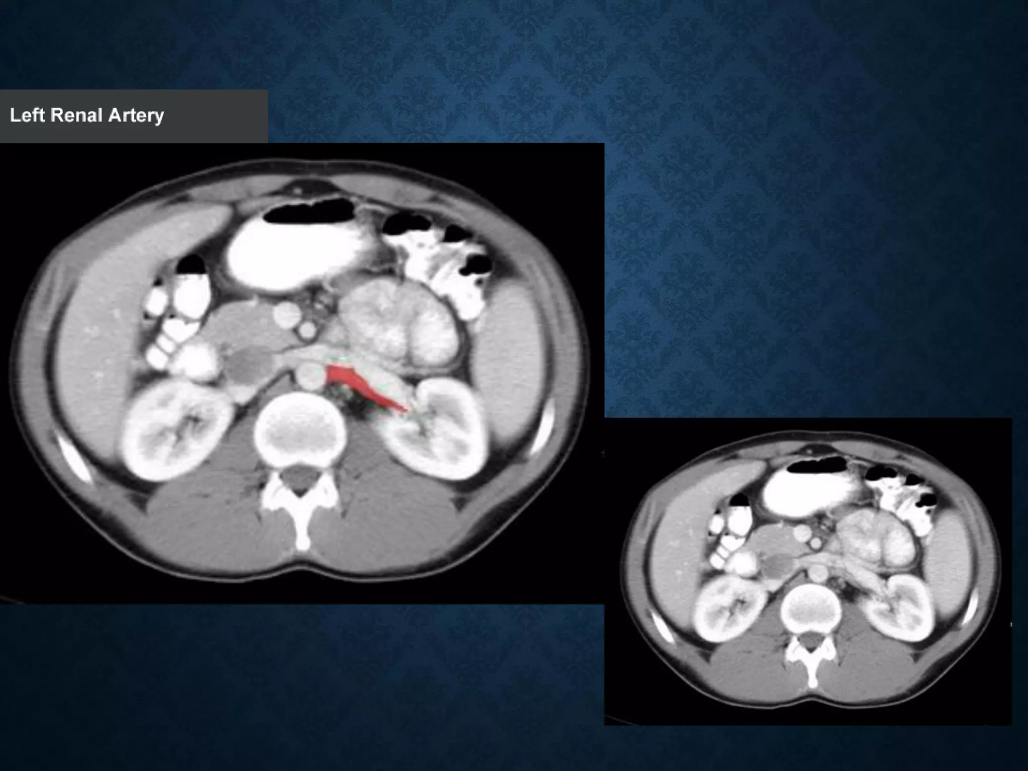
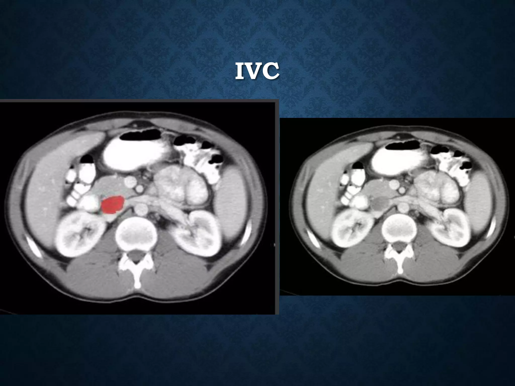
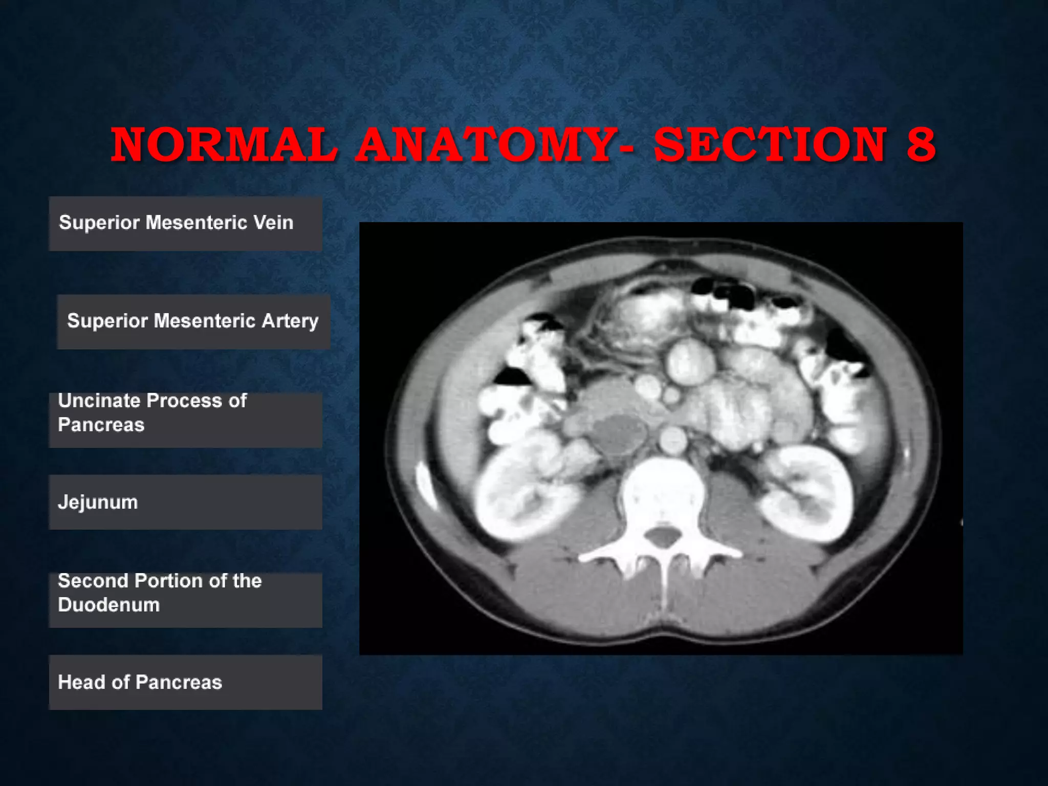
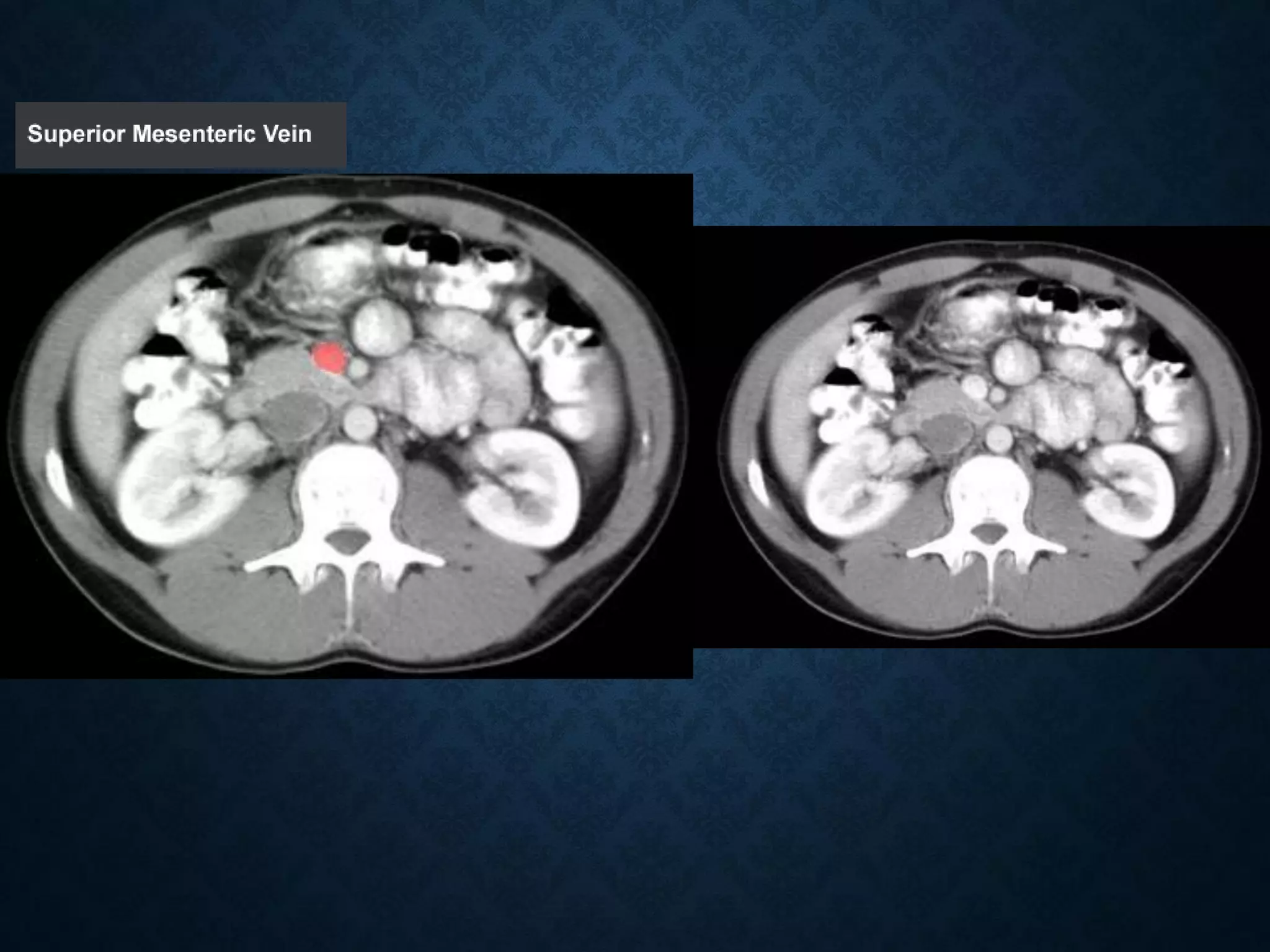
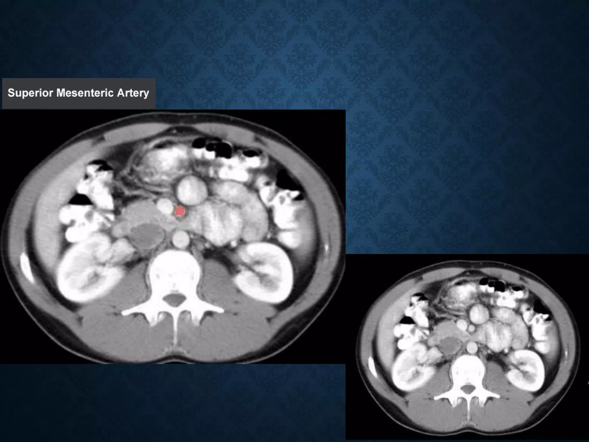
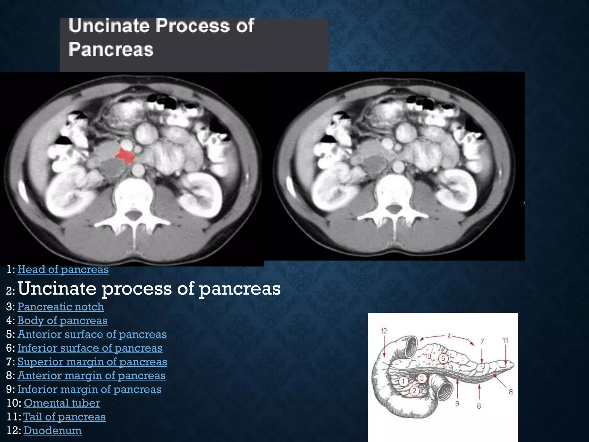
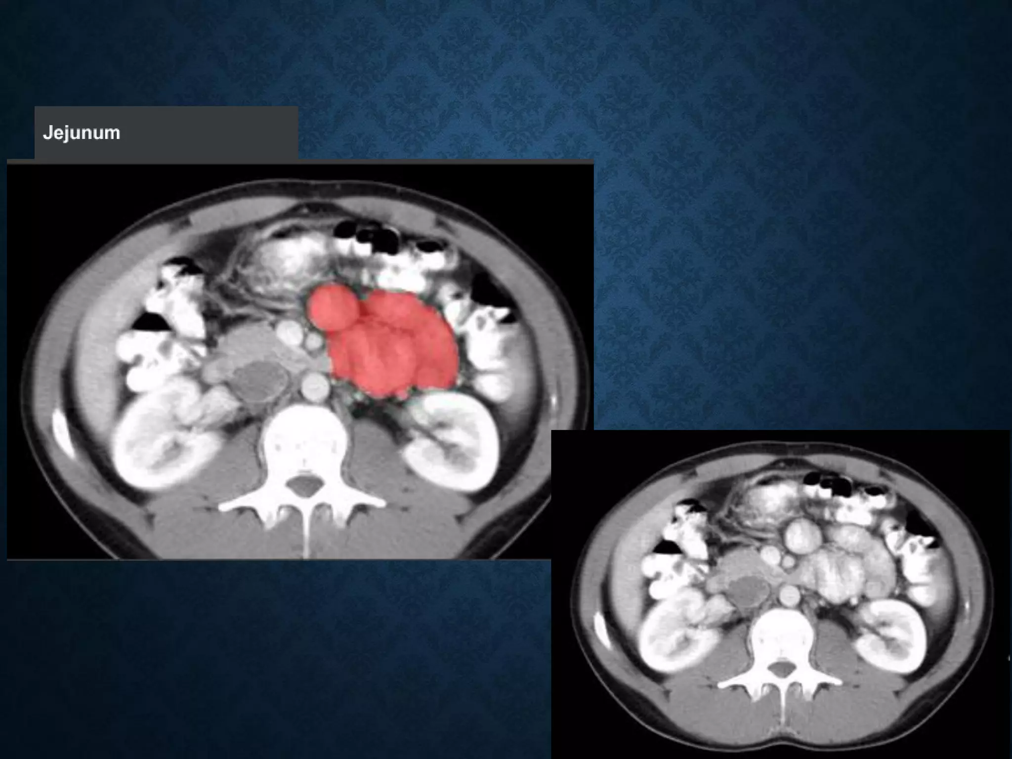
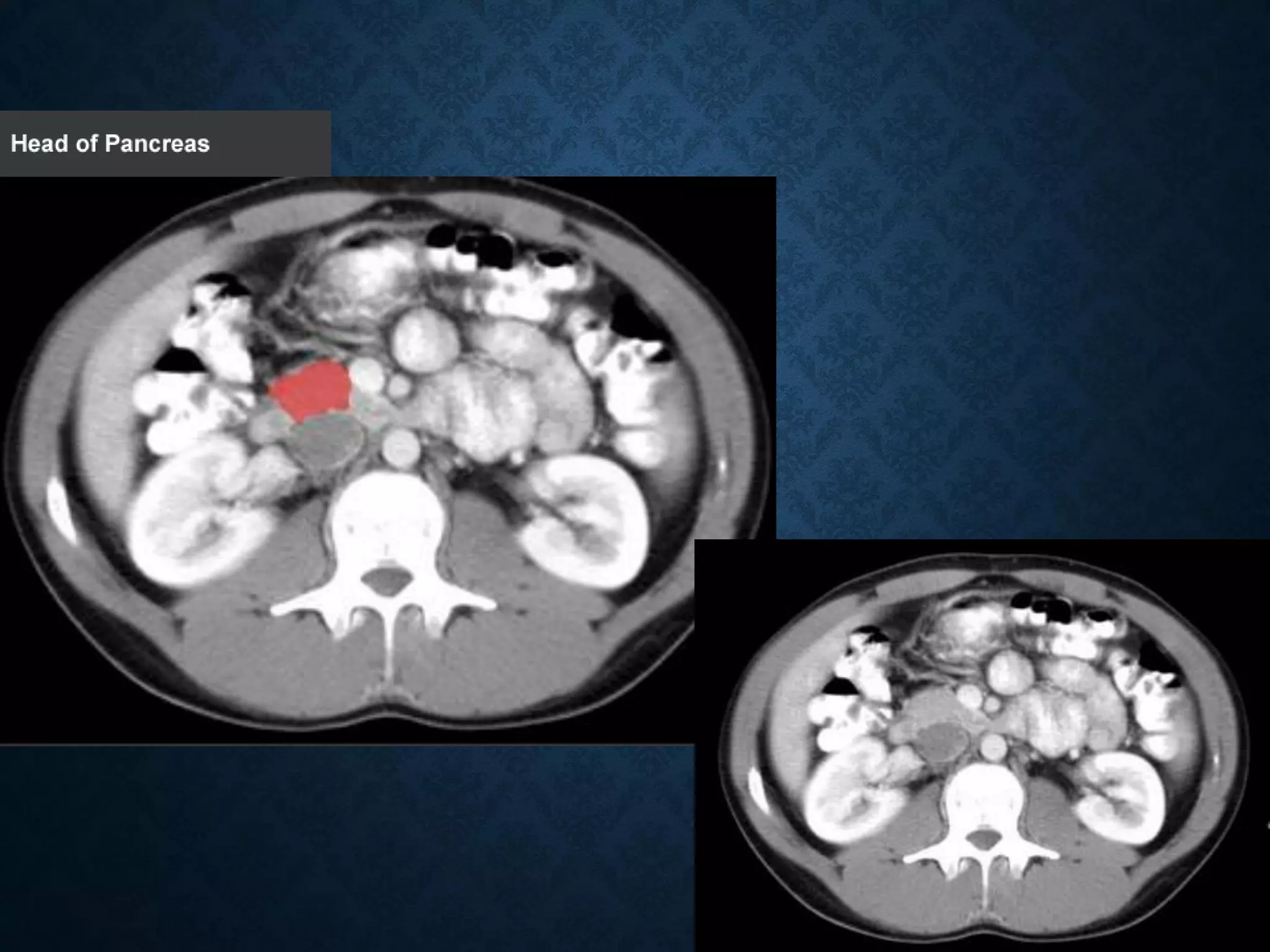
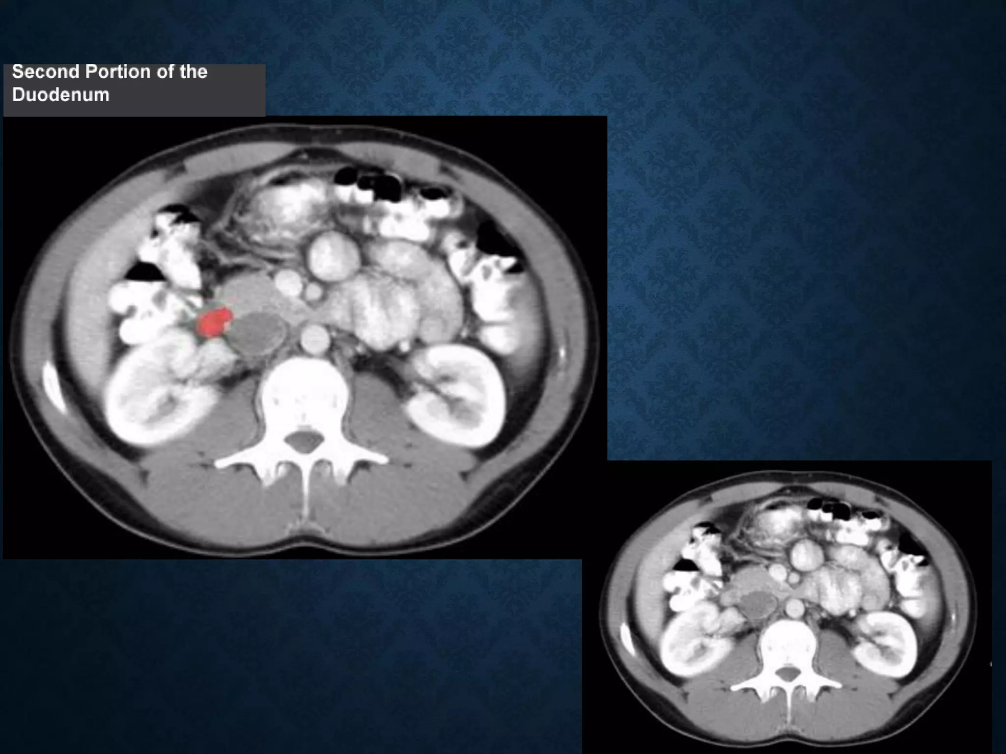
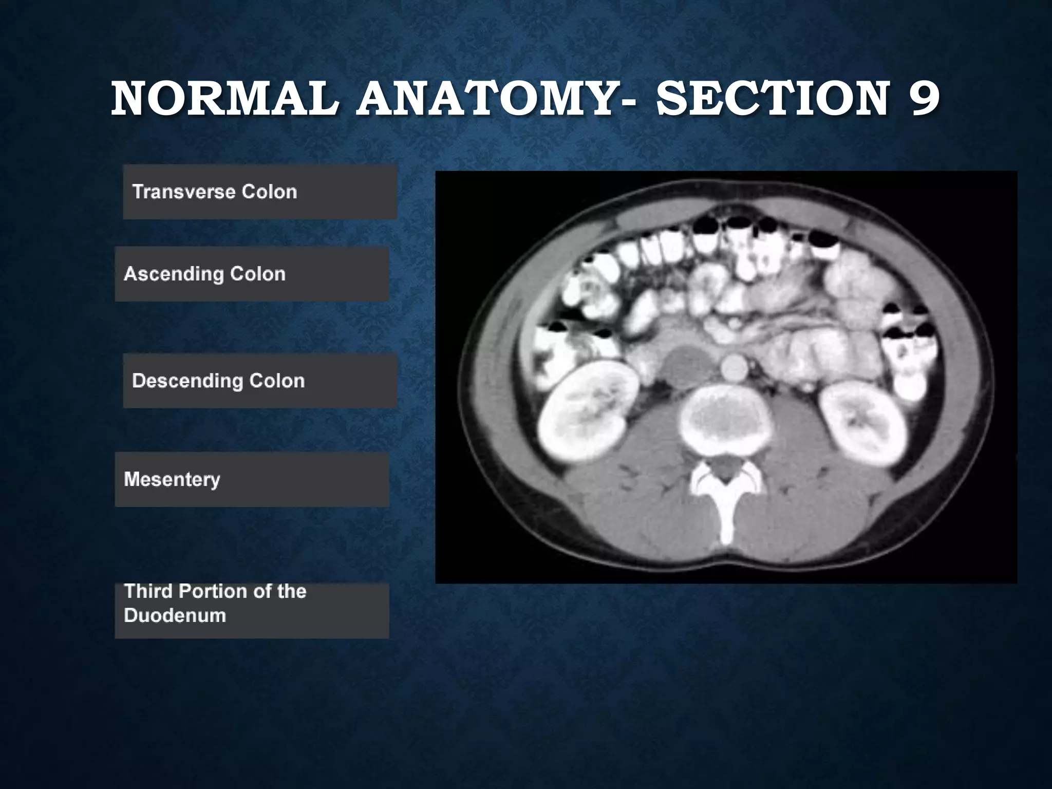
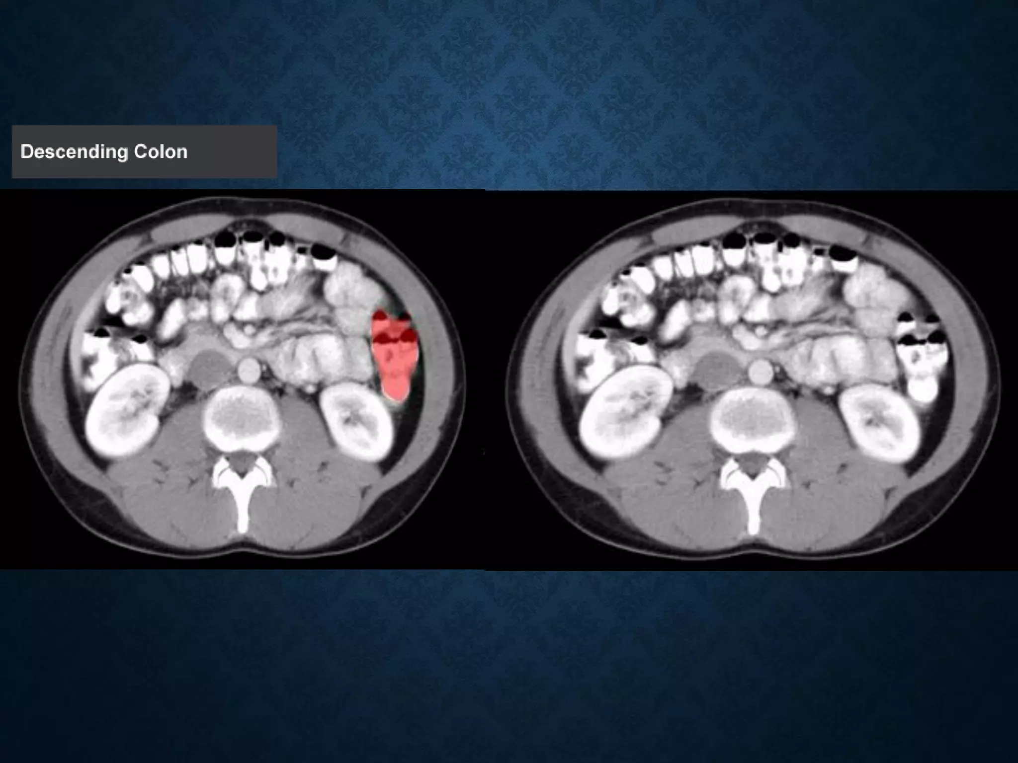
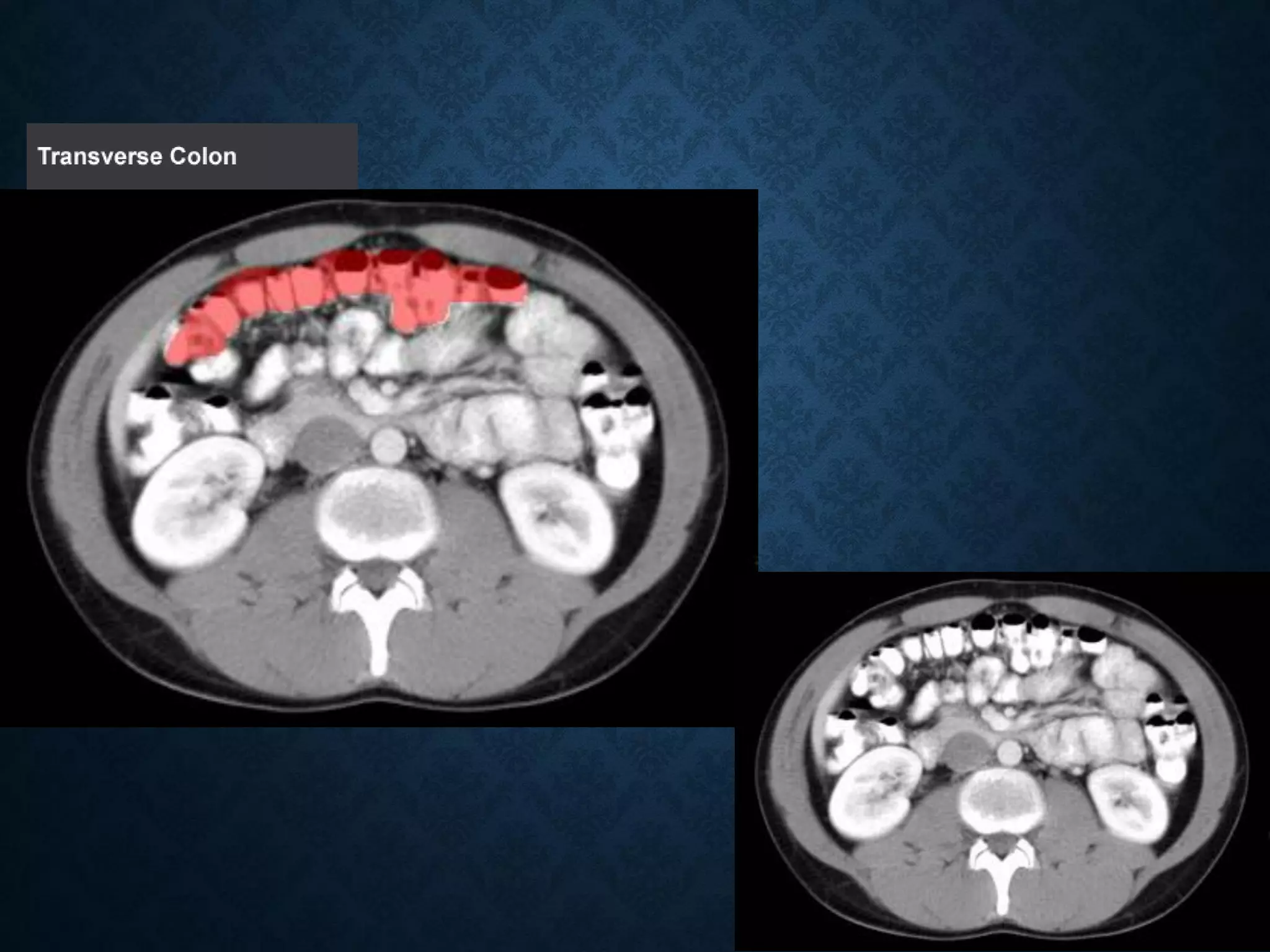
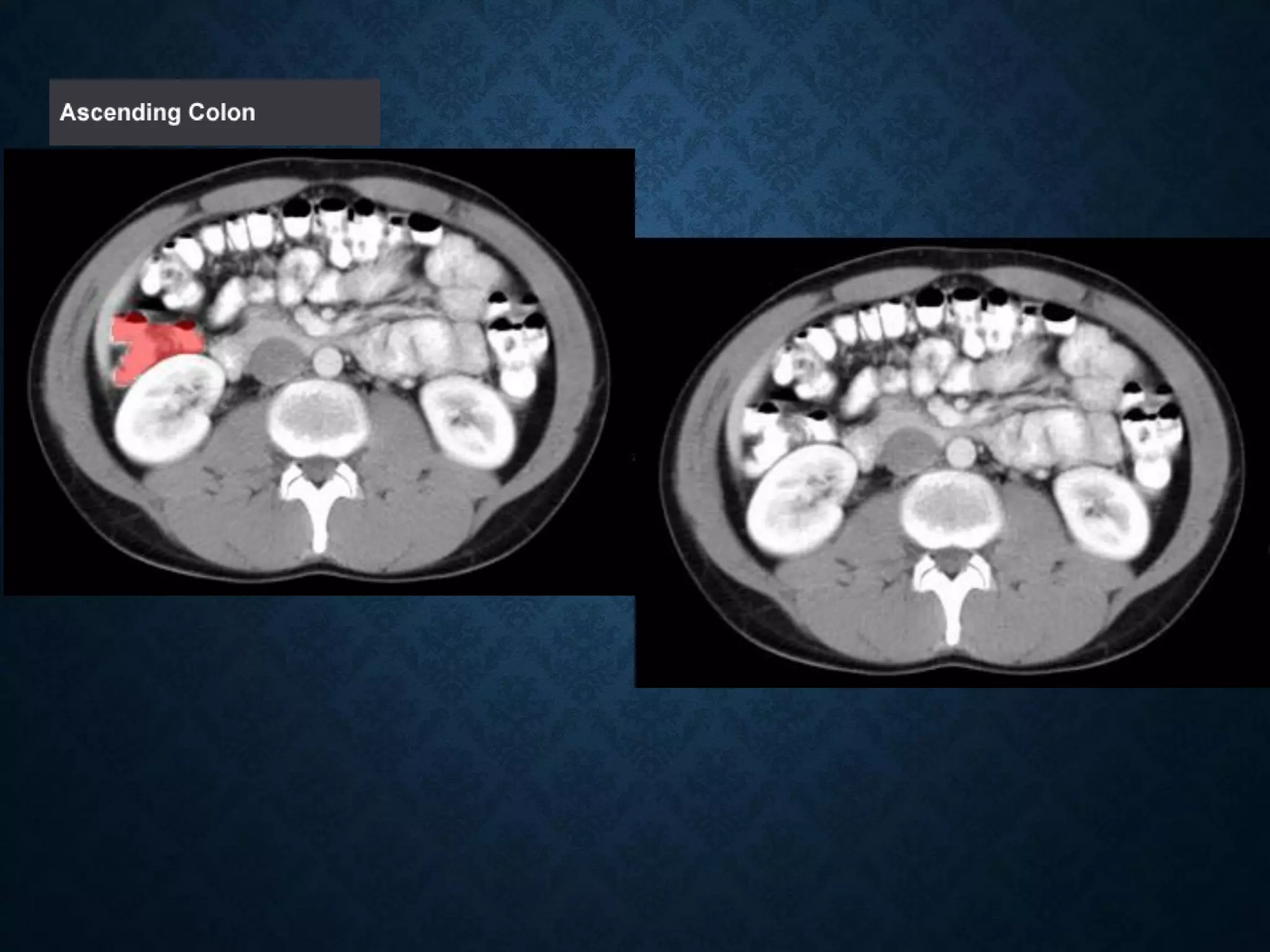
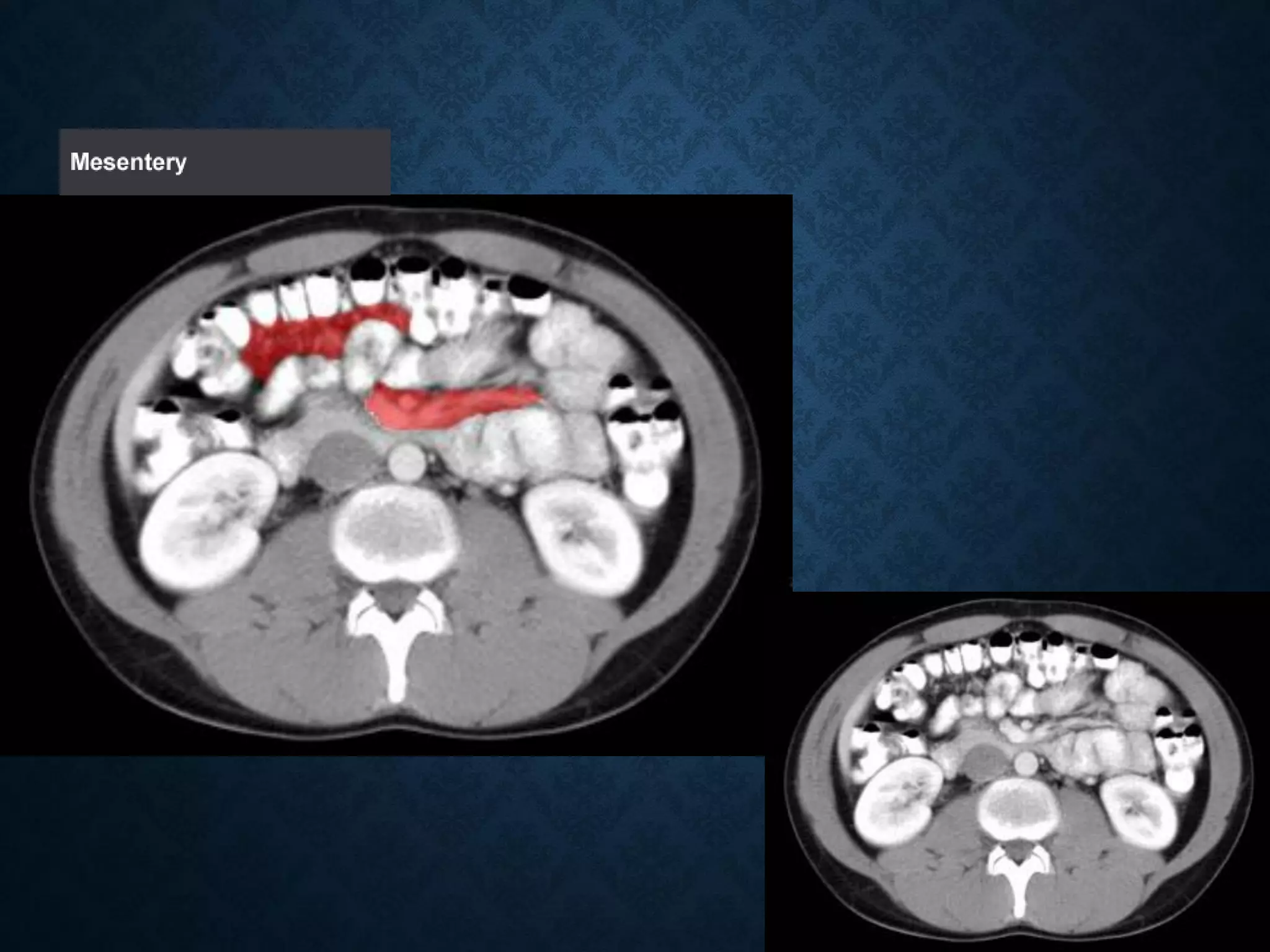
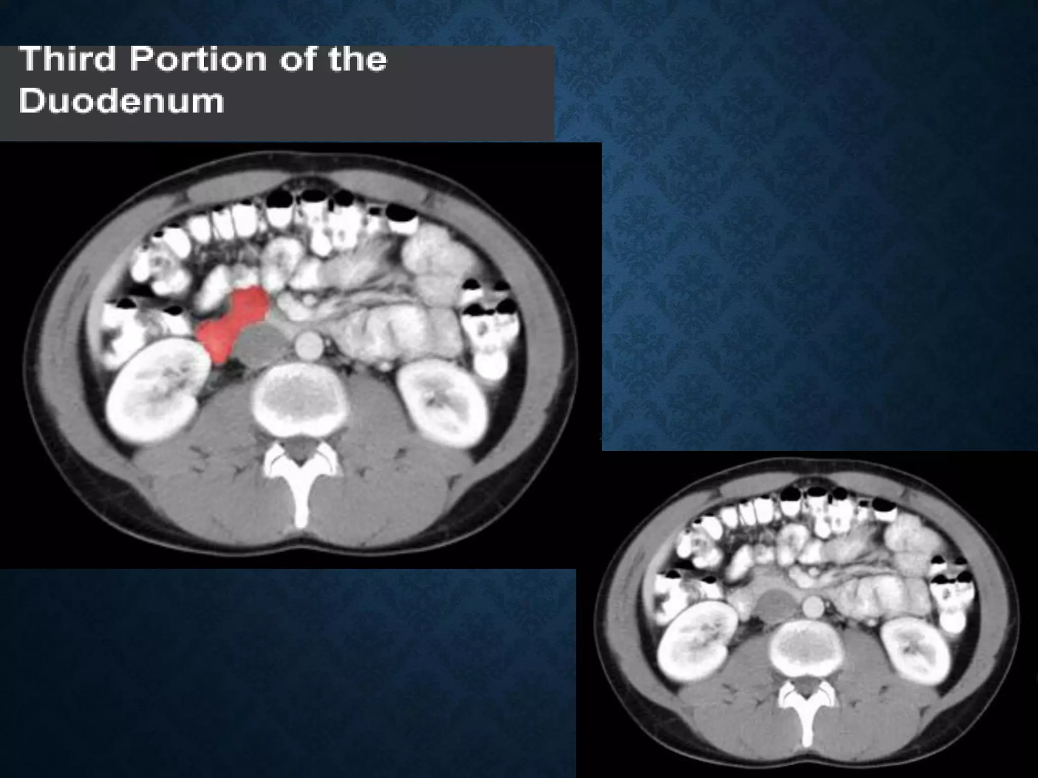
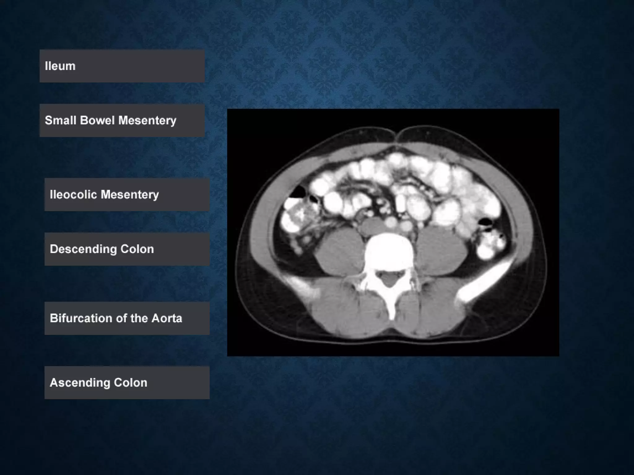
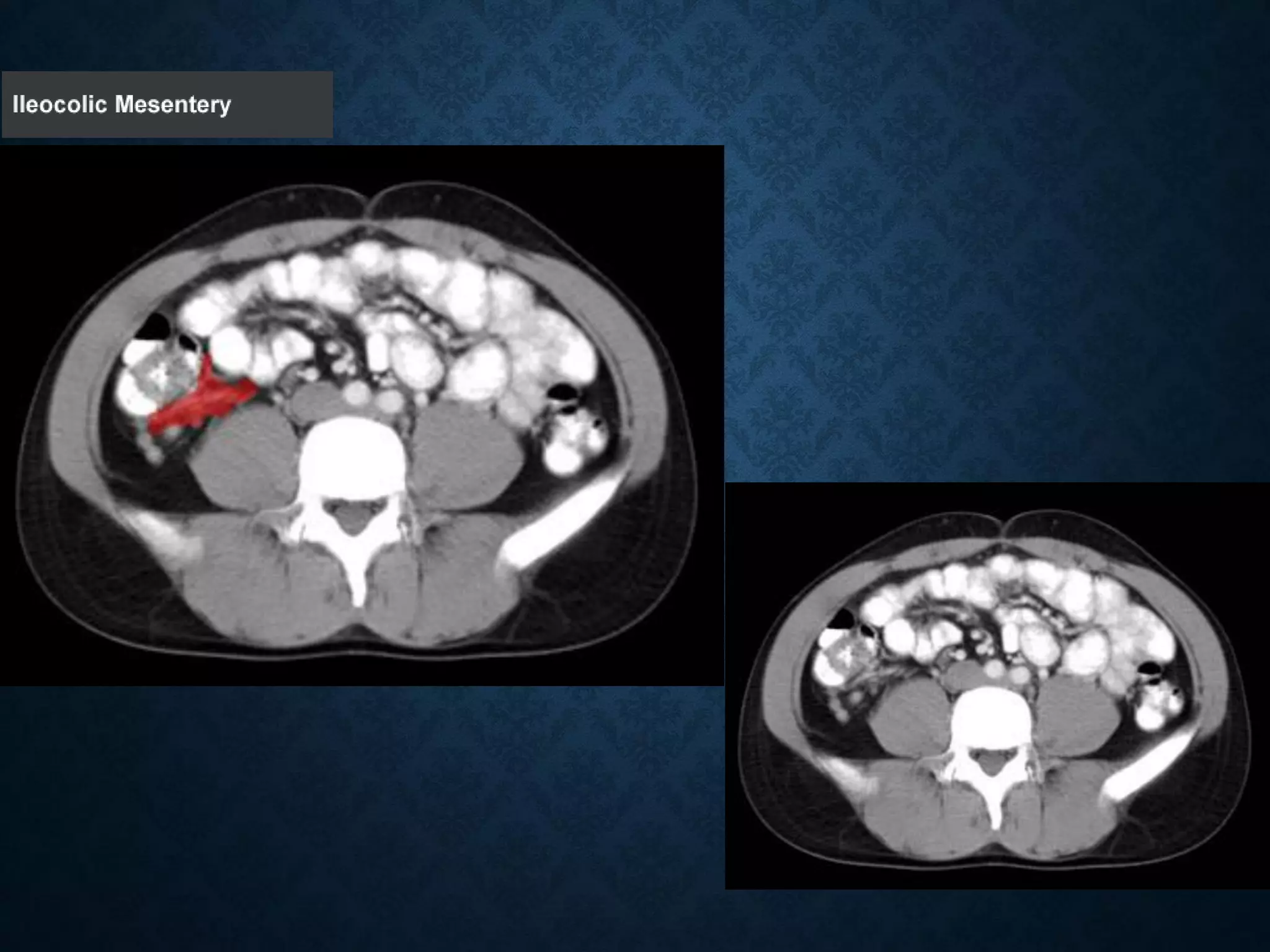
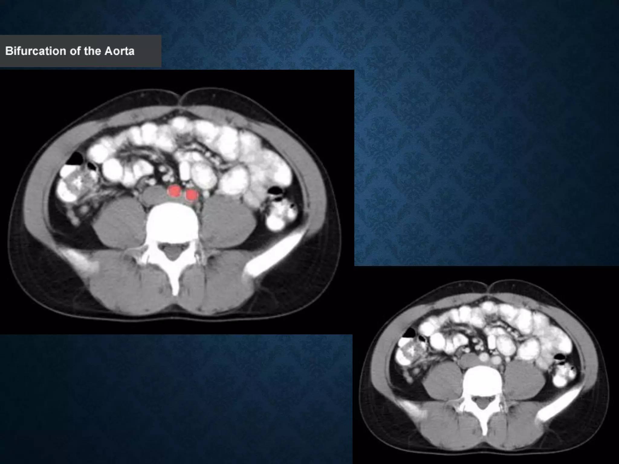
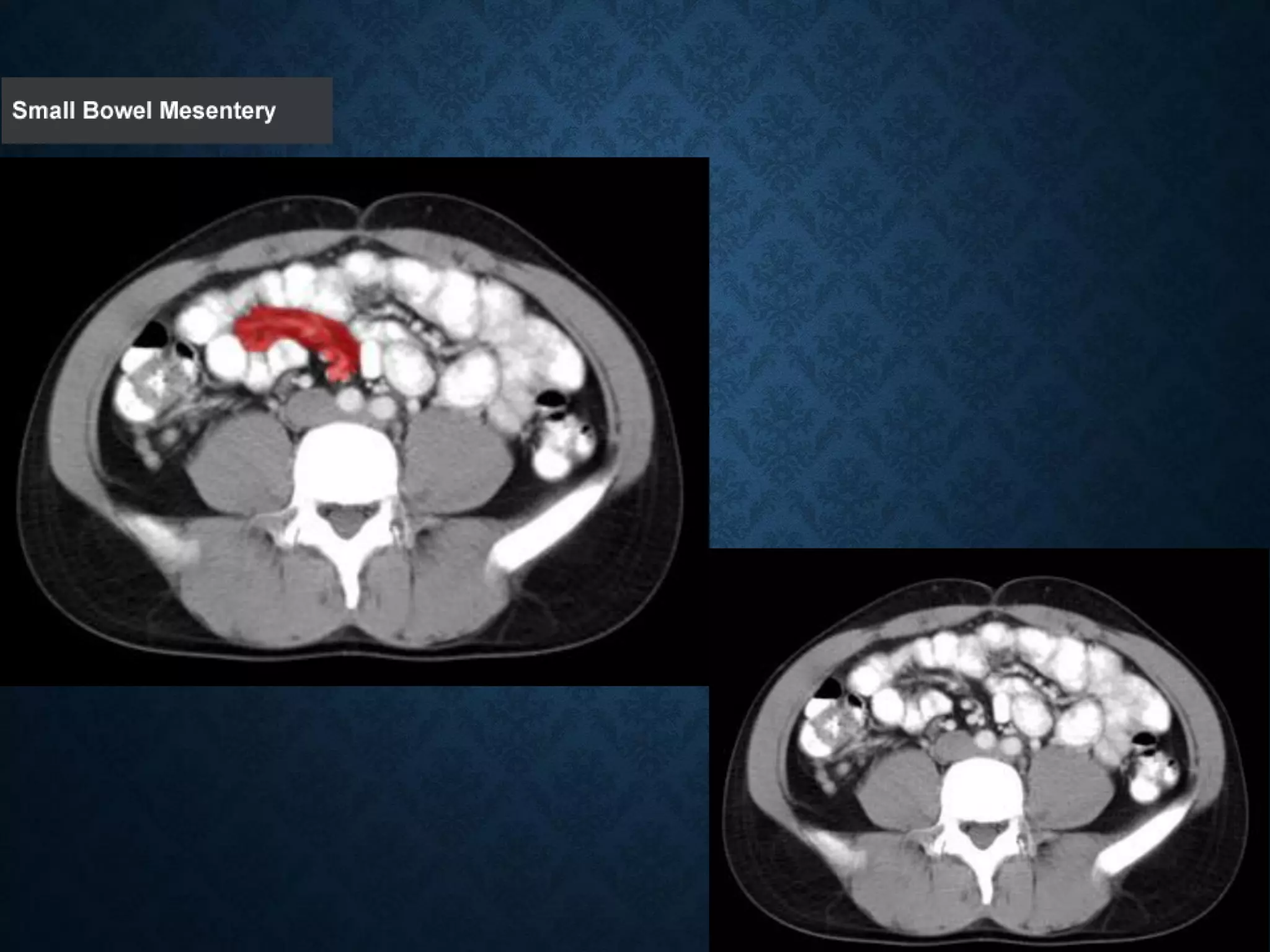
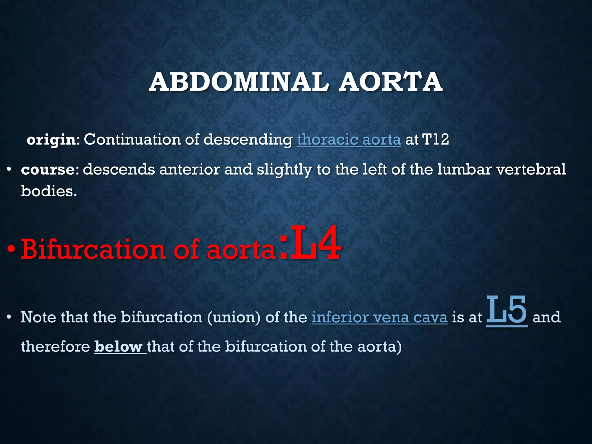
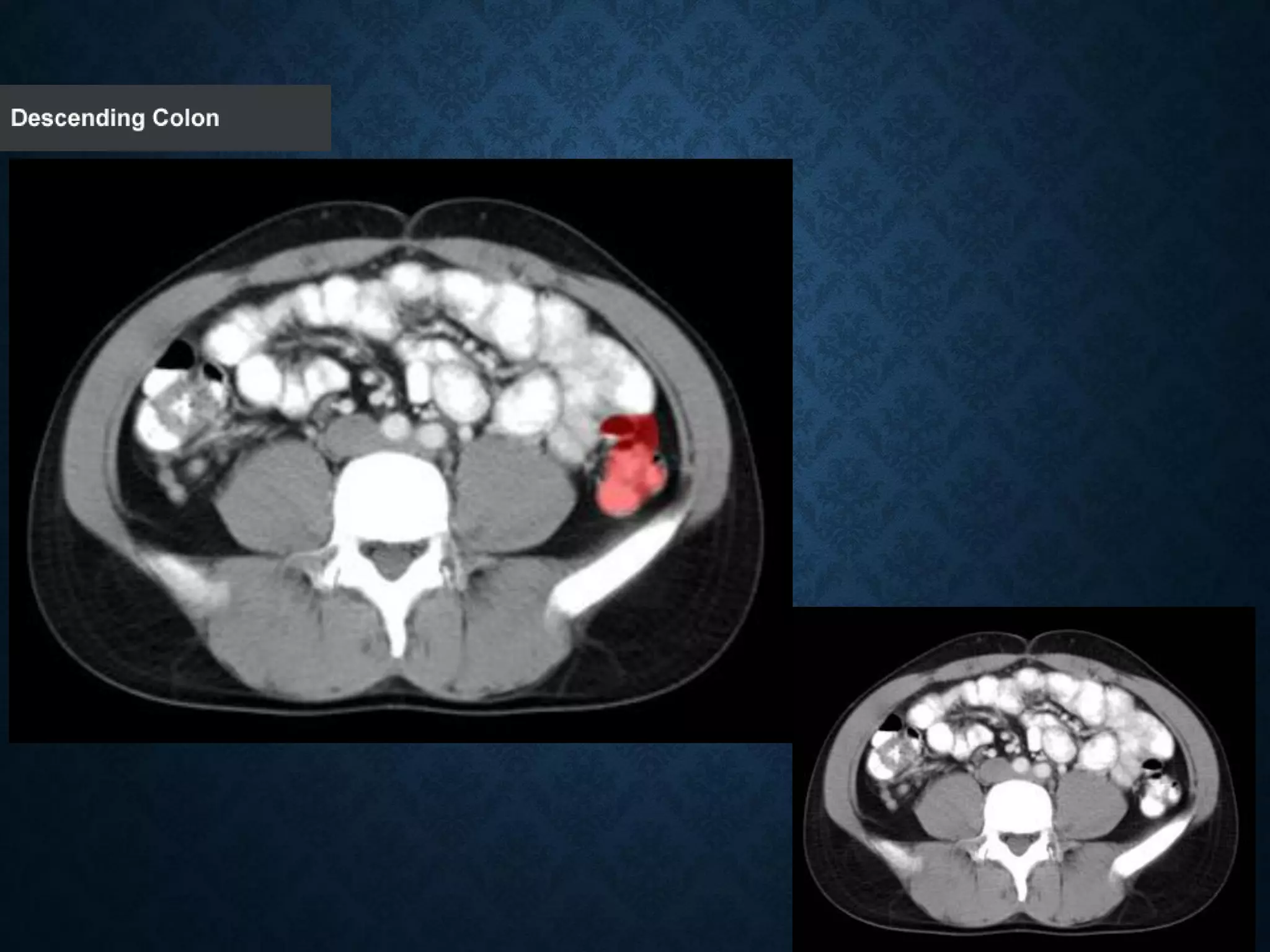
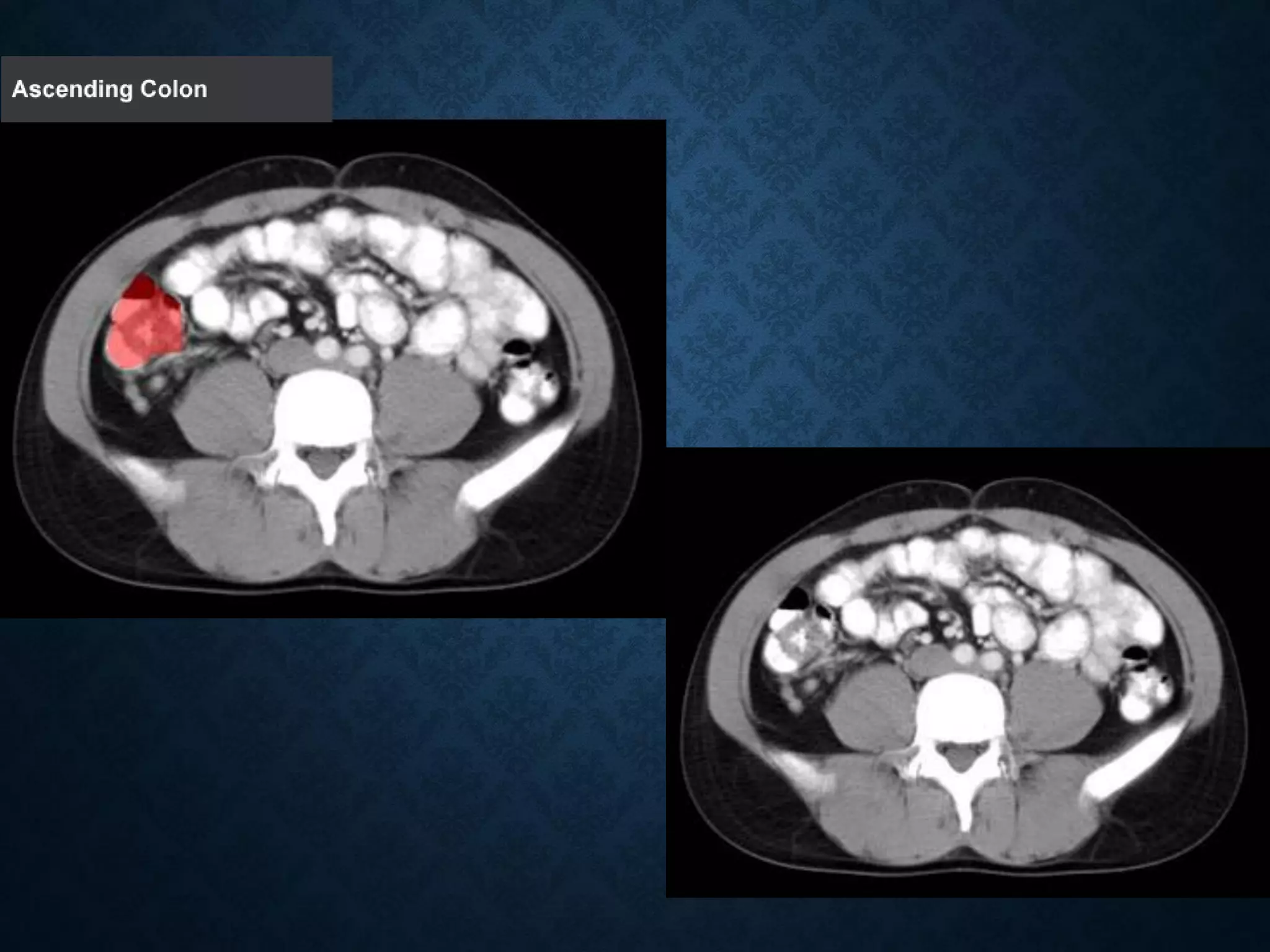
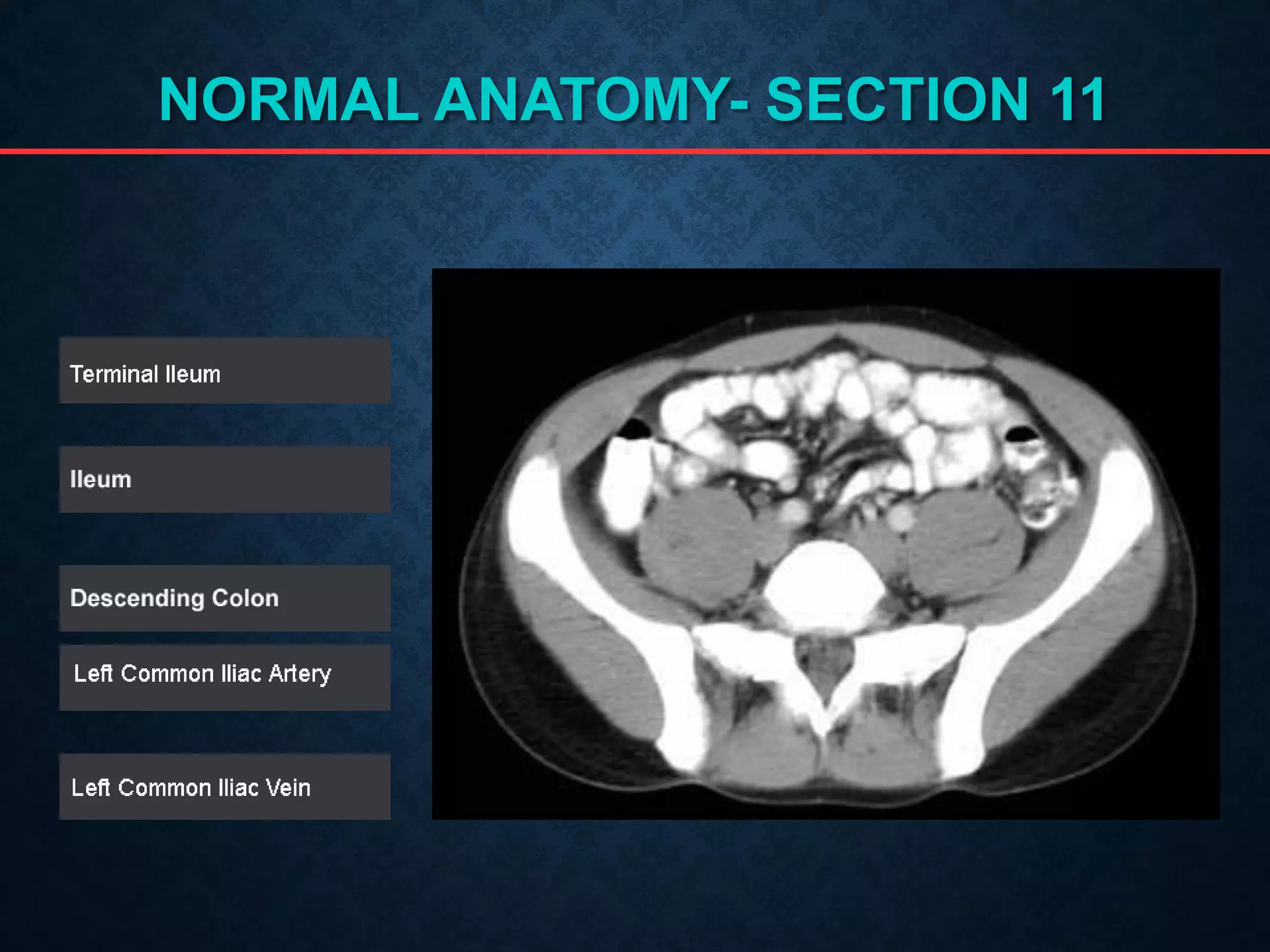
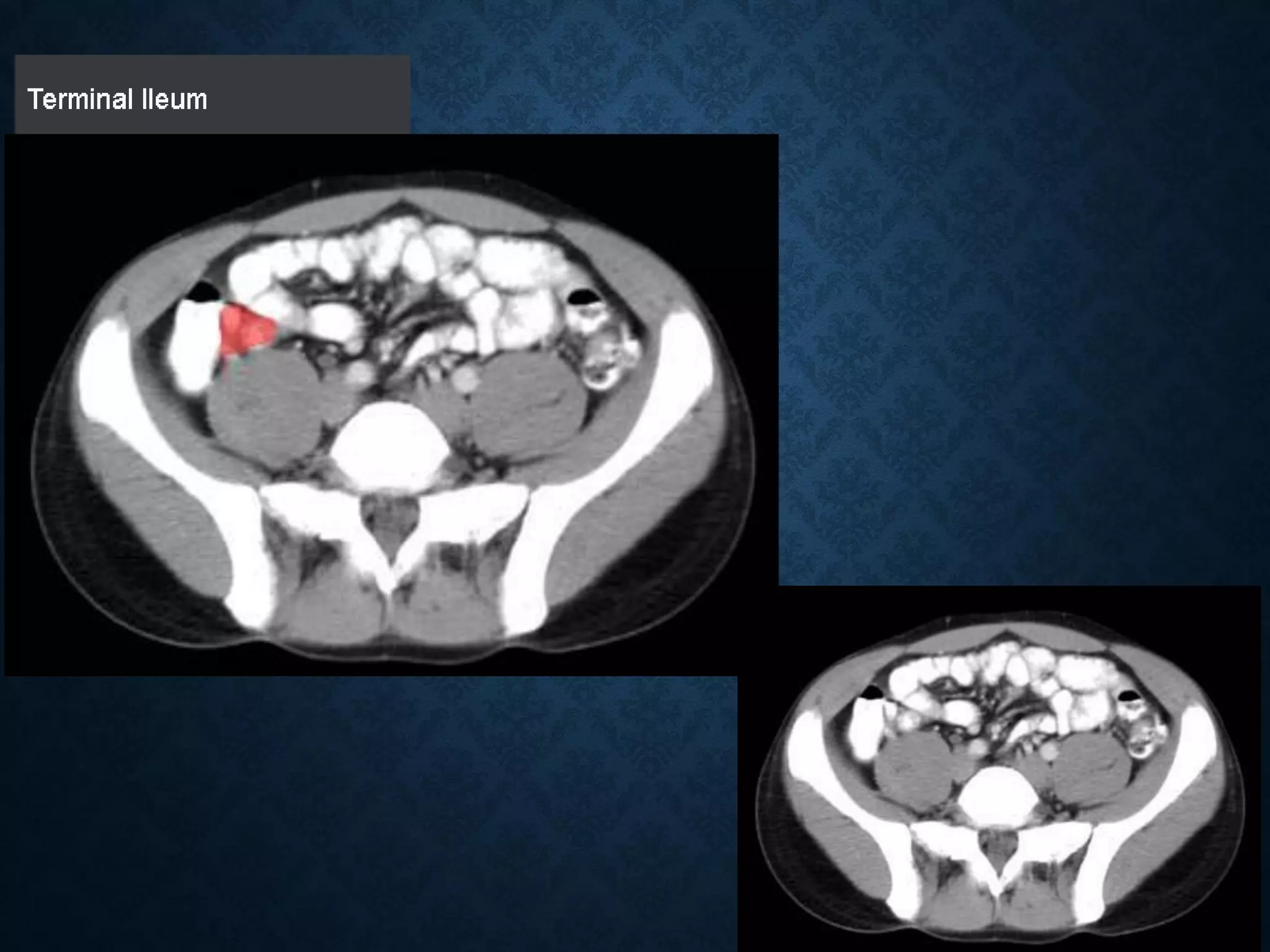
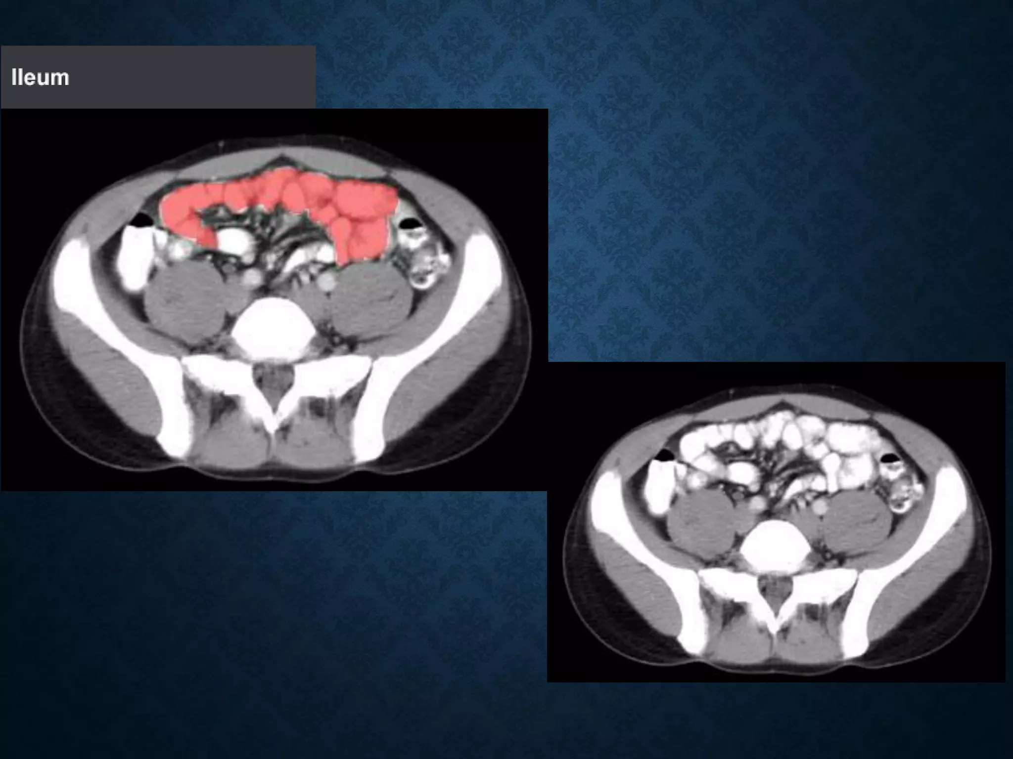
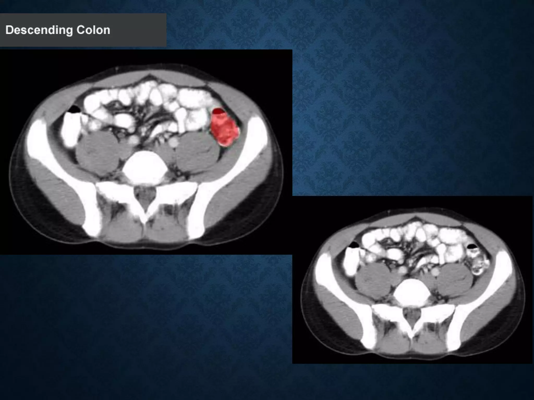
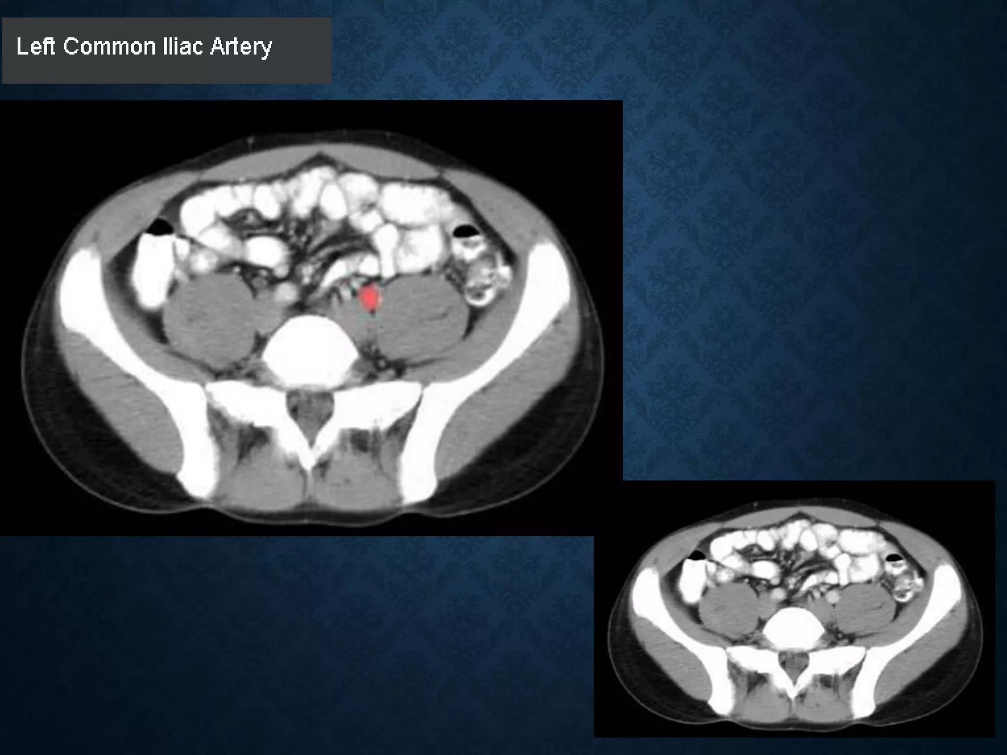
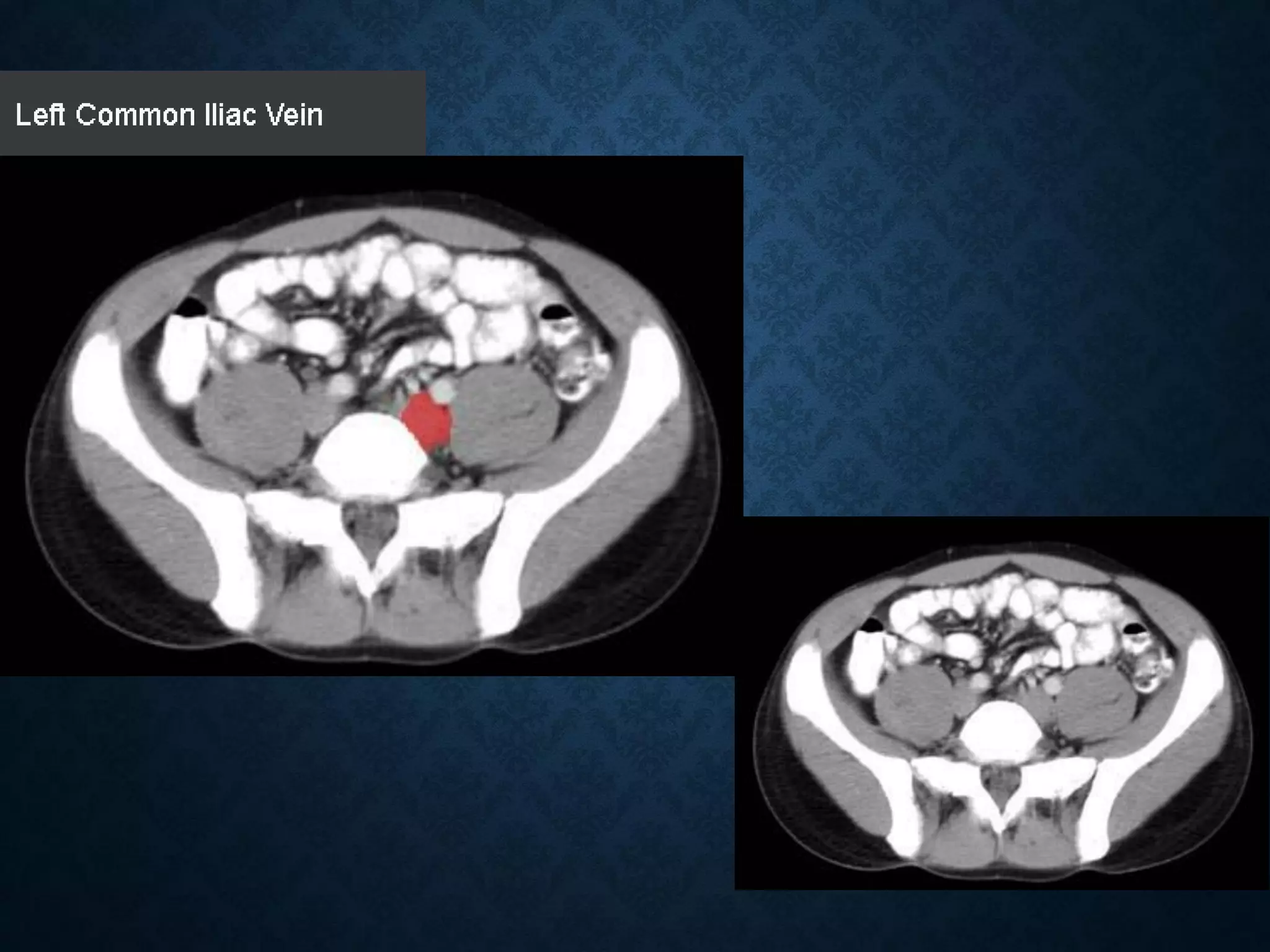
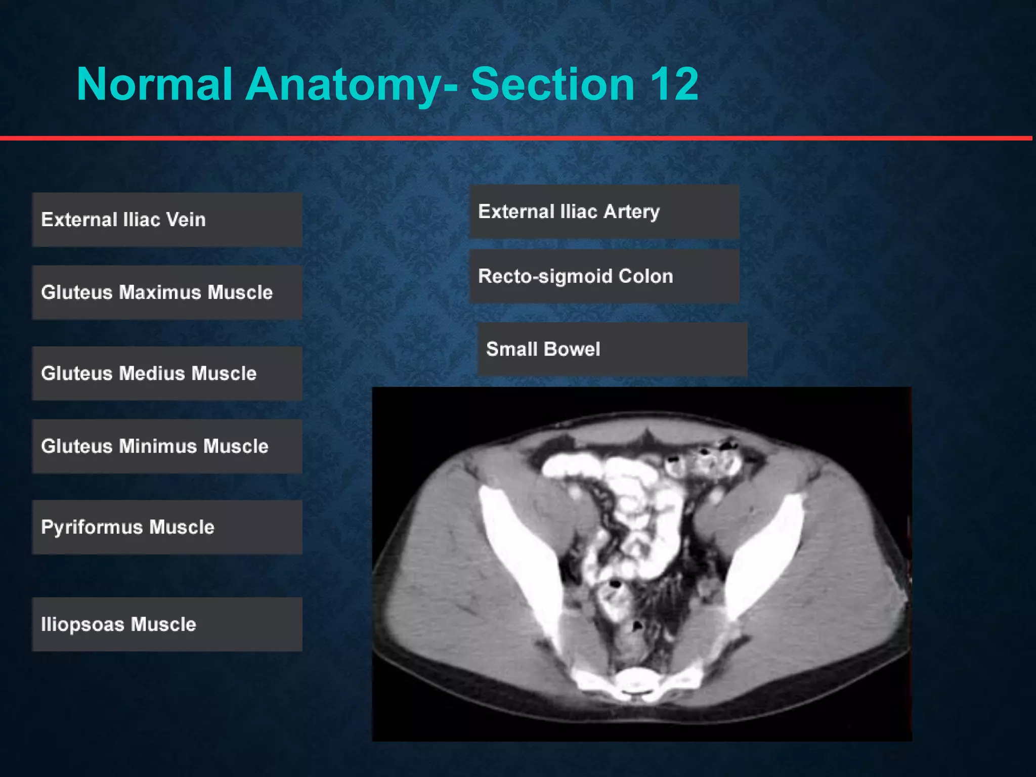
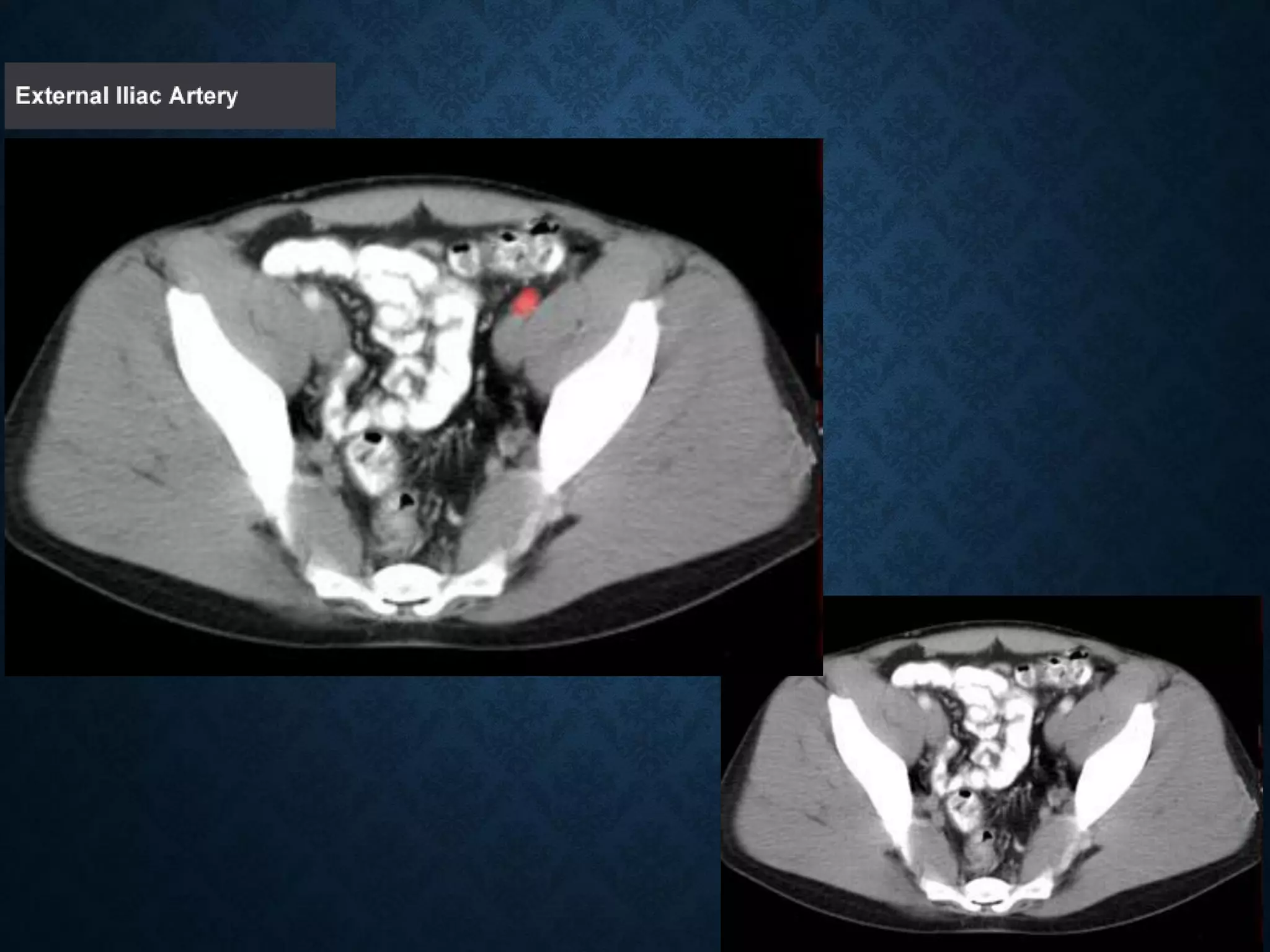
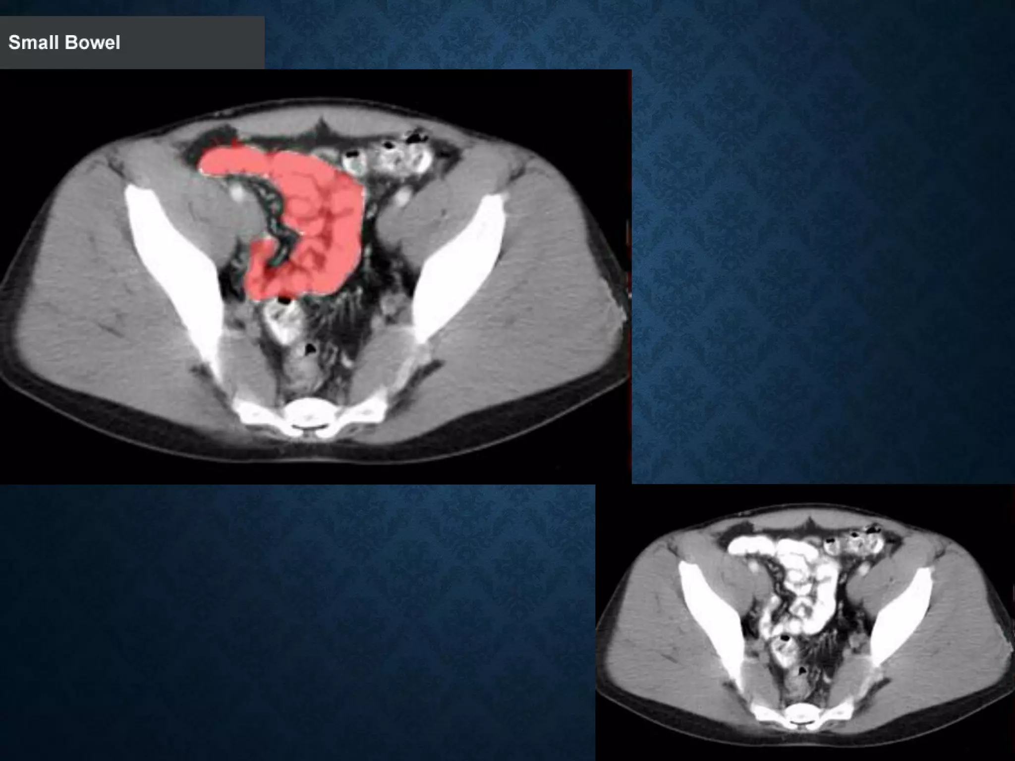
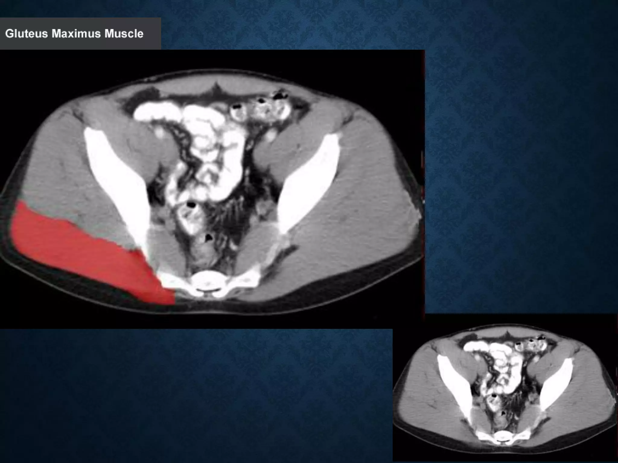
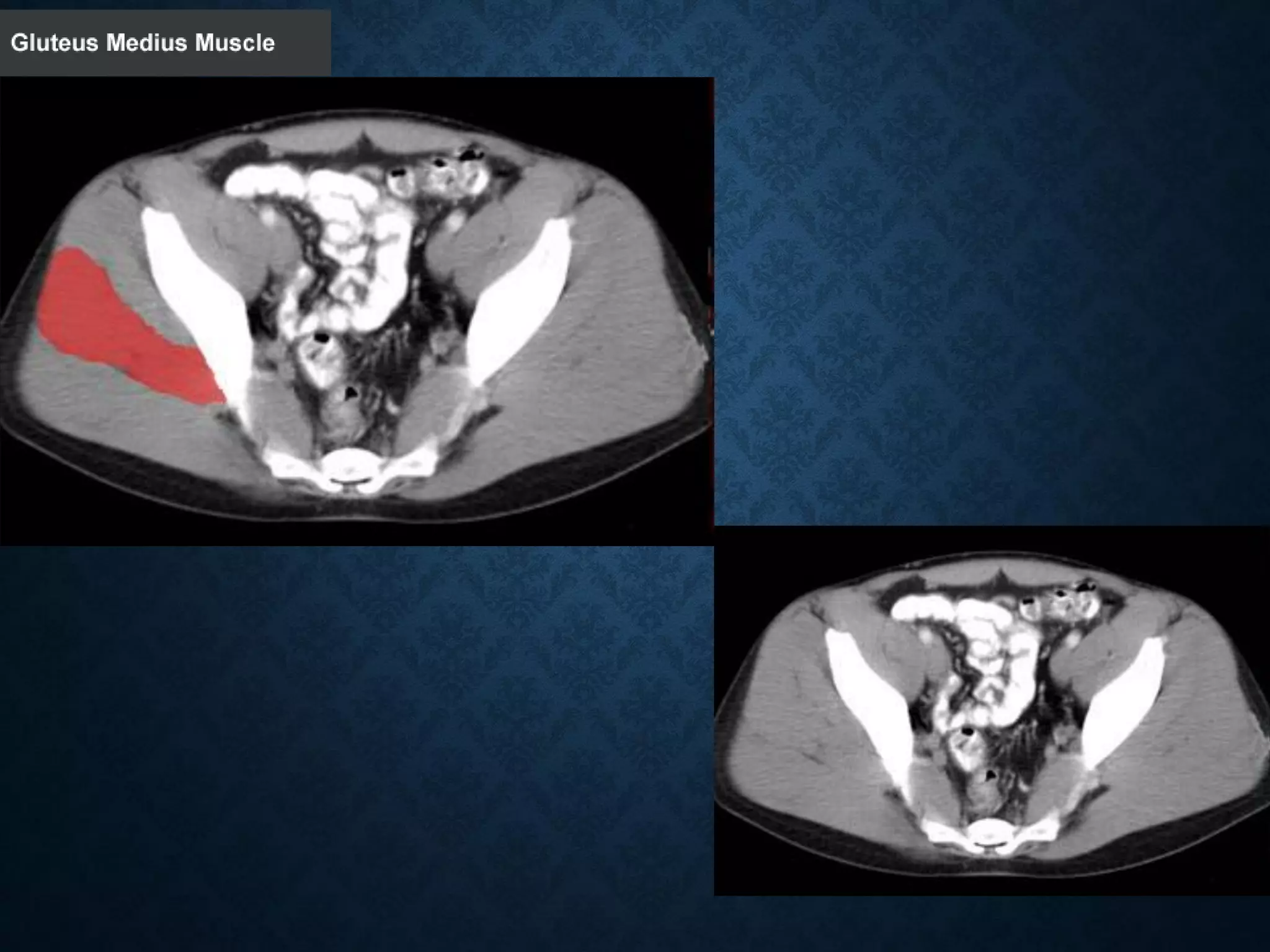
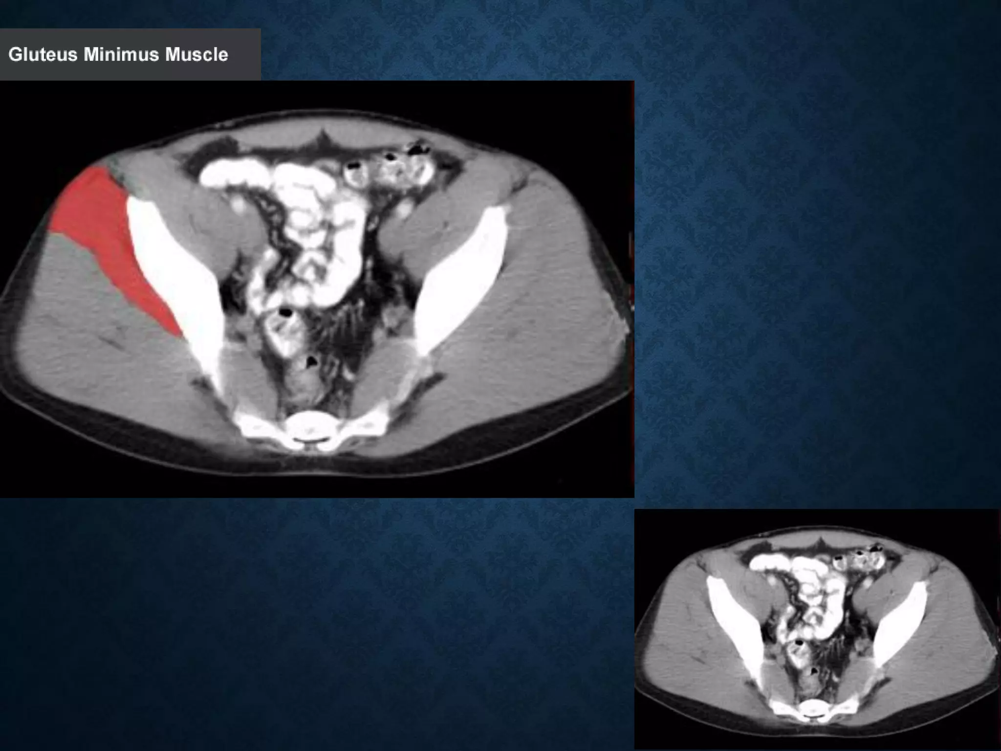
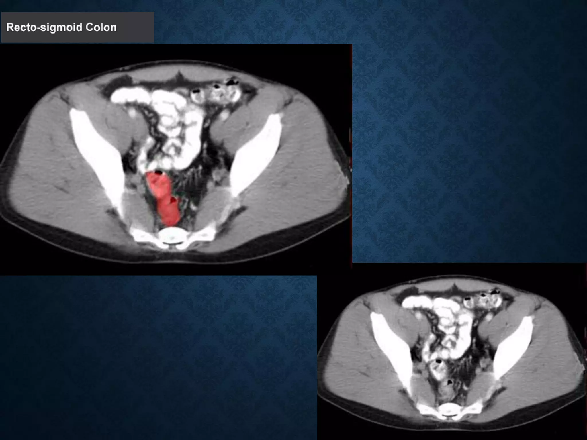
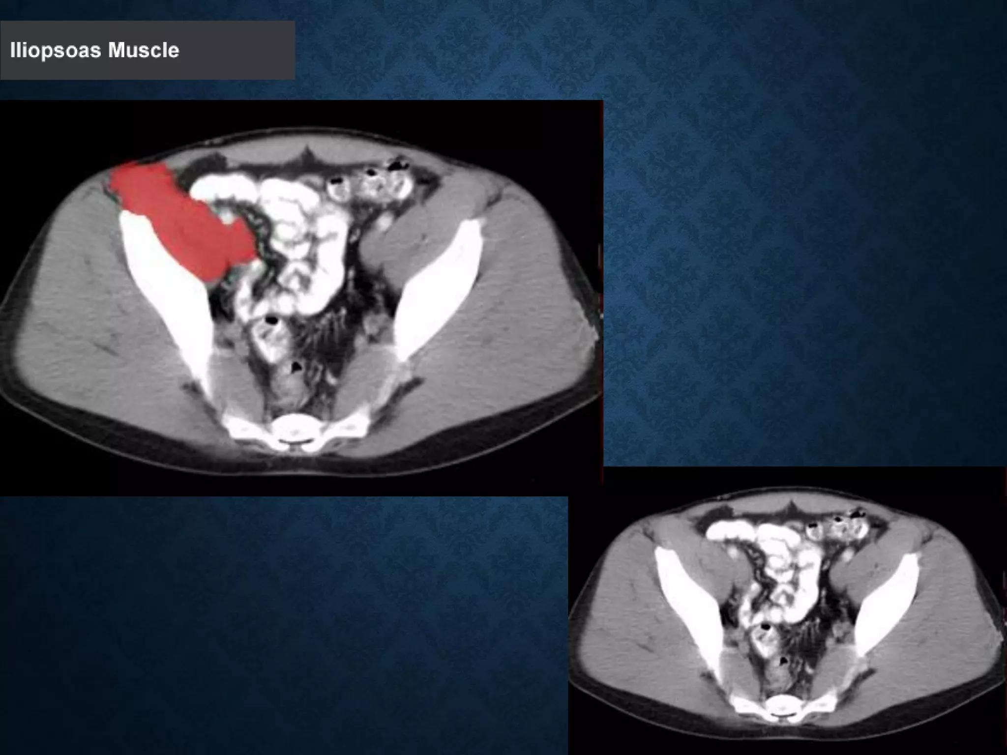
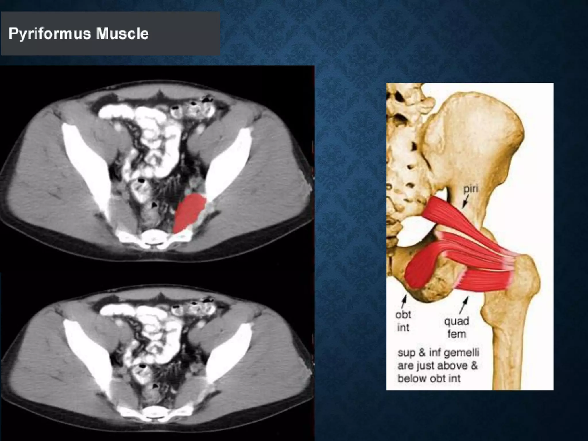
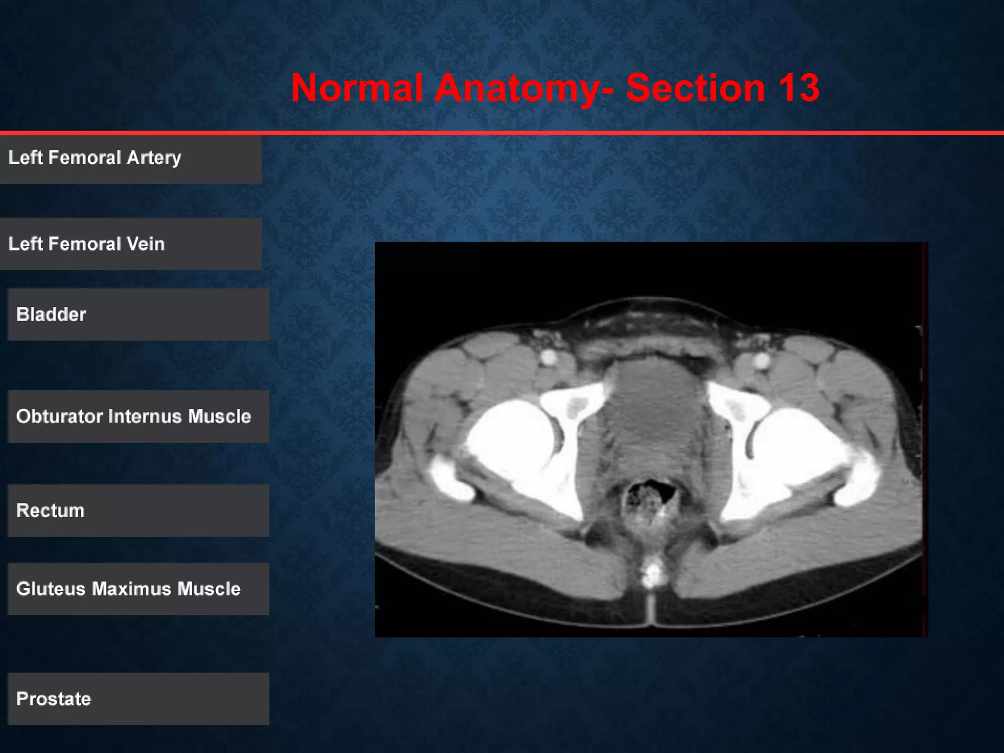
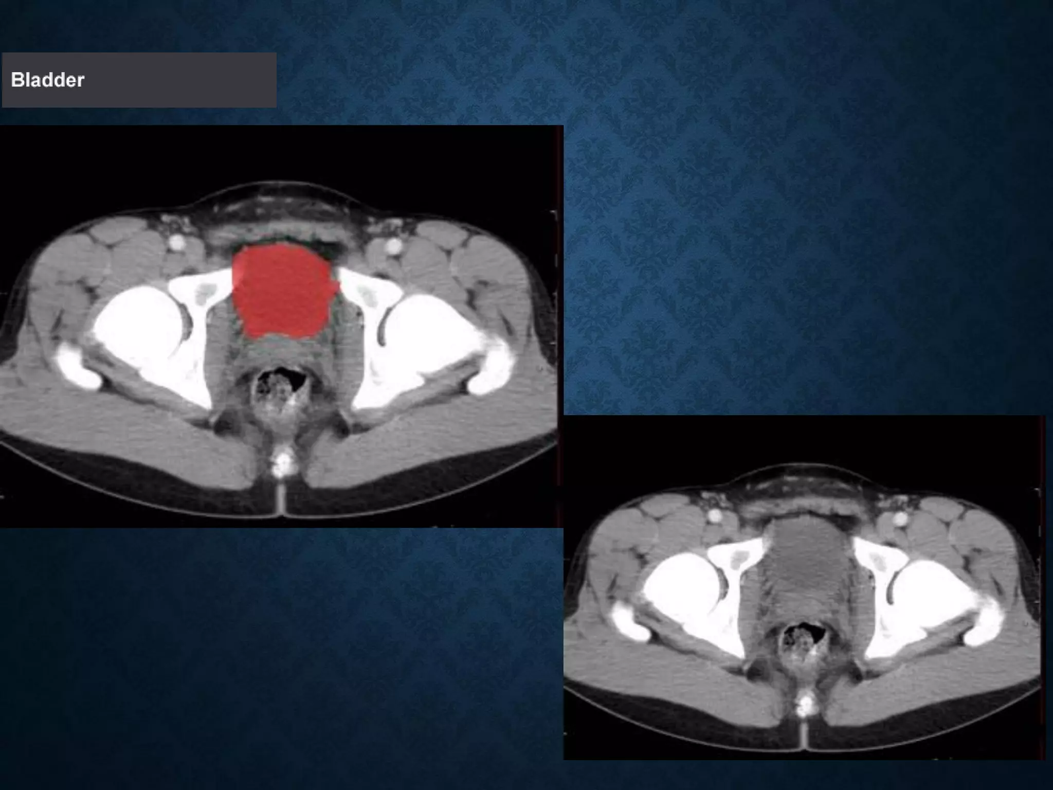
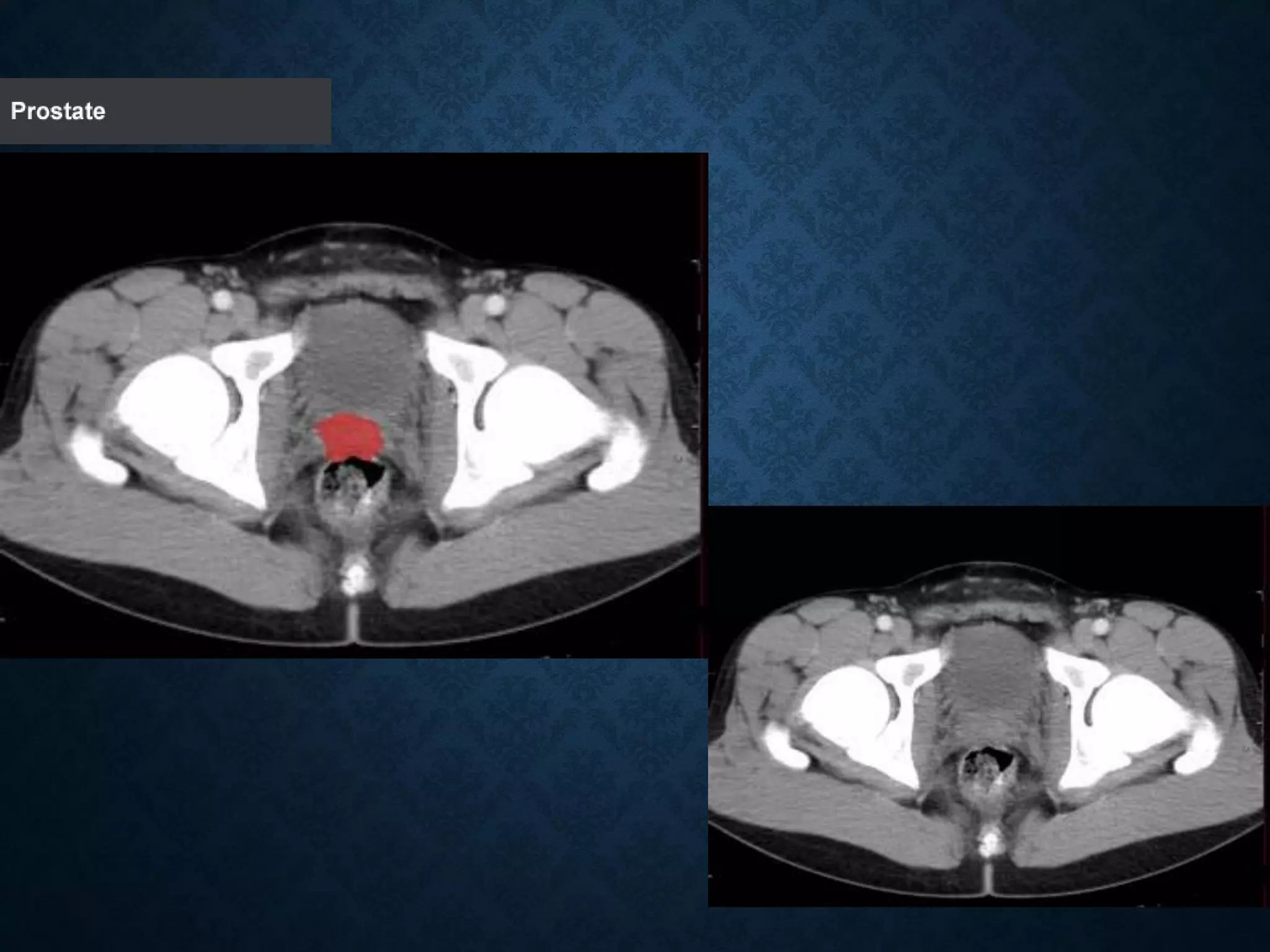
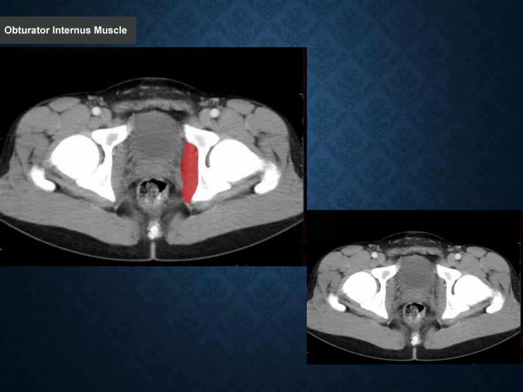
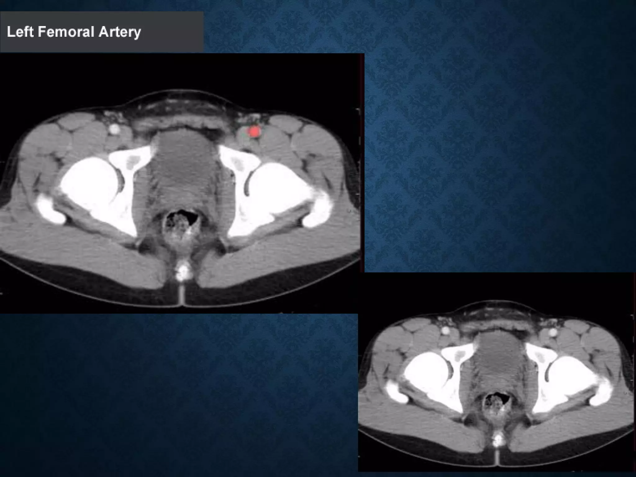
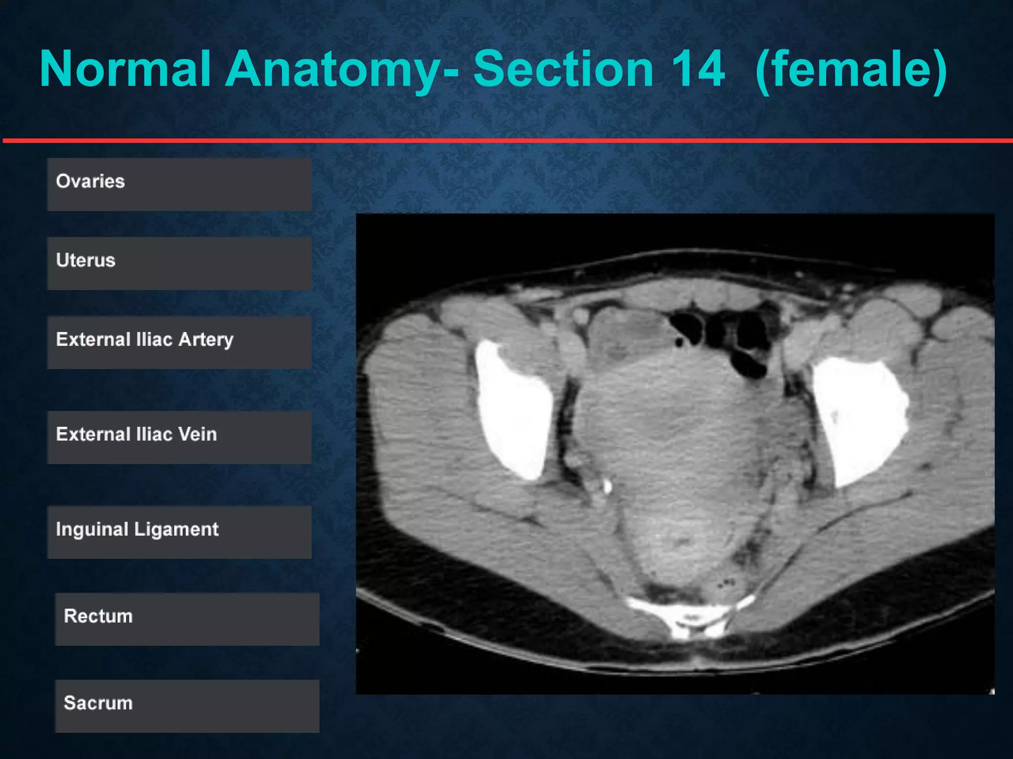
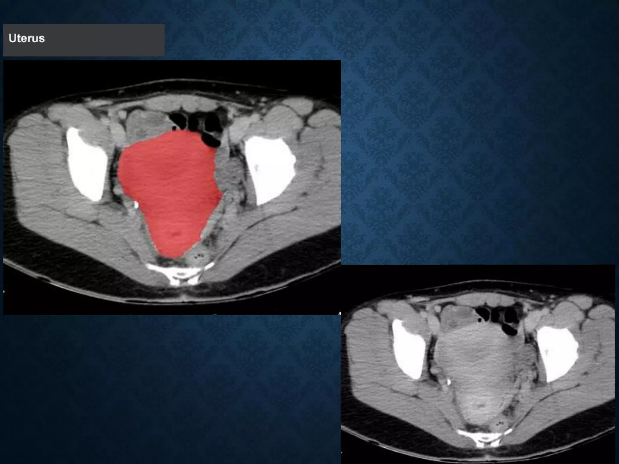
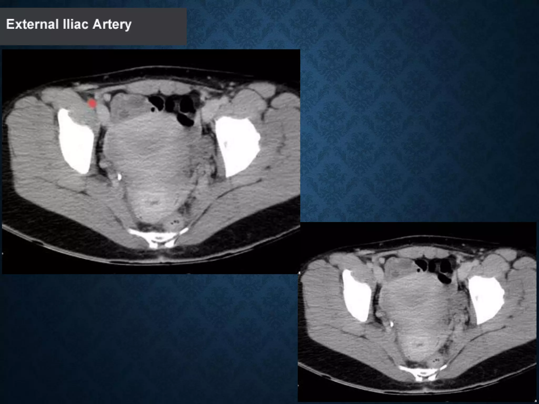
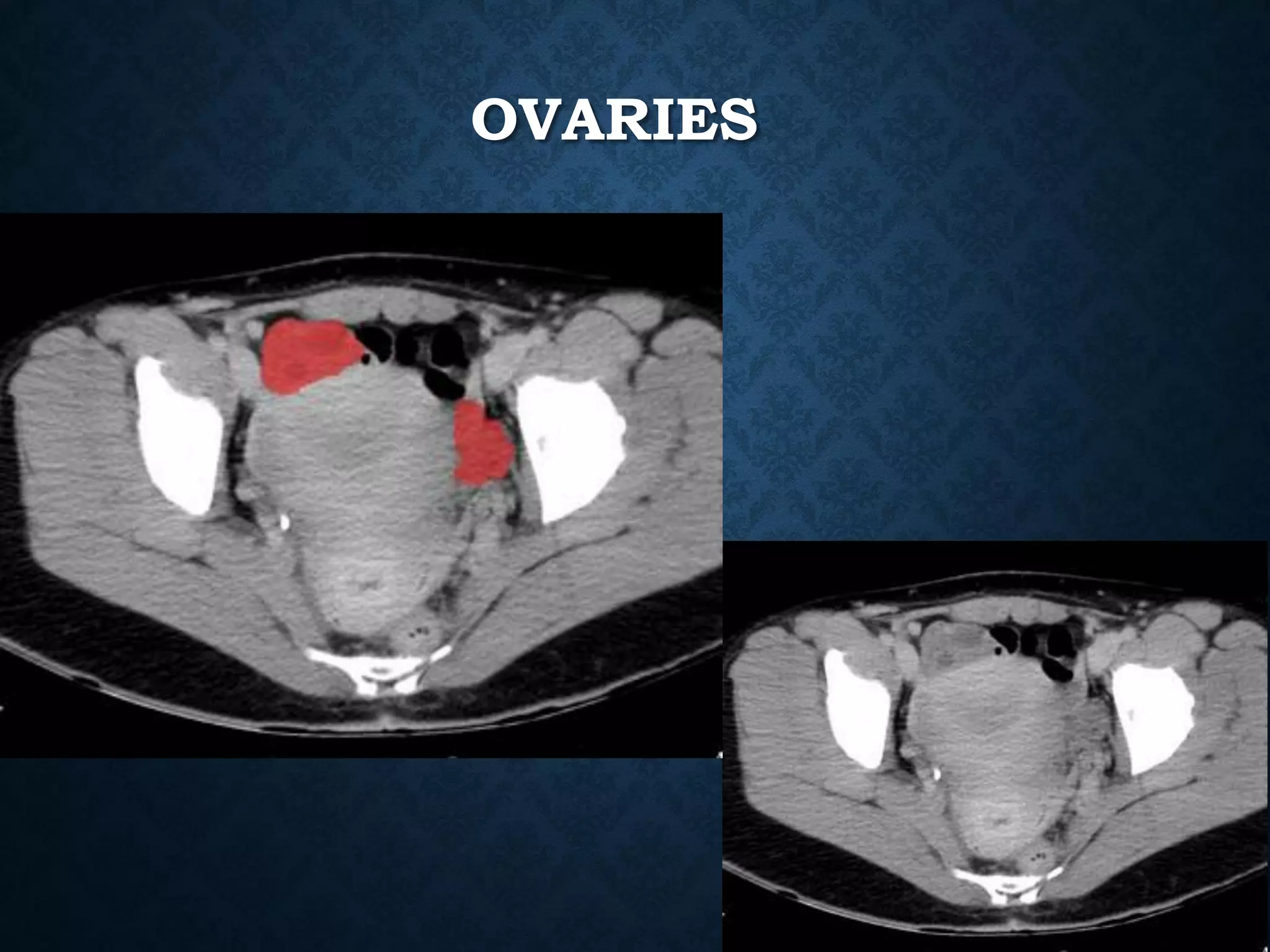
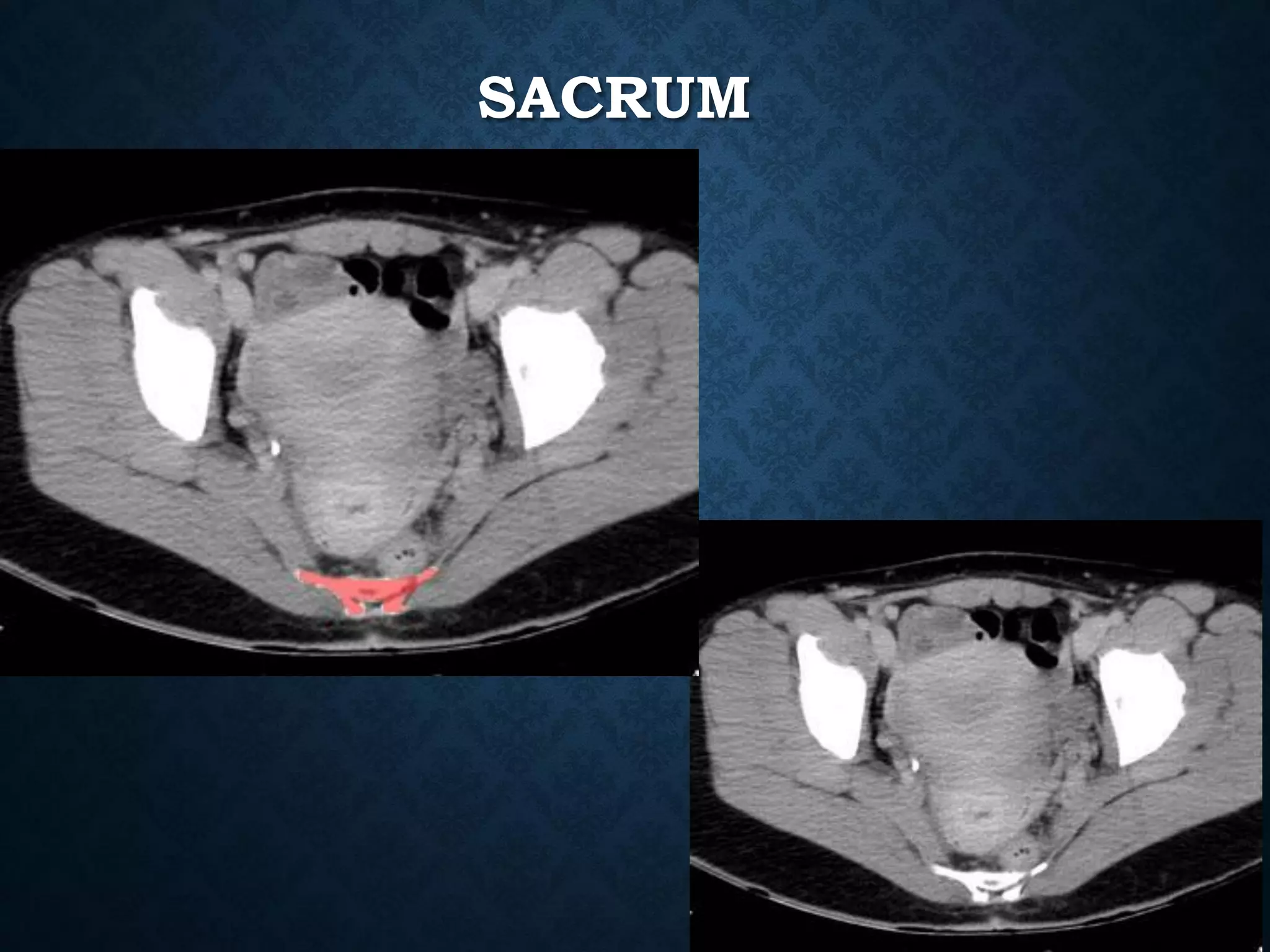
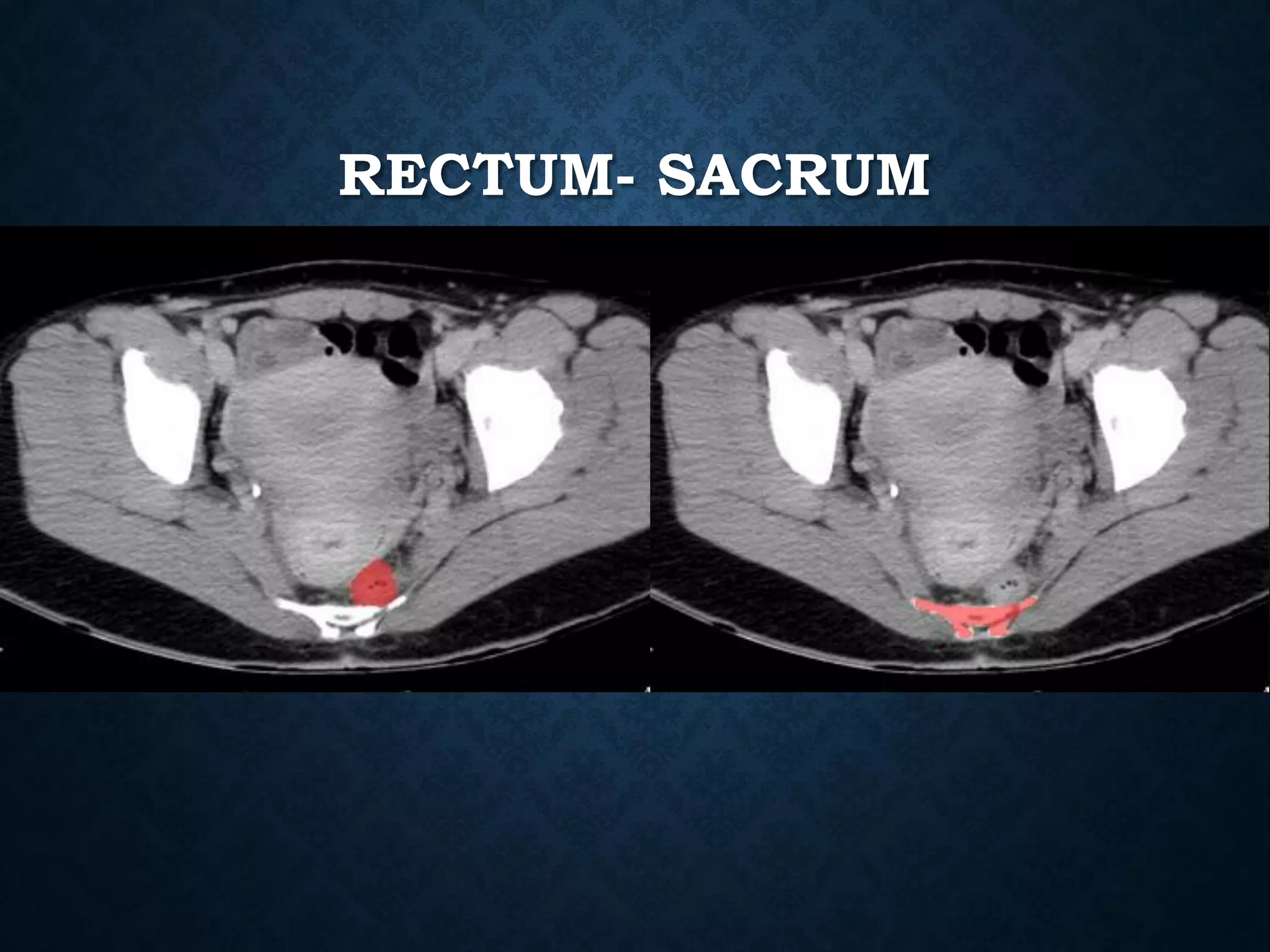
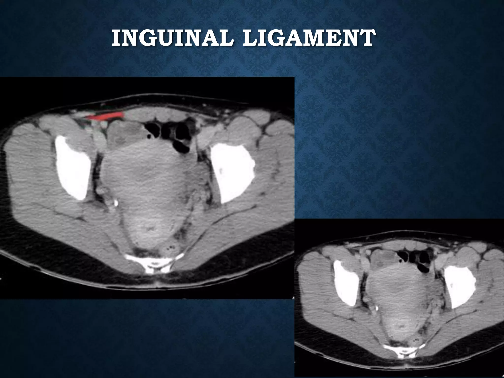
ABDOMEN CT TUTORIAL SLIDES
Mustafa Ahmed Al-Thabhawee, MD
Identify the following structures in the body CT to the right. To view the location of the structure in the image click on
the label at the left and the structure will be indicated in the image. Abdominal CT scans typically begin just above
the diaphragm, so the first slice you see is of the lower chest.
PYLORUS
STOMACH
PANCREAS
1: Head of pancreas
2: Uncinate process of pancreas
3: Pancreatic notch
4: Body of pancreas
5: Anterior surface of pancreas
6: Inferior surface of pancreas
7: Superior margin of pancreas
8: Anterior margin of pancreas
9: Inferior margin of pancreas
10: Omental tuber
11:Tail of pancreas
12: Duodenum
IVC
CELIAC
• Branching from the aorta 'anterior to the
T12‘ one of three anterior/ midline branches of the
abdominal aorta (the others are the superior and inferior
mesenteric arteries).
CELIAC ARTERY BRANCHES
• Left gastric artery
• Common hepatic artery
• Splenic artery
SUPERIOR MESENTERIC VEIN
• In anatomy, the superior mesenteric vein (SMV) is a blood vessel
that drains blood from the small intestine (jejunum and ileum).
• At its termination behind the neck of the pancreas,
• the SMV combines with the splenic vein to form the hepatic portal vein.
• The SMV lies to the right of the similarly named artery, the superior
mesenteric artery, which originates from the abdominal aorta.
POSTERIOR VIEW
IVC
IVC
NORMAL ANATOMY- SECTION 8
1: Head of pancreas
2: Uncinate process of pancreas
3: Pancreatic notch
4: Body of pancreas
5: Anterior surface of pancreas
6: Inferior surface of pancreas
7: Superior margin of pancreas
8: Anterior margin of pancreas
9: Inferior margin of pancreas
10: Omental tuber
11:Tail of pancreas
12: Duodenum
NORMAL ANATOMY- SECTION 9
NORMAL ANATOMY- SECTION 10
ABDOMINAL AORTA
origin: Continuation of descending thoracic aorta at T12
• course: descends anterior and slightly to the left of the lumbar vertebral
bodies.
•Bifurcation of aorta:L4
• Note that the bifurcation (union) of the inferior vena cava is at L5and
therefore below that of the bifurcation of the aorta)
NORMAL ANATOMY- SECTION 11
Normal Anatomy- Section 12
Normal Anatomy- Section 13
Normal Anatomy- Section 14 (female)
OVARIES
SACRUM
RECTUM- SACRUM
INGUINAL LIGAMENT
Tutorial Abdominal CT scan test.pdf

Tutorial Abdominal CT scan test.pdf

  • 1.
    ABDOMEN CT TUTORIALSLIDES Mustafa Ahmed Al-Thabhawee, MD
  • 3.
    Identify the followingstructures in the body CT to the right. To view the location of the structure in the image click on the label at the left and the structure will be indicated in the image. Abdominal CT scans typically begin just above the diaphragm, so the first slice you see is of the lower chest.
  • 21.
  • 22.
  • 25.
    PANCREAS 1: Head ofpancreas 2: Uncinate process of pancreas 3: Pancreatic notch 4: Body of pancreas 5: Anterior surface of pancreas 6: Inferior surface of pancreas 7: Superior margin of pancreas 8: Anterior margin of pancreas 9: Inferior margin of pancreas 10: Omental tuber 11:Tail of pancreas 12: Duodenum
  • 28.
  • 34.
    CELIAC • Branching fromthe aorta 'anterior to the T12‘ one of three anterior/ midline branches of the abdominal aorta (the others are the superior and inferior mesenteric arteries).
  • 35.
    CELIAC ARTERY BRANCHES •Left gastric artery • Common hepatic artery • Splenic artery
  • 46.
    SUPERIOR MESENTERIC VEIN •In anatomy, the superior mesenteric vein (SMV) is a blood vessel that drains blood from the small intestine (jejunum and ileum). • At its termination behind the neck of the pancreas, • the SMV combines with the splenic vein to form the hepatic portal vein. • The SMV lies to the right of the similarly named artery, the superior mesenteric artery, which originates from the abdominal aorta.
  • 48.
  • 51.
  • 62.
  • 63.
  • 66.
    1: Head ofpancreas 2: Uncinate process of pancreas 3: Pancreatic notch 4: Body of pancreas 5: Anterior surface of pancreas 6: Inferior surface of pancreas 7: Superior margin of pancreas 8: Anterior margin of pancreas 9: Inferior margin of pancreas 10: Omental tuber 11:Tail of pancreas 12: Duodenum
  • 70.
  • 76.
  • 81.
    ABDOMINAL AORTA origin: Continuationof descending thoracic aorta at T12 • course: descends anterior and slightly to the left of the lumbar vertebral bodies. •Bifurcation of aorta:L4 • Note that the bifurcation (union) of the inferior vena cava is at L5and therefore below that of the bifurcation of the aorta)
  • 85.
  • 91.
  • 101.
  • 111.
  • 115.
  • 116.
  • 117.
  • 118.