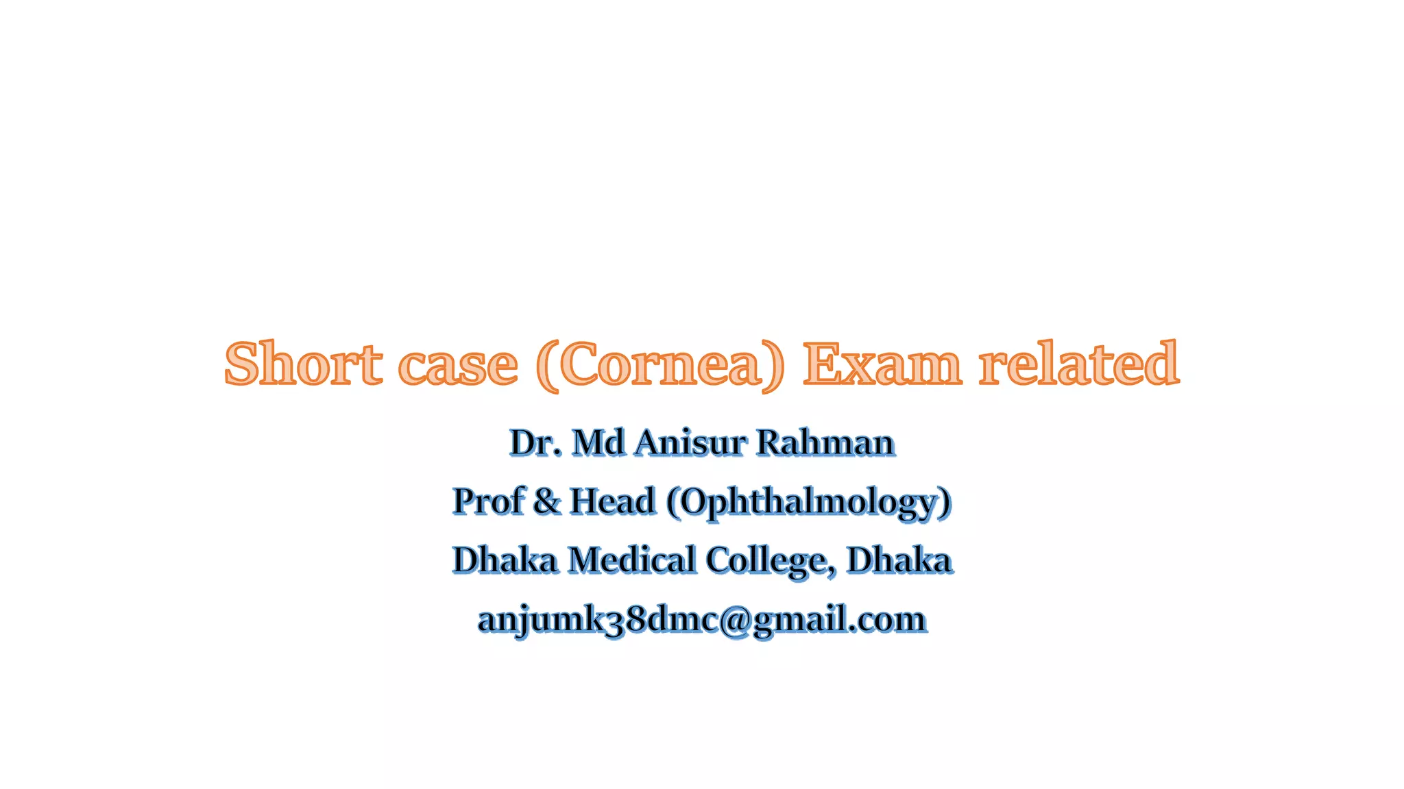
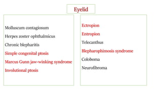
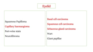
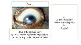
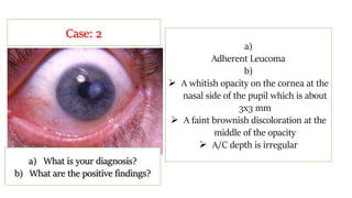
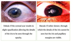
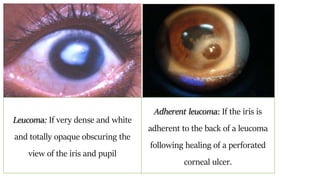
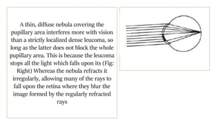
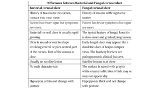
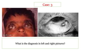
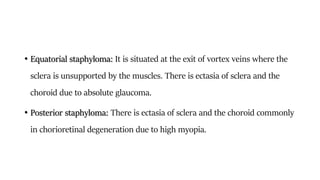
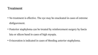
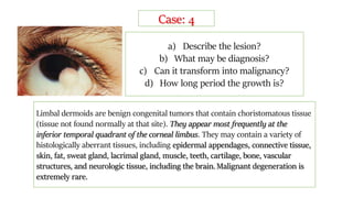
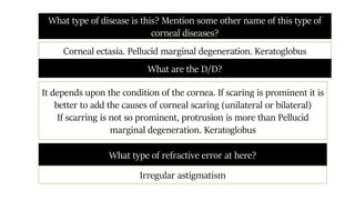
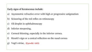
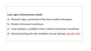
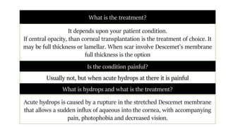
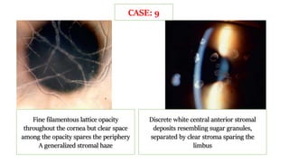
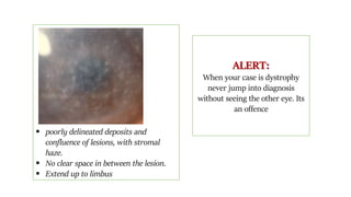
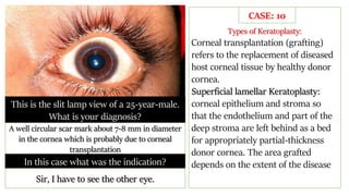
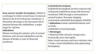
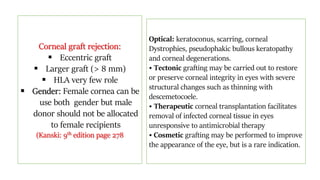
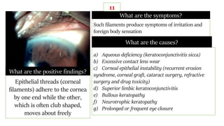
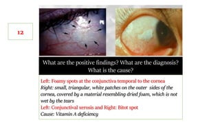
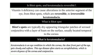
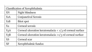
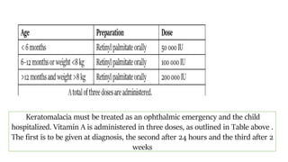
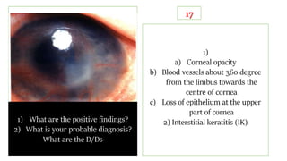
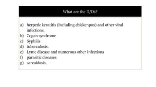
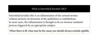
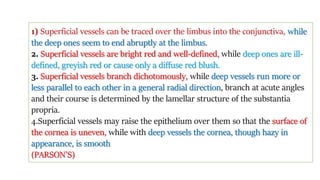
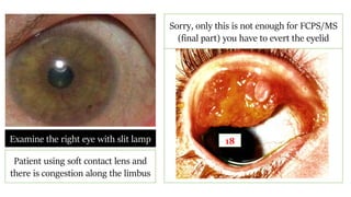
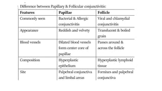
SlideShare a Scribd company logo
Eyelid
Molluscum contagiosum
Herpes zoster ophthalmicus
Chronic blepharitis
Simple congenital ptosis
Marcus Gunn jaw-winking syndrome
Involutional ptosis
Ectropion
Entropion
Telecanthus
Blepharophimosis syndrome
Coloboma
Neurofibroma
Eyelid
Squamous Papilloma
Capillary haemangioma
Port-wine stain
Neurofibroma
Basal cell carcinoma
Squamous cell carcinoma
Sebaceous gland carcinoma
Wart
Giant papillae
Orbit
1) Under the heading of proptosis
a) Thyroid
b) Optic nerve glioma
c) Optic nerve sheath meningioma
d) Capillary haemangioma
e) Cavernous haemangioma
f) Lacrimal gland tumor
2) Orbit floor fracture
3) Crouzon syndrome
4) Prosthetic eye
Cornea
1) Corneal opacity
2) Adherent leucoma
3) Staphyloma (post type in post seg)
4) Dystrophy
5) Keratoconus
6) Vascularized cornea
7) Subepithelial keratitis: Nummular
8) Disciform Keratitis
9) Filamentary keratopathy
10) Mooren ulcer
11) K-F ring
12) Megalocornea
13) Microcornea
14) Post PKP
Complications of surgery
Sand’s of Sahara: LASIK
Vascularization around knot
Loose knot: PKP
Cataract Surgery
Descemet’s fold
Displacement of IOL
Optic capture
Haptic capture
Lens in A/C
Iris prolapse
Filtration surgery:
Bleb failure
Over filtration
Cystic bleb
Polycoria: Iatrogenic
VKC
PCO
Ectopia lentis
Posterior synechiae
Festooned
K P: Different types
Iris coloboma
 Lacrimal fistula
 Mucocele
 Growth at sac region
 Pterygium
 Pseudo-pterygium
 Chemical burn causes disfiguration
Miscellaneous
This is the slit lamp view
a) What are the positive findings at here?
b) What may be the cause of iris hole?
a)
Optical iridectomy
Central corneal opacity
b)
Surgical
Case: 1
a) What is your diagnosis?
b) What are the positive findings?
a)
Adherent Leucoma
b)
 A whitish opacity on the cornea at the
nasal side of the pupil which is about
3x3 mm
 A faint brownish discoloration at the
middle of the opacity
 A/C depth is irregular
Case: 2
The opacity cover only the nasal side of the pupillary aperture what VA
you deserve?
If VA is counting finger what may be the probable cause? And how will
you examine it at here, without additional device?
c) What are the causes of unilateral and bilateral corneal opacity?
d) What are the nebula, macula and leucoma?
e) Which one hamper VA more nebula or macula if pupil is free? And why?
• Causes of unilateral corneal opacity
• Corneal ulcer
• Trauma
• Congenital
• Keratoconus
• Dystrophy (Usually bilateral)
Causes of bilateral corneal opacity:
• Steven-Johnson syndrome
• Dystrophy
• Chemical injury
• Nutritional
• KC
• Congenital
• Corneal ulcer
Nebula: If the corneal scar results in
slight opacification allowing the details
of the iris to be seen through the
opacity.
Macula: If rather denser, through
which the details of the iris cannot be
seen but the iris and pupillary
margins are visible.
Leucoma: If very dense and white
and totally opaque obscuring the
view of the iris and pupil
Adherent leucoma: If the iris is
adherent to the back of a leucoma
following healing of a perforated
corneal ulcer.
A thin, diffuse nebula covering the
pupillary area interferes more with vision
than a strictly localized dense leucoma, so
long as the latter does not block the whole
pupillary area. This is because the leucoma
stops all the light which falls upon its (Fig:
Right) Whereas the nebula refracts it
irregularly, allowing many of the rays to
fall upon the retina where they blur the
image formed by the regularly refracted
rays
What is the diagnosis in left and right pictures?
Case: 3
Types of staphyloma
• A staphyloma is a clinical condition characterized by an ectasia of the outer coats
(cornea, or sclera or both) of the eye with an incarceration of the uveal tissue.
• The basic underlying pathology is a weakening of the eye wall, which can be
caused by many inflammatory or degenerative diseases involving these
structures. Most commonly the diseases causing a weakening of the globe are
accompanied by a raised intraocular pressure and both contribute to the
development of the staphyloma.
Different types of staphyloma
• Anterior staphyloma: It is associated with ectasia of the cornea and iris. The
most common cause is perforating corneal ulcer or injury.
• Intercalary staphyloma: It lies between the iris and the ciliary body. There is
ectasia of sclera and root of iris. The most common cause is absolute glaucoma.
• Ciliary staphyloma: There is ectasia of sclera and the ciliary body. The common
causes are absolute glaucoma and scleritis.
• Equatorial staphyloma: It is situated at the exit of vortex veins where the
sclera is unsupported by the muscles. There is ectasia of sclera and the
choroid due to absolute glaucoma.
• Posterior staphyloma: There is ectasia of sclera and the choroid commonly
in chorioretinal degeneration due to high myopia.
Treatment
• No treatment is effective. The eye may be enucleated in cases of extreme
disfigurement.
• Posterior staphyloma can be treated by reinforcement surgery by fascia
lata or silicon band in cases of high myopia.
• Evisceration is indicated in cases of bleeding anterior staphyloma.
Case: 4
a) Describe the lesion?
b) What may be diagnosis?
c) Can it transform into malignancy?
d) How long period the growth is?
Limbal dermoids are benign congenital tumors that contain choristomatous tissue
(tissue not found normally at that site). They appear most frequently at the
inferior temporal quadrant of the corneal limbus. They may contain a variety of
histologically aberrant tissues, including epidermal appendages, connective tissue,
skin, fat, sweat gland, lacrimal gland, muscle, teeth, cartilage, bone, vascular
structures, and neurologic tissue, including the brain. Malignant degeneration is
extremely rare.
What are the D/Ds? (But the D/D will be according to the case)
• Foreign body granuloma
• Sclerocornea
• Staphyloma
• Corneal scar (from trauma or infection)
Treatment:
• Treatment is indicated for cosmesis, chronic irritation, dellen formation
and amblyopia from astigmatism or involvement of the visual axis. Small
dermoids can undergo simple excision, but lamellar keratosclerectomy
may be required for large lesions.
Case: 5
 What are the positive findings?
 What is the diagnosis?
 What may be the cause?
 A disc/coin shaped lesion at the
center part of cornea which involve--
-- layer
 Disciform keratitis
 Herpes
Keratitis is the descriptive term used for any type of corneal inflammation.
Inflammation anterior to Bowman’s membrane (involving the epithelium and
Bowman’s membrane) is called superficial keratitis.
If only occurring in discrete patches, the latter is further categorized as
superficial punctate keratitis (SPK)
Inflammation in the stroma is called deep keratitis. Deep keratitis is further
categorized as stromal or interstitial keratitis or endothelitis, depending on
the direct involvement of the endothelium in the inflammatory process.
Ref: Parson’s
Disciform keratitis
• The aetiopathogenesis of disciform keratitis (endotheliitis) is controversial.
• It may be the result of active HSV infection of keratocytes or endothelium,
or a hypersensitivity reaction to viral antigen in the cornea
Ref: Kanski
Symptoms:
Blurred vision of gradual onset, which may be associated with haloes
around lights. Discomfort and redness are common, but tend to be milder
than in purely epithelial disease. A clear past history of epithelial ulceration
is not always present, and the possibility of a mimicking infection such as
acanthamoeba or fungal keratitis should be borne in mind.
A central zone of stromal oedema, often with overlying epithelial oedema
occasionally the lesion is eccentric.
Large (granulomatous) keratic precipitates underlying the oedema
Folds in Descemet membrane in severe cases. A surrounding (Wessely) immune
ring of deep stromal haze signifies deposition of viral antigen and host antibody
complexes.
The IOP may be elevated.
 Reduced corneal sensation; this may aid in distinguishing other forms of
infection.
Healed lesions often have a faint ring of stromal or subepithelial opacification
and thinning.
Consecutive episodes may be associated with gradually worsening subepithelial
and/or stromal scarring and superficial or deep vascularization. Mid-stromal
scarring from disciform keratitis is a cause of interstitial keratitis.
Treatment
A broad approach to management is set out below, but in practice regimens
should be tailored individually. Careful monitoring and adequate treatment,
dependent on severity of inflammation, is critical to minimize progression of
scarring. Patients should be cautioned to seek treatment at the first
suggestion of recurrence, though some authorities feel that minimal
inflammation may not warrant treatment or can be addressed with
cycloplegia alone.
Initial treatment is with topical steroids (prednisolone 1% or
dexamethasone 0.1%) with antiviral cover, both 5 times daily. As
improvement occurs, the frequency of administration of both is reduced
in parallel over not less than 4 weeks. It is prudent to keep steroid
intensity and duration to the minimum required for effective control of
inflammation.
IOP should be monitored.
Cycloplegia can be used to improve comfort if necessary, and some
practitioners recommend topical antibacterial prophylaxis.
a) What is the positive finding?
b) What clinical test will you do?
c) What may be probable diagnosis?
d) What is the cause?
e) What is the treatment?
f) What may happen if not properly treated?
a) Multiple tiny granular deposits
surrounded by a halo of stromal
haze.
b) Corneal sensation
c) Nummular keratitis
d) adenoviral keratoconjunctivitis &
HZO
e) Steroid
f) Disciform Keratitis
Case: 6
Nummular keratitis: usually develops at the site of epithelial lesions
about 10 days after the onset of the rash. It is characterized by fine
granular subepithelial deposits surrounded by a halo of stromal haze.
The lesions fade in response to topical steroids but recur if
treatment is discontinued prematurely. (Kanski)
Slit lamp view 3 days after LASIK.
a) What is the diagnosis?
b) After how many days it occur?
c) Why this happened?
d) Treatment?
e) How will u differentiate from infective?
a) Diffuse lamellar keratitis (‘sands of the
Sahara’)
b) 1 – 7 days
c) Next slide
d) Steroid in stage I & II. In stage III Lifting the
flap and irrigating the interface with
balanced salt solution may wash out
proteolytic enzymes produced by
neutrophilic cells, resulting in limitation of
tissue destruction
e) Next slide
Case: 7
• The etiology of DLK is still a mystery. Although it has been related to an
immunologic or toxic reaction a single agent has not yet been detected.
Various agents have been suspected to cause DLK.
a) debris from the microkeratome blades,
b) meibomian gland secretion,
c) debris from the eyelid,
d) cleaning or lubricating solution used on blades or microkeratomes,
e) polyvidone iodine solution,
f) heat-stable endotoxins released from sterilizer reservoir biofilms,
g) epithelial defects during or after surgery,
h) and a host-immune response
Differentiation between DLK and infectious keratitis
Diffuse lamellar keratitis is scattered through a large area of the interface. It
is confined to the interface not extending anteriorly or posteriorly into the
stroma. A dominant focus is absent with no anterior chamber reaction or
ciliary injection.
In contrast, microbial keratitis does not respect the boundaries of the flap
interface in terms of a single dominant focus with either extension
anteriorly into the flap or posteriorly into the stroma frequently present.
There is also often a conjunctival and ciliary injection and in some cases
inflammatory cells in the anterior chamber, all of which are not present in
DLK
Figure 1. Demonstrates central Vogt's striae, or parallel, vertical lines
from posterior stromal stress. A Fleischer ring, or epithelial iron
deposition around the base of the cone is visible superiorly. The
horizontally-oriented opacities are areas of stromal scarring.
Figure 2. Significant apical scarring in the contralateral eye
What are the positive findings? What is the diagnosis?
CASE:8
What type of disease is this? Mention some other name of this type of
corneal diseases?
Corneal ectasia. Pellucid marginal degeneration. Keratoglobus
What are the D/D?
It depends upon the condition of the cornea. If scaring is prominent it is
better to add the causes of corneal scaring (unilateral or bilateral)
If scarring is not so prominent, protrusion is more than Pellucid
marginal degeneration. Keratoglobus
What type of refractive error at here?
Irregular astigmatism
Early signs of Keratoconus include:
a) Asymmetric refractive error with high or progressive astigmatism
b) Scissoring of the red reflex on retinoscopy
c) Oil droplet in ophthalmoscopy
d) Inferior steepening,
e) Corneal thinning, especially in the inferior cornea.
f) Rizutti’s sign or a conical reflection on the nasal cornea
g) Vogt’s striae, (Eyewiki: AAO)
Later signs of Keratoconus include:
a) Munson’s sign, a protrusion of the lower eyelid in downgaze.
b) Breaks in Bowman’s membrane
c) Acute hydrops, a condition where a break in Descemet’s membrane
d) Stromal scarring after the resolution of acute hydrops, (Eyewiki: AAO)
Is the condition painful?
What is the treatment?
It depends upon your patient condition.
If central opacity, than corneal transplantation is the treatment of choice. It
may be full thickness or lamellar. When scar involve Descemet’s membrane
full thickness is the option
Usually not, but when acute hydrops at there it is painful
What is hydrops and what is the treatment?
Acute hydrops is caused by a rupture in the stretched Descemet membrane
that allows a sudden influx of aqueous into the cornea, with accompanying
pain, photophobia and decreased vision.
Acute episodes are initially treated with
 cycloplegia,
 hypertonic (5%) saline ointment and
 patching or a soft bandage contact lens.
 Accelerated resolution has been reported with intracameral gas injection in
the acute stage. (Kanski)
In short case treatment depends upon the condition of patient
a) Eye rubbing should be avoided.
b) Spectacles or soft contact lenses are generally sufficient in early
c) Rigid contact lenses: usually corneal sometimes scleral, are
required for higher degrees of astigmatism
d) Corneal collagen cross-linking (CXL), using riboflavin drops to
photosensitize the eye followed by exposure to ultraviolet-A light,
may stabilize or even reverse ectasia, CXL is commonly used only
after progression has been documented.
What are the treatments?
e) Corneal collagen cross-linking (CXL), using riboflavin drops to
photosensitize the eye followed by exposure to ultraviolet-A light,
may stabilize or even reverse ectasia, CXL is commonly used only
after progression has been documented.
f) Intracorneal ring segment implantation
g) Keratoplasty, either penetrating or deep anterior lamellar (DALK),
may be necessary in patients with severe disease. Outcomes may be
compromised by residual astigmatism and by anisometropia,
necessitating contact lens correction for optimal acuity. (Kanski)
Treatment (continued)
Pellucid marginal degeneration is a rare progressive peripheral corneal
thinning disorder, typically involving the inferior cornea in both eyes.
Presentation is usually in adulthood.
Keratoglobus: In contrast to keratoconus, the cornea develops globular
rather than conical ectasia; corneal thinning is generalized Keratoglobus is
an extremely rare condition that can be present at birth when differential
diagnosis is from congenital glaucoma and megalocornea and associations
may be present, or acquired, with onset in adulthood. (Kanski)
CASE: 9
Fine filamentous lattice opacity
throughout the cornea but clear space
among the opacity spares the periphery
A generalized stromal haze
Discrete white central anterior stromal
deposits resembling sugar granules,
separated by clear stroma sparing the
limbus
 poorly delineated deposits and
confluence of lesions, with stromal
haze.
 No clear space in between the lesion.
 Extend up to limbus
ALERT:
When your case is dystrophy
never jump into diagnosis
without seeing the other eye. Its
an offence
All 3 are corneal dystrophy, Lattice, granular and macular. All the dystrophies (corneal)
are AD but only macular variety is AR
This is the slit lamp view of a 25-year-male.
What is your diagnosis?
A well circular scar mark about 7-8 mm in diameter
in the cornea which is probably due to corneal
transplantation
In this case what was the indication?
Sir, I have to see the other eye.
Types of Keratoplasty:
Corneal transplantation (grafting)
refers to the replacement of diseased
host corneal tissue by healthy donor
cornea.
Superficial lamellar Keratoplasty:
corneal epithelium and stroma so
that the endothelium and part of the
deep stroma are left behind as a bed
for appropriately partial-thickness
donor cornea. The area grafted
depends on the extent of the disease
CASE: 10
Deep anterior lamellar Keratoplasty: (DALK) is
a technique in which corneal tissue is removed
almost to the level of Descemet membrane. A
theoretical advantage is the decreased risk of
rejection because the endothelium, a major
target for rejection
Indications
Disease involving the anterior 95% of corneal
thickness with normal endothelium and the
absence of breaks or scars in Descemet
membrane
Endothelial Keratoplasty:
Endothelial Keratoplasty involves removal only
of diseased endothelium along with Descemet
membrane (DM) through a corneoscleral or
corneal incision. Descemet stripping
(automated) endothelial Keratoplasty (DSAEK),
• Indications include endothelial disease such
as Fuchs
endothelial corneal dystrophy.
• Advantages
○ Relatively little refractive change and a
structurally more intact globe.
better visual outcomes and lower rejection rates
○ Faster visual rehabilitation than penetrating
Keratoplasty
Corneal graft rejection:
 Eccentric graft
 Larger graft (> 8 mm)
 HLA very few role
 Gender: Female cornea can be
use both gender but male
donor should not be allocated
to female recipients
(Kanski: 9th edition page 278
Optical: keratoconus, scarring, corneal
Dystrophies, pseudophakic bullous keratopathy
and corneal degenerations.
• Tectonic grafting may be carried out to restore
or preserve corneal integrity in eyes with severe
structural changes such as thinning with
descemetocoele.
• Therapeutic corneal transplantation facilitates
removal of infected corneal tissue in eyes
unresponsive to antimicrobial therapy
• Cosmetic grafting may be performed to improve
the appearance of the eye, but is a rare indication.
Epithelial threads (corneal
filaments) adhere to the cornea
by one end while the other,
which is often club shaped,
moves about freely
What are the positive findings?
Such filaments produce symptoms of irritation and
foreign body sensation
a) Aqueous deficiency (keratoconjunctivitis sicca)
b) Excessive contact lens wear
c) Corneal epithelial instability (recurrent erosion
syndrome, corneal graft, cataract surgery, refractive
surgery and drug toxicity)
d) Superior limbic keratoconjunctivitis
e) Bullous keratopathy
f) Neurotrophic keratopathy
g) Prolonged or frequent eye closure
What are the symptoms?
What are the causes?
11
What are the positive findings? What are the diagnosis?
What is the cause?
Left: Foamy spots at the conjunctiva temporal to the cornea
Right: small, triangular, white patches on the outer sides of the
cornea, covered by a material resembling dried foam, which is not
wet by the tears
Left: Conjunctival xerosis and Right: Bitot spot
Cause: Vitamin A deficiency
12
Is Bitot spots, and keratomalacia reversible?.
Vitamin A deficiency can cause alterations in the anterior segment of the
eye, from Bitot spots, which are reversible, to irreversible
keratomalacia.
Bitot's spots are typically dry-appearing triangular patches of xerosed
conjunctiva with a layer of foam on the surface, usually located temporal
to the cornea
What is Bitot spot
Keratomalacia is an eye condition in which the cornea, the clear front part of the eye,
gets cloudy and softens. This eye disease often starts as xerophthalmia, which is
severe dryness of the cornea and conjunctiva.
What is keratomalacia?
Keratomalacia must be treated as an ophthalmic emergency and the child
hospitalized. Vitamin A is administered in three doses, as outlined in Table above .
The first is to be given at diagnosis, the second after 24 hours and the third after 2
weeks
This is the slit lamp view of cornea
What is the positive finding at here?
There is a complete dark-brown ring at
the periphery of the cornea at
Descemet’s membrane level
What is the name of the diseases?
What is other name of the diseases?
Which structures it involves?
 Wilson's disease
 Hepatolenticular degeneration
 Liver, caudate nucleas of brain and
cornea mainly
13
What deposited at here?
When the level is low what is the name of the diseases?
Copper (Ceruloplasmin)
Low ceruloplasmin might also mean Menkes disease
What is the name of this ring?
How many percentage will you get this ring?
2 4 hour urinary copper
Only morning urine is sufficient and result is same
How urinary copper measure?
What Bangladesh modify the test?
Kayser-Fleischer ring.
95% (according to Jay Kracher)
a) What is the diagnosis?
b) What may be the D/D?
c) What type of genetic disorder is this?
d) What may be the condition of lens and
why?
e) What may be refractive status?
a) Megalocornea
b) Congenital glaucoma
c) X-Linked
d) Lens subluxation may occur
due to zonular stretching
e) Myopia and astigmatism
14
15
This is the slit lamp view
a) What is the positive finding?
b) What may be the cause?
a) Prolapse of uveal tissue through sclera. The
sclera has blanched
b) It is due to MMC after pterygium surgery
(bare sclera technique)
How can you manage it?
What is the concentration of MMC we used?
By scleral patch graft
0.02% (0.2 mg/ml) or 0.04% (0.4
mg/ml)
16
What is the positive finding?
What may be the cause?
Vascularization (superficial) of cornea at
the upper portion which extend up to the
pupillary margin
a) Contact lens
b) Trachoma
c) Chronic irritation
d) Herpes Keratitis
e) Chemical burn
f) VKC
(Other cause may be: corneal ulcers, phylctenular
keratoconjunctivitis, rosacea keratitis, interstitial
keratitis, sclerosing keratitis)
1) What are the positive findings?
2) What is your probable diagnosis?
What are the D/Ds
17
1)
a) Corneal opacity
b) Blood vessels about 360 degree
from the limbus towards the
centre of cornea
c) Loss of epithelium at the upper
part of cornea
2) Interstitial keratitis (IK)
What are the D/Ds?
a) herpetic keratitis (including chickenpox) and other viral
infections,
b) Cogan syndrome
c) Syphilis
d) tuberculosis,
e) Lyme disease and numerous other infections
f) parasitic diseases
g) sarcoidosis,
What is Interstitial keratitis (IK)?
Interstitial keratitis (IK) is an inflammation of the corneal stroma
without primary involvement of the epithelium or endothelium.
In most cases, the inflammation is thought to be an immune mediated
process triggered by an appropriate antigen.
When there is IK what may be the cause you should always exclude syphilis
1) Superficial vessels can be traced over the limbus into the conjunctiva, while
the deep ones seem to end abruptly at the limbus.
2. Superficial vessels are bright red and well-defined, while deep ones are ill-
defined, greyish red or cause only a diffuse red blush.
3. Superficial vessels branch dichotomously, while deep vessels run more or
less parallel to each other in a general radial direction, branch at acute angles
and their course is determined by the lamellar structure of the substantia
propria.
4.Superficial vessels may raise the epithelium over them so that the surface of
the cornea is uneven, while with deep vessels the cornea, though hazy in
appearance, is smooth
(PARSON’S)
Examine the right eye with slit lamp
Patient using soft contact lens and
there is congestion along the limbus
Sorry, only this is not enough for FCPS/MS
(final part) you have to evert the eyelid
18
What you have seen after everting the lid?
Why this occur?
What are causes?
ADD ANSWER
Short case Cornea

More Related Content

What's hot

CRAO
CRAOCRAO
Macular hole
Macular holeMacular hole
Macular hole
Dr Samarth Mishra
 
Branch retinal vein occlusion (BRVO)
Branch retinal vein occlusion (BRVO)Branch retinal vein occlusion (BRVO)
Branch retinal vein occlusion (BRVO)
NIKHIL GOTMARE
 
Peripheral fundus & its disorders
Peripheral fundus & its disordersPeripheral fundus & its disorders
Peripheral fundus & its disorders
Rohit Rao
 
Mooren’s ulcer
Mooren’s ulcerMooren’s ulcer
Mooren’s ulcer
ikramdr01
 
Keratoconus
KeratoconusKeratoconus
Keratoconus
Laya Pillai
 
Posterior segment manifestations of blunt trauma
Posterior segment manifestations of blunt traumaPosterior segment manifestations of blunt trauma
Posterior segment manifestations of blunt trauma
SSSIHMS-PG
 
Diabetic macular edema
Diabetic macular edemaDiabetic macular edema
Diabetic macular edema
drkvasantha
 
Cystoid macular oedema
Cystoid macular oedemaCystoid macular oedema
Cystoid macular oedema
Laxmi Eye Institute
 
Central retinal vein occlusion CRVO
Central retinal vein occlusion CRVOCentral retinal vein occlusion CRVO
Central retinal vein occlusion CRVO
Dr. Md. Suzon Islam
 
Mooren’s ulcer
Mooren’s ulcerMooren’s ulcer
Mooren’s ulcer
Jagdish Dukre
 
Iridocorneal endothelial syndrome
Iridocorneal endothelial syndromeIridocorneal endothelial syndrome
Iridocorneal endothelial syndrome
SSSIHMS-PG
 
Anterior ischemic optic neuropathy
Anterior ischemic optic neuropathyAnterior ischemic optic neuropathy
Anterior ischemic optic neuropathyJagdish Dukre
 
Intermediate uveitis
Intermediate uveitisIntermediate uveitis
Intermediate uveitis
Barun Garg
 
Trabeculectomy surgical procedure
Trabeculectomy surgical procedureTrabeculectomy surgical procedure
Trabeculectomy surgical procedure
Iddi Ndyabawe
 
Corneal degeneration & depos
Corneal degeneration & deposCorneal degeneration & depos
Corneal degeneration & depos
Niwar Ameen
 
Papilloedema presentation1
Papilloedema presentation1Papilloedema presentation1
Papilloedema presentation1
shovon2026
 
Hypertensive Retinopathy
Hypertensive RetinopathyHypertensive Retinopathy
Hypertensive RetinopathyHossein Mirzaie
 
Differential Diagnosis of Disc Edema
Differential Diagnosis of Disc EdemaDifferential Diagnosis of Disc Edema
Differential Diagnosis of Disc Edema
Sahil Thakur
 
Polypoidal choroidal vasculopathy
Polypoidal choroidal vasculopathyPolypoidal choroidal vasculopathy
Polypoidal choroidal vasculopathy
Sujay Chauhan
 

What's hot (20)

CRAO
CRAOCRAO
CRAO
 
Macular hole
Macular holeMacular hole
Macular hole
 
Branch retinal vein occlusion (BRVO)
Branch retinal vein occlusion (BRVO)Branch retinal vein occlusion (BRVO)
Branch retinal vein occlusion (BRVO)
 
Peripheral fundus & its disorders
Peripheral fundus & its disordersPeripheral fundus & its disorders
Peripheral fundus & its disorders
 
Mooren’s ulcer
Mooren’s ulcerMooren’s ulcer
Mooren’s ulcer
 
Keratoconus
KeratoconusKeratoconus
Keratoconus
 
Posterior segment manifestations of blunt trauma
Posterior segment manifestations of blunt traumaPosterior segment manifestations of blunt trauma
Posterior segment manifestations of blunt trauma
 
Diabetic macular edema
Diabetic macular edemaDiabetic macular edema
Diabetic macular edema
 
Cystoid macular oedema
Cystoid macular oedemaCystoid macular oedema
Cystoid macular oedema
 
Central retinal vein occlusion CRVO
Central retinal vein occlusion CRVOCentral retinal vein occlusion CRVO
Central retinal vein occlusion CRVO
 
Mooren’s ulcer
Mooren’s ulcerMooren’s ulcer
Mooren’s ulcer
 
Iridocorneal endothelial syndrome
Iridocorneal endothelial syndromeIridocorneal endothelial syndrome
Iridocorneal endothelial syndrome
 
Anterior ischemic optic neuropathy
Anterior ischemic optic neuropathyAnterior ischemic optic neuropathy
Anterior ischemic optic neuropathy
 
Intermediate uveitis
Intermediate uveitisIntermediate uveitis
Intermediate uveitis
 
Trabeculectomy surgical procedure
Trabeculectomy surgical procedureTrabeculectomy surgical procedure
Trabeculectomy surgical procedure
 
Corneal degeneration & depos
Corneal degeneration & deposCorneal degeneration & depos
Corneal degeneration & depos
 
Papilloedema presentation1
Papilloedema presentation1Papilloedema presentation1
Papilloedema presentation1
 
Hypertensive Retinopathy
Hypertensive RetinopathyHypertensive Retinopathy
Hypertensive Retinopathy
 
Differential Diagnosis of Disc Edema
Differential Diagnosis of Disc EdemaDifferential Diagnosis of Disc Edema
Differential Diagnosis of Disc Edema
 
Polypoidal choroidal vasculopathy
Polypoidal choroidal vasculopathyPolypoidal choroidal vasculopathy
Polypoidal choroidal vasculopathy
 

Similar to Short case Cornea

opthalmology.Cornea&sclera.(dr.tara)
opthalmology.Cornea&sclera.(dr.tara)opthalmology.Cornea&sclera.(dr.tara)
opthalmology.Cornea&sclera.(dr.tara)student
 
Ophthalmology 5th year, 4th lecture (Dr. Bakhtyar)
Ophthalmology 5th year, 4th lecture (Dr. Bakhtyar)Ophthalmology 5th year, 4th lecture (Dr. Bakhtyar)
Ophthalmology 5th year, 4th lecture (Dr. Bakhtyar)
College of Medicine, Sulaymaniyah
 
Cornea
CorneaCornea
Ophthalmology 5th year, 3rd lecture (Dr. Bakhtyar)
Ophthalmology 5th year, 3rd lecture (Dr. Bakhtyar)Ophthalmology 5th year, 3rd lecture (Dr. Bakhtyar)
Ophthalmology 5th year, 3rd lecture (Dr. Bakhtyar)
College of Medicine, Sulaymaniyah
 
CORNEAL DYSTROPHY and degen.pptx
CORNEAL DYSTROPHY and degen.pptxCORNEAL DYSTROPHY and degen.pptx
CORNEAL DYSTROPHY and degen.pptx
ManoAarthi1
 
Depositions and Degenerations of Conjuctiva and Cornea.docx
Depositions and Degenerations of Conjuctiva and Cornea.docxDepositions and Degenerations of Conjuctiva and Cornea.docx
Depositions and Degenerations of Conjuctiva and Cornea.docx
Iddi Ndyabawe
 
Diseases of Cornea Keratitis
Diseases of Cornea KeratitisDiseases of Cornea Keratitis
Diseases of Cornea Keratitis
Arslan Chaudhry
 
KERATOCONUS
KERATOCONUSKERATOCONUS
KERATOCONUS
MadhumitaBooks
 
Corneal Ectasias
Corneal Ectasias Corneal Ectasias
Corneal Ectasias
Vineela Cherukuri
 
Ocular Manifestations of Leprosy - EYE
Ocular Manifestations of Leprosy - EYEOcular Manifestations of Leprosy - EYE
Ocular Manifestations of Leprosy - EYE
Chetan Ganteppanavar
 
DISEASES OF SHUKLAMANDALA-MODERN PART
DISEASES OF SHUKLAMANDALA-MODERN PARTDISEASES OF SHUKLAMANDALA-MODERN PART
DISEASES OF SHUKLAMANDALA-MODERN PART
Dr Veeresh Adoor
 
EPISCLERITIS AND SCLERITIS.pptx bbbbbbbb
EPISCLERITIS AND SCLERITIS.pptx bbbbbbbbEPISCLERITIS AND SCLERITIS.pptx bbbbbbbb
EPISCLERITIS AND SCLERITIS.pptx bbbbbbbb
Harshika Malik
 
Clinical 5th Grade Dr Ahmed Osama Hashem Ophthalmology.pptx
Clinical 5th Grade Dr Ahmed Osama Hashem Ophthalmology.pptxClinical 5th Grade Dr Ahmed Osama Hashem Ophthalmology.pptx
Clinical 5th Grade Dr Ahmed Osama Hashem Ophthalmology.pptx
Ahmed Osama Hashem
 
Eyelid Disorders.pptx
Eyelid Disorders.pptxEyelid Disorders.pptx
Eyelid Disorders.pptx
dratulkranand
 
Peripheral Ulcerative Keratitis ( PUK )
Peripheral Ulcerative Keratitis ( PUK )Peripheral Ulcerative Keratitis ( PUK )
Peripheral Ulcerative Keratitis ( PUK )
DiyarAlzubaidy
 
Keratoconus 2016
Keratoconus 2016Keratoconus 2016
Keratoconus 2016
DINESH and SONALEE
 
disorders of eyelids
disorders of eyelidsdisorders of eyelids
disorders of eyelids
kalpanabhandari19
 
Degenerative condition of eye
Degenerative condition of eyeDegenerative condition of eye
Degenerative condition of eye
OPTOM FASLU MUHAMMED
 
Glaucoma & lens
Glaucoma & lensGlaucoma & lens
Glaucoma & lens
Anisur Rahman
 
KERATITIS.pptx
KERATITIS.pptxKERATITIS.pptx
KERATITIS.pptx
NSHM Knowledge Campus
 

Similar to Short case Cornea (20)

opthalmology.Cornea&sclera.(dr.tara)
opthalmology.Cornea&sclera.(dr.tara)opthalmology.Cornea&sclera.(dr.tara)
opthalmology.Cornea&sclera.(dr.tara)
 
Ophthalmology 5th year, 4th lecture (Dr. Bakhtyar)
Ophthalmology 5th year, 4th lecture (Dr. Bakhtyar)Ophthalmology 5th year, 4th lecture (Dr. Bakhtyar)
Ophthalmology 5th year, 4th lecture (Dr. Bakhtyar)
 
Cornea
CorneaCornea
Cornea
 
Ophthalmology 5th year, 3rd lecture (Dr. Bakhtyar)
Ophthalmology 5th year, 3rd lecture (Dr. Bakhtyar)Ophthalmology 5th year, 3rd lecture (Dr. Bakhtyar)
Ophthalmology 5th year, 3rd lecture (Dr. Bakhtyar)
 
CORNEAL DYSTROPHY and degen.pptx
CORNEAL DYSTROPHY and degen.pptxCORNEAL DYSTROPHY and degen.pptx
CORNEAL DYSTROPHY and degen.pptx
 
Depositions and Degenerations of Conjuctiva and Cornea.docx
Depositions and Degenerations of Conjuctiva and Cornea.docxDepositions and Degenerations of Conjuctiva and Cornea.docx
Depositions and Degenerations of Conjuctiva and Cornea.docx
 
Diseases of Cornea Keratitis
Diseases of Cornea KeratitisDiseases of Cornea Keratitis
Diseases of Cornea Keratitis
 
KERATOCONUS
KERATOCONUSKERATOCONUS
KERATOCONUS
 
Corneal Ectasias
Corneal Ectasias Corneal Ectasias
Corneal Ectasias
 
Ocular Manifestations of Leprosy - EYE
Ocular Manifestations of Leprosy - EYEOcular Manifestations of Leprosy - EYE
Ocular Manifestations of Leprosy - EYE
 
DISEASES OF SHUKLAMANDALA-MODERN PART
DISEASES OF SHUKLAMANDALA-MODERN PARTDISEASES OF SHUKLAMANDALA-MODERN PART
DISEASES OF SHUKLAMANDALA-MODERN PART
 
EPISCLERITIS AND SCLERITIS.pptx bbbbbbbb
EPISCLERITIS AND SCLERITIS.pptx bbbbbbbbEPISCLERITIS AND SCLERITIS.pptx bbbbbbbb
EPISCLERITIS AND SCLERITIS.pptx bbbbbbbb
 
Clinical 5th Grade Dr Ahmed Osama Hashem Ophthalmology.pptx
Clinical 5th Grade Dr Ahmed Osama Hashem Ophthalmology.pptxClinical 5th Grade Dr Ahmed Osama Hashem Ophthalmology.pptx
Clinical 5th Grade Dr Ahmed Osama Hashem Ophthalmology.pptx
 
Eyelid Disorders.pptx
Eyelid Disorders.pptxEyelid Disorders.pptx
Eyelid Disorders.pptx
 
Peripheral Ulcerative Keratitis ( PUK )
Peripheral Ulcerative Keratitis ( PUK )Peripheral Ulcerative Keratitis ( PUK )
Peripheral Ulcerative Keratitis ( PUK )
 
Keratoconus 2016
Keratoconus 2016Keratoconus 2016
Keratoconus 2016
 
disorders of eyelids
disorders of eyelidsdisorders of eyelids
disorders of eyelids
 
Degenerative condition of eye
Degenerative condition of eyeDegenerative condition of eye
Degenerative condition of eye
 
Glaucoma & lens
Glaucoma & lensGlaucoma & lens
Glaucoma & lens
 
KERATITIS.pptx
KERATITIS.pptxKERATITIS.pptx
KERATITIS.pptx
 

More from Anisur Rahman

Hypertensive retinopathy
Hypertensive retinopathyHypertensive retinopathy
Hypertensive retinopathy
Anisur Rahman
 
LASER
LASERLASER
Goldman Applanation Tonometer
Goldman Applanation TonometerGoldman Applanation Tonometer
Goldman Applanation Tonometer
Anisur Rahman
 
Neuro-ophthalmology
Neuro-ophthalmologyNeuro-ophthalmology
Neuro-ophthalmology
Anisur Rahman
 
Central tendency and dispersion
Central tendency and dispersionCentral tendency and dispersion
Central tendency and dispersion
Anisur Rahman
 
Ophthalmoscope direct and indirect
Ophthalmoscope direct and indirectOphthalmoscope direct and indirect
Ophthalmoscope direct and indirect
Anisur Rahman
 
Neuro ophthalmology
Neuro ophthalmologyNeuro ophthalmology
Neuro ophthalmology
Anisur Rahman
 
Refractive error
Refractive error Refractive error
Refractive error
Anisur Rahman
 
04 lecture Neuro-ophthalmology
04 lecture Neuro-ophthalmology04 lecture Neuro-ophthalmology
04 lecture Neuro-ophthalmology
Anisur Rahman
 
04 prism
04 prism 04 prism
04 prism
Anisur Rahman
 
06 lecture
06 lecture06 lecture
06 lecture
Anisur Rahman
 
03 mirror and lens
03 mirror and lens03 mirror and lens
03 mirror and lens
Anisur Rahman
 
03 lecture neuro
03 lecture neuro03 lecture neuro
03 lecture neuro
Anisur Rahman
 
5th lecture on research methodology
5th lecture on research methodology5th lecture on research methodology
5th lecture on research methodology
Anisur Rahman
 
02 lecture 16 april
02 lecture 16 april02 lecture 16 april
02 lecture 16 april
Anisur Rahman
 
Second lecture neuro ophthalmology
Second lecture neuro ophthalmologySecond lecture neuro ophthalmology
Second lecture neuro ophthalmology
Anisur Rahman
 
Sample and Sampling Technique 3rd Lecture
Sample and Sampling Technique 3rd LectureSample and Sampling Technique 3rd Lecture
Sample and Sampling Technique 3rd Lecture
Anisur Rahman
 
Optics 09 april 2021
Optics 09 april 2021Optics 09 april 2021
Optics 09 april 2021
Anisur Rahman
 
0 protocol
0 protocol 0 protocol
0 protocol
Anisur Rahman
 
Ospe mbbs
Ospe mbbsOspe mbbs
Ospe mbbs
Anisur Rahman
 

More from Anisur Rahman (20)

Hypertensive retinopathy
Hypertensive retinopathyHypertensive retinopathy
Hypertensive retinopathy
 
LASER
LASERLASER
LASER
 
Goldman Applanation Tonometer
Goldman Applanation TonometerGoldman Applanation Tonometer
Goldman Applanation Tonometer
 
Neuro-ophthalmology
Neuro-ophthalmologyNeuro-ophthalmology
Neuro-ophthalmology
 
Central tendency and dispersion
Central tendency and dispersionCentral tendency and dispersion
Central tendency and dispersion
 
Ophthalmoscope direct and indirect
Ophthalmoscope direct and indirectOphthalmoscope direct and indirect
Ophthalmoscope direct and indirect
 
Neuro ophthalmology
Neuro ophthalmologyNeuro ophthalmology
Neuro ophthalmology
 
Refractive error
Refractive error Refractive error
Refractive error
 
04 lecture Neuro-ophthalmology
04 lecture Neuro-ophthalmology04 lecture Neuro-ophthalmology
04 lecture Neuro-ophthalmology
 
04 prism
04 prism 04 prism
04 prism
 
06 lecture
06 lecture06 lecture
06 lecture
 
03 mirror and lens
03 mirror and lens03 mirror and lens
03 mirror and lens
 
03 lecture neuro
03 lecture neuro03 lecture neuro
03 lecture neuro
 
5th lecture on research methodology
5th lecture on research methodology5th lecture on research methodology
5th lecture on research methodology
 
02 lecture 16 april
02 lecture 16 april02 lecture 16 april
02 lecture 16 april
 
Second lecture neuro ophthalmology
Second lecture neuro ophthalmologySecond lecture neuro ophthalmology
Second lecture neuro ophthalmology
 
Sample and Sampling Technique 3rd Lecture
Sample and Sampling Technique 3rd LectureSample and Sampling Technique 3rd Lecture
Sample and Sampling Technique 3rd Lecture
 
Optics 09 april 2021
Optics 09 april 2021Optics 09 april 2021
Optics 09 april 2021
 
0 protocol
0 protocol 0 protocol
0 protocol
 
Ospe mbbs
Ospe mbbsOspe mbbs
Ospe mbbs
 

Recently uploaded

24 Upakrama.pptx class ppt useful in all
24 Upakrama.pptx class ppt useful in all24 Upakrama.pptx class ppt useful in all
24 Upakrama.pptx class ppt useful in all
DrSathishMS1
 
Maxilla, Mandible & Hyoid Bone & Clinical Correlations by Dr. RIG.pptx
Maxilla, Mandible & Hyoid Bone & Clinical Correlations by Dr. RIG.pptxMaxilla, Mandible & Hyoid Bone & Clinical Correlations by Dr. RIG.pptx
Maxilla, Mandible & Hyoid Bone & Clinical Correlations by Dr. RIG.pptx
Dr. Rabia Inam Gandapore
 
Antiulcer drugs Advance Pharmacology .pptx
Antiulcer drugs Advance Pharmacology .pptxAntiulcer drugs Advance Pharmacology .pptx
Antiulcer drugs Advance Pharmacology .pptx
Rohit chaurpagar
 
Charaka Samhita Sutra sthana Chapter 15 Upakalpaniyaadhyaya
Charaka Samhita Sutra sthana Chapter 15 UpakalpaniyaadhyayaCharaka Samhita Sutra sthana Chapter 15 Upakalpaniyaadhyaya
Charaka Samhita Sutra sthana Chapter 15 Upakalpaniyaadhyaya
Dr KHALID B.M
 
Are There Any Natural Remedies To Treat Syphilis.pdf
Are There Any Natural Remedies To Treat Syphilis.pdfAre There Any Natural Remedies To Treat Syphilis.pdf
Are There Any Natural Remedies To Treat Syphilis.pdf
Little Cross Family Clinic
 
ARTHROLOGY PPT NCISM SYLLABUS AYURVEDA STUDENTS
ARTHROLOGY PPT NCISM SYLLABUS AYURVEDA STUDENTSARTHROLOGY PPT NCISM SYLLABUS AYURVEDA STUDENTS
ARTHROLOGY PPT NCISM SYLLABUS AYURVEDA STUDENTS
Dr. Vinay Pareek
 
ARTIFICIAL INTELLIGENCE IN HEALTHCARE.pdf
ARTIFICIAL INTELLIGENCE IN  HEALTHCARE.pdfARTIFICIAL INTELLIGENCE IN  HEALTHCARE.pdf
ARTIFICIAL INTELLIGENCE IN HEALTHCARE.pdf
Anujkumaranit
 
MANAGEMENT OF ATRIOVENTRICULAR CONDUCTION BLOCK.pdf
MANAGEMENT OF ATRIOVENTRICULAR CONDUCTION BLOCK.pdfMANAGEMENT OF ATRIOVENTRICULAR CONDUCTION BLOCK.pdf
MANAGEMENT OF ATRIOVENTRICULAR CONDUCTION BLOCK.pdf
Jim Jacob Roy
 
Surat @ℂall @Girls ꧁❤8527049040❤꧂@ℂall @Girls Service Vip Top Model Safe
Surat @ℂall @Girls ꧁❤8527049040❤꧂@ℂall @Girls Service Vip Top Model SafeSurat @ℂall @Girls ꧁❤8527049040❤꧂@ℂall @Girls Service Vip Top Model Safe
Surat @ℂall @Girls ꧁❤8527049040❤꧂@ℂall @Girls Service Vip Top Model Safe
Savita Shen $i11
 
Tom Selleck Health: A Comprehensive Look at the Iconic Actor’s Wellness Journey
Tom Selleck Health: A Comprehensive Look at the Iconic Actor’s Wellness JourneyTom Selleck Health: A Comprehensive Look at the Iconic Actor’s Wellness Journey
Tom Selleck Health: A Comprehensive Look at the Iconic Actor’s Wellness Journey
greendigital
 
Ocular injury ppt Upendra pal optometrist upums saifai etawah
Ocular injury  ppt  Upendra pal  optometrist upums saifai etawahOcular injury  ppt  Upendra pal  optometrist upums saifai etawah
Ocular injury ppt Upendra pal optometrist upums saifai etawah
pal078100
 
Couples presenting to the infertility clinic- Do they really have infertility...
Couples presenting to the infertility clinic- Do they really have infertility...Couples presenting to the infertility clinic- Do they really have infertility...
Couples presenting to the infertility clinic- Do they really have infertility...
Sujoy Dasgupta
 
Hemodialysis: Chapter 3, Dialysis Water Unit - Dr.Gawad
Hemodialysis: Chapter 3, Dialysis Water Unit - Dr.GawadHemodialysis: Chapter 3, Dialysis Water Unit - Dr.Gawad
Hemodialysis: Chapter 3, Dialysis Water Unit - Dr.Gawad
NephroTube - Dr.Gawad
 
Charaka Samhita Sutra Sthana 9 Chapter khuddakachatuspadadhyaya
Charaka Samhita Sutra Sthana 9 Chapter khuddakachatuspadadhyayaCharaka Samhita Sutra Sthana 9 Chapter khuddakachatuspadadhyaya
Charaka Samhita Sutra Sthana 9 Chapter khuddakachatuspadadhyaya
Dr KHALID B.M
 
HOT NEW PRODUCT! BIG SALES FAST SHIPPING NOW FROM CHINA!! EU KU DB BK substit...
HOT NEW PRODUCT! BIG SALES FAST SHIPPING NOW FROM CHINA!! EU KU DB BK substit...HOT NEW PRODUCT! BIG SALES FAST SHIPPING NOW FROM CHINA!! EU KU DB BK substit...
HOT NEW PRODUCT! BIG SALES FAST SHIPPING NOW FROM CHINA!! EU KU DB BK substit...
GL Anaacs
 
basicmodesofventilation2022-220313203758.pdf
basicmodesofventilation2022-220313203758.pdfbasicmodesofventilation2022-220313203758.pdf
basicmodesofventilation2022-220313203758.pdf
aljamhori teaching hospital
 
micro teaching on communication m.sc nursing.pdf
micro teaching on communication m.sc nursing.pdfmicro teaching on communication m.sc nursing.pdf
micro teaching on communication m.sc nursing.pdf
Anurag Sharma
 
ANATOMY AND PHYSIOLOGY OF URINARY SYSTEM.pptx
ANATOMY AND PHYSIOLOGY OF URINARY SYSTEM.pptxANATOMY AND PHYSIOLOGY OF URINARY SYSTEM.pptx
ANATOMY AND PHYSIOLOGY OF URINARY SYSTEM.pptx
Swetaba Besh
 
Ozempic: Preoperative Management of Patients on GLP-1 Receptor Agonists
Ozempic: Preoperative Management of Patients on GLP-1 Receptor Agonists  Ozempic: Preoperative Management of Patients on GLP-1 Receptor Agonists
Ozempic: Preoperative Management of Patients on GLP-1 Receptor Agonists
Saeid Safari
 
POST OPERATIVE OLIGURIA and its management
POST OPERATIVE OLIGURIA and its managementPOST OPERATIVE OLIGURIA and its management
POST OPERATIVE OLIGURIA and its management
touseefaziz1
 

Recently uploaded (20)

24 Upakrama.pptx class ppt useful in all
24 Upakrama.pptx class ppt useful in all24 Upakrama.pptx class ppt useful in all
24 Upakrama.pptx class ppt useful in all
 
Maxilla, Mandible & Hyoid Bone & Clinical Correlations by Dr. RIG.pptx
Maxilla, Mandible & Hyoid Bone & Clinical Correlations by Dr. RIG.pptxMaxilla, Mandible & Hyoid Bone & Clinical Correlations by Dr. RIG.pptx
Maxilla, Mandible & Hyoid Bone & Clinical Correlations by Dr. RIG.pptx
 
Antiulcer drugs Advance Pharmacology .pptx
Antiulcer drugs Advance Pharmacology .pptxAntiulcer drugs Advance Pharmacology .pptx
Antiulcer drugs Advance Pharmacology .pptx
 
Charaka Samhita Sutra sthana Chapter 15 Upakalpaniyaadhyaya
Charaka Samhita Sutra sthana Chapter 15 UpakalpaniyaadhyayaCharaka Samhita Sutra sthana Chapter 15 Upakalpaniyaadhyaya
Charaka Samhita Sutra sthana Chapter 15 Upakalpaniyaadhyaya
 
Are There Any Natural Remedies To Treat Syphilis.pdf
Are There Any Natural Remedies To Treat Syphilis.pdfAre There Any Natural Remedies To Treat Syphilis.pdf
Are There Any Natural Remedies To Treat Syphilis.pdf
 
ARTHROLOGY PPT NCISM SYLLABUS AYURVEDA STUDENTS
ARTHROLOGY PPT NCISM SYLLABUS AYURVEDA STUDENTSARTHROLOGY PPT NCISM SYLLABUS AYURVEDA STUDENTS
ARTHROLOGY PPT NCISM SYLLABUS AYURVEDA STUDENTS
 
ARTIFICIAL INTELLIGENCE IN HEALTHCARE.pdf
ARTIFICIAL INTELLIGENCE IN  HEALTHCARE.pdfARTIFICIAL INTELLIGENCE IN  HEALTHCARE.pdf
ARTIFICIAL INTELLIGENCE IN HEALTHCARE.pdf
 
MANAGEMENT OF ATRIOVENTRICULAR CONDUCTION BLOCK.pdf
MANAGEMENT OF ATRIOVENTRICULAR CONDUCTION BLOCK.pdfMANAGEMENT OF ATRIOVENTRICULAR CONDUCTION BLOCK.pdf
MANAGEMENT OF ATRIOVENTRICULAR CONDUCTION BLOCK.pdf
 
Surat @ℂall @Girls ꧁❤8527049040❤꧂@ℂall @Girls Service Vip Top Model Safe
Surat @ℂall @Girls ꧁❤8527049040❤꧂@ℂall @Girls Service Vip Top Model SafeSurat @ℂall @Girls ꧁❤8527049040❤꧂@ℂall @Girls Service Vip Top Model Safe
Surat @ℂall @Girls ꧁❤8527049040❤꧂@ℂall @Girls Service Vip Top Model Safe
 
Tom Selleck Health: A Comprehensive Look at the Iconic Actor’s Wellness Journey
Tom Selleck Health: A Comprehensive Look at the Iconic Actor’s Wellness JourneyTom Selleck Health: A Comprehensive Look at the Iconic Actor’s Wellness Journey
Tom Selleck Health: A Comprehensive Look at the Iconic Actor’s Wellness Journey
 
Ocular injury ppt Upendra pal optometrist upums saifai etawah
Ocular injury  ppt  Upendra pal  optometrist upums saifai etawahOcular injury  ppt  Upendra pal  optometrist upums saifai etawah
Ocular injury ppt Upendra pal optometrist upums saifai etawah
 
Couples presenting to the infertility clinic- Do they really have infertility...
Couples presenting to the infertility clinic- Do they really have infertility...Couples presenting to the infertility clinic- Do they really have infertility...
Couples presenting to the infertility clinic- Do they really have infertility...
 
Hemodialysis: Chapter 3, Dialysis Water Unit - Dr.Gawad
Hemodialysis: Chapter 3, Dialysis Water Unit - Dr.GawadHemodialysis: Chapter 3, Dialysis Water Unit - Dr.Gawad
Hemodialysis: Chapter 3, Dialysis Water Unit - Dr.Gawad
 
Charaka Samhita Sutra Sthana 9 Chapter khuddakachatuspadadhyaya
Charaka Samhita Sutra Sthana 9 Chapter khuddakachatuspadadhyayaCharaka Samhita Sutra Sthana 9 Chapter khuddakachatuspadadhyaya
Charaka Samhita Sutra Sthana 9 Chapter khuddakachatuspadadhyaya
 
HOT NEW PRODUCT! BIG SALES FAST SHIPPING NOW FROM CHINA!! EU KU DB BK substit...
HOT NEW PRODUCT! BIG SALES FAST SHIPPING NOW FROM CHINA!! EU KU DB BK substit...HOT NEW PRODUCT! BIG SALES FAST SHIPPING NOW FROM CHINA!! EU KU DB BK substit...
HOT NEW PRODUCT! BIG SALES FAST SHIPPING NOW FROM CHINA!! EU KU DB BK substit...
 
basicmodesofventilation2022-220313203758.pdf
basicmodesofventilation2022-220313203758.pdfbasicmodesofventilation2022-220313203758.pdf
basicmodesofventilation2022-220313203758.pdf
 
micro teaching on communication m.sc nursing.pdf
micro teaching on communication m.sc nursing.pdfmicro teaching on communication m.sc nursing.pdf
micro teaching on communication m.sc nursing.pdf
 
ANATOMY AND PHYSIOLOGY OF URINARY SYSTEM.pptx
ANATOMY AND PHYSIOLOGY OF URINARY SYSTEM.pptxANATOMY AND PHYSIOLOGY OF URINARY SYSTEM.pptx
ANATOMY AND PHYSIOLOGY OF URINARY SYSTEM.pptx
 
Ozempic: Preoperative Management of Patients on GLP-1 Receptor Agonists
Ozempic: Preoperative Management of Patients on GLP-1 Receptor Agonists  Ozempic: Preoperative Management of Patients on GLP-1 Receptor Agonists
Ozempic: Preoperative Management of Patients on GLP-1 Receptor Agonists
 
POST OPERATIVE OLIGURIA and its management
POST OPERATIVE OLIGURIA and its managementPOST OPERATIVE OLIGURIA and its management
POST OPERATIVE OLIGURIA and its management
 

Short case Cornea

  • 1.
  • 2. Eyelid Molluscum contagiosum Herpes zoster ophthalmicus Chronic blepharitis Simple congenital ptosis Marcus Gunn jaw-winking syndrome Involutional ptosis Ectropion Entropion Telecanthus Blepharophimosis syndrome Coloboma Neurofibroma
  • 3. Eyelid Squamous Papilloma Capillary haemangioma Port-wine stain Neurofibroma Basal cell carcinoma Squamous cell carcinoma Sebaceous gland carcinoma Wart Giant papillae
  • 4. Orbit 1) Under the heading of proptosis a) Thyroid b) Optic nerve glioma c) Optic nerve sheath meningioma d) Capillary haemangioma e) Cavernous haemangioma f) Lacrimal gland tumor 2) Orbit floor fracture 3) Crouzon syndrome 4) Prosthetic eye
  • 5. Cornea 1) Corneal opacity 2) Adherent leucoma 3) Staphyloma (post type in post seg) 4) Dystrophy 5) Keratoconus 6) Vascularized cornea 7) Subepithelial keratitis: Nummular 8) Disciform Keratitis 9) Filamentary keratopathy 10) Mooren ulcer 11) K-F ring 12) Megalocornea 13) Microcornea 14) Post PKP
  • 6. Complications of surgery Sand’s of Sahara: LASIK Vascularization around knot Loose knot: PKP Cataract Surgery Descemet’s fold Displacement of IOL Optic capture Haptic capture Lens in A/C Iris prolapse Filtration surgery: Bleb failure Over filtration Cystic bleb Polycoria: Iatrogenic
  • 7. VKC PCO Ectopia lentis Posterior synechiae Festooned K P: Different types Iris coloboma  Lacrimal fistula  Mucocele  Growth at sac region  Pterygium  Pseudo-pterygium  Chemical burn causes disfiguration Miscellaneous
  • 8. This is the slit lamp view a) What are the positive findings at here? b) What may be the cause of iris hole? a) Optical iridectomy Central corneal opacity b) Surgical Case: 1
  • 9. a) What is your diagnosis? b) What are the positive findings? a) Adherent Leucoma b)  A whitish opacity on the cornea at the nasal side of the pupil which is about 3x3 mm  A faint brownish discoloration at the middle of the opacity  A/C depth is irregular Case: 2
  • 10. The opacity cover only the nasal side of the pupillary aperture what VA you deserve? If VA is counting finger what may be the probable cause? And how will you examine it at here, without additional device?
  • 11. c) What are the causes of unilateral and bilateral corneal opacity? d) What are the nebula, macula and leucoma? e) Which one hamper VA more nebula or macula if pupil is free? And why?
  • 12. • Causes of unilateral corneal opacity • Corneal ulcer • Trauma • Congenital • Keratoconus • Dystrophy (Usually bilateral)
  • 13. Causes of bilateral corneal opacity: • Steven-Johnson syndrome • Dystrophy • Chemical injury • Nutritional • KC • Congenital • Corneal ulcer
  • 14. Nebula: If the corneal scar results in slight opacification allowing the details of the iris to be seen through the opacity. Macula: If rather denser, through which the details of the iris cannot be seen but the iris and pupillary margins are visible.
  • 15. Leucoma: If very dense and white and totally opaque obscuring the view of the iris and pupil Adherent leucoma: If the iris is adherent to the back of a leucoma following healing of a perforated corneal ulcer.
  • 16. A thin, diffuse nebula covering the pupillary area interferes more with vision than a strictly localized dense leucoma, so long as the latter does not block the whole pupillary area. This is because the leucoma stops all the light which falls upon its (Fig: Right) Whereas the nebula refracts it irregularly, allowing many of the rays to fall upon the retina where they blur the image formed by the regularly refracted rays
  • 17.
  • 18. What is the diagnosis in left and right pictures? Case: 3
  • 20. • A staphyloma is a clinical condition characterized by an ectasia of the outer coats (cornea, or sclera or both) of the eye with an incarceration of the uveal tissue. • The basic underlying pathology is a weakening of the eye wall, which can be caused by many inflammatory or degenerative diseases involving these structures. Most commonly the diseases causing a weakening of the globe are accompanied by a raised intraocular pressure and both contribute to the development of the staphyloma.
  • 21. Different types of staphyloma
  • 22. • Anterior staphyloma: It is associated with ectasia of the cornea and iris. The most common cause is perforating corneal ulcer or injury. • Intercalary staphyloma: It lies between the iris and the ciliary body. There is ectasia of sclera and root of iris. The most common cause is absolute glaucoma. • Ciliary staphyloma: There is ectasia of sclera and the ciliary body. The common causes are absolute glaucoma and scleritis.
  • 23. • Equatorial staphyloma: It is situated at the exit of vortex veins where the sclera is unsupported by the muscles. There is ectasia of sclera and the choroid due to absolute glaucoma. • Posterior staphyloma: There is ectasia of sclera and the choroid commonly in chorioretinal degeneration due to high myopia.
  • 24. Treatment • No treatment is effective. The eye may be enucleated in cases of extreme disfigurement. • Posterior staphyloma can be treated by reinforcement surgery by fascia lata or silicon band in cases of high myopia. • Evisceration is indicated in cases of bleeding anterior staphyloma.
  • 25. Case: 4 a) Describe the lesion? b) What may be diagnosis? c) Can it transform into malignancy? d) How long period the growth is? Limbal dermoids are benign congenital tumors that contain choristomatous tissue (tissue not found normally at that site). They appear most frequently at the inferior temporal quadrant of the corneal limbus. They may contain a variety of histologically aberrant tissues, including epidermal appendages, connective tissue, skin, fat, sweat gland, lacrimal gland, muscle, teeth, cartilage, bone, vascular structures, and neurologic tissue, including the brain. Malignant degeneration is extremely rare.
  • 26. What are the D/Ds? (But the D/D will be according to the case) • Foreign body granuloma • Sclerocornea • Staphyloma • Corneal scar (from trauma or infection)
  • 27. Treatment: • Treatment is indicated for cosmesis, chronic irritation, dellen formation and amblyopia from astigmatism or involvement of the visual axis. Small dermoids can undergo simple excision, but lamellar keratosclerectomy may be required for large lesions.
  • 28. Case: 5  What are the positive findings?  What is the diagnosis?  What may be the cause?  A disc/coin shaped lesion at the center part of cornea which involve-- -- layer  Disciform keratitis  Herpes
  • 29. Keratitis is the descriptive term used for any type of corneal inflammation. Inflammation anterior to Bowman’s membrane (involving the epithelium and Bowman’s membrane) is called superficial keratitis. If only occurring in discrete patches, the latter is further categorized as superficial punctate keratitis (SPK) Inflammation in the stroma is called deep keratitis. Deep keratitis is further categorized as stromal or interstitial keratitis or endothelitis, depending on the direct involvement of the endothelium in the inflammatory process. Ref: Parson’s
  • 30. Disciform keratitis • The aetiopathogenesis of disciform keratitis (endotheliitis) is controversial. • It may be the result of active HSV infection of keratocytes or endothelium, or a hypersensitivity reaction to viral antigen in the cornea Ref: Kanski
  • 31. Symptoms: Blurred vision of gradual onset, which may be associated with haloes around lights. Discomfort and redness are common, but tend to be milder than in purely epithelial disease. A clear past history of epithelial ulceration is not always present, and the possibility of a mimicking infection such as acanthamoeba or fungal keratitis should be borne in mind.
  • 32. A central zone of stromal oedema, often with overlying epithelial oedema occasionally the lesion is eccentric. Large (granulomatous) keratic precipitates underlying the oedema Folds in Descemet membrane in severe cases. A surrounding (Wessely) immune ring of deep stromal haze signifies deposition of viral antigen and host antibody complexes. The IOP may be elevated.
  • 33.  Reduced corneal sensation; this may aid in distinguishing other forms of infection. Healed lesions often have a faint ring of stromal or subepithelial opacification and thinning. Consecutive episodes may be associated with gradually worsening subepithelial and/or stromal scarring and superficial or deep vascularization. Mid-stromal scarring from disciform keratitis is a cause of interstitial keratitis.
  • 34. Treatment A broad approach to management is set out below, but in practice regimens should be tailored individually. Careful monitoring and adequate treatment, dependent on severity of inflammation, is critical to minimize progression of scarring. Patients should be cautioned to seek treatment at the first suggestion of recurrence, though some authorities feel that minimal inflammation may not warrant treatment or can be addressed with cycloplegia alone.
  • 35. Initial treatment is with topical steroids (prednisolone 1% or dexamethasone 0.1%) with antiviral cover, both 5 times daily. As improvement occurs, the frequency of administration of both is reduced in parallel over not less than 4 weeks. It is prudent to keep steroid intensity and duration to the minimum required for effective control of inflammation. IOP should be monitored. Cycloplegia can be used to improve comfort if necessary, and some practitioners recommend topical antibacterial prophylaxis.
  • 36. a) What is the positive finding? b) What clinical test will you do? c) What may be probable diagnosis? d) What is the cause? e) What is the treatment? f) What may happen if not properly treated? a) Multiple tiny granular deposits surrounded by a halo of stromal haze. b) Corneal sensation c) Nummular keratitis d) adenoviral keratoconjunctivitis & HZO e) Steroid f) Disciform Keratitis Case: 6
  • 37. Nummular keratitis: usually develops at the site of epithelial lesions about 10 days after the onset of the rash. It is characterized by fine granular subepithelial deposits surrounded by a halo of stromal haze. The lesions fade in response to topical steroids but recur if treatment is discontinued prematurely. (Kanski)
  • 38. Slit lamp view 3 days after LASIK. a) What is the diagnosis? b) After how many days it occur? c) Why this happened? d) Treatment? e) How will u differentiate from infective? a) Diffuse lamellar keratitis (‘sands of the Sahara’) b) 1 – 7 days c) Next slide d) Steroid in stage I & II. In stage III Lifting the flap and irrigating the interface with balanced salt solution may wash out proteolytic enzymes produced by neutrophilic cells, resulting in limitation of tissue destruction e) Next slide Case: 7
  • 39. • The etiology of DLK is still a mystery. Although it has been related to an immunologic or toxic reaction a single agent has not yet been detected. Various agents have been suspected to cause DLK.
  • 40. a) debris from the microkeratome blades, b) meibomian gland secretion, c) debris from the eyelid, d) cleaning or lubricating solution used on blades or microkeratomes, e) polyvidone iodine solution, f) heat-stable endotoxins released from sterilizer reservoir biofilms, g) epithelial defects during or after surgery, h) and a host-immune response
  • 41. Differentiation between DLK and infectious keratitis Diffuse lamellar keratitis is scattered through a large area of the interface. It is confined to the interface not extending anteriorly or posteriorly into the stroma. A dominant focus is absent with no anterior chamber reaction or ciliary injection. In contrast, microbial keratitis does not respect the boundaries of the flap interface in terms of a single dominant focus with either extension anteriorly into the flap or posteriorly into the stroma frequently present. There is also often a conjunctival and ciliary injection and in some cases inflammatory cells in the anterior chamber, all of which are not present in DLK
  • 42. Figure 1. Demonstrates central Vogt's striae, or parallel, vertical lines from posterior stromal stress. A Fleischer ring, or epithelial iron deposition around the base of the cone is visible superiorly. The horizontally-oriented opacities are areas of stromal scarring. Figure 2. Significant apical scarring in the contralateral eye What are the positive findings? What is the diagnosis? CASE:8
  • 43. What type of disease is this? Mention some other name of this type of corneal diseases? Corneal ectasia. Pellucid marginal degeneration. Keratoglobus What are the D/D? It depends upon the condition of the cornea. If scaring is prominent it is better to add the causes of corneal scaring (unilateral or bilateral) If scarring is not so prominent, protrusion is more than Pellucid marginal degeneration. Keratoglobus What type of refractive error at here? Irregular astigmatism
  • 44. Early signs of Keratoconus include: a) Asymmetric refractive error with high or progressive astigmatism b) Scissoring of the red reflex on retinoscopy c) Oil droplet in ophthalmoscopy d) Inferior steepening, e) Corneal thinning, especially in the inferior cornea. f) Rizutti’s sign or a conical reflection on the nasal cornea g) Vogt’s striae, (Eyewiki: AAO)
  • 45. Later signs of Keratoconus include: a) Munson’s sign, a protrusion of the lower eyelid in downgaze. b) Breaks in Bowman’s membrane c) Acute hydrops, a condition where a break in Descemet’s membrane d) Stromal scarring after the resolution of acute hydrops, (Eyewiki: AAO)
  • 46. Is the condition painful? What is the treatment? It depends upon your patient condition. If central opacity, than corneal transplantation is the treatment of choice. It may be full thickness or lamellar. When scar involve Descemet’s membrane full thickness is the option Usually not, but when acute hydrops at there it is painful What is hydrops and what is the treatment? Acute hydrops is caused by a rupture in the stretched Descemet membrane that allows a sudden influx of aqueous into the cornea, with accompanying pain, photophobia and decreased vision.
  • 47. Acute episodes are initially treated with  cycloplegia,  hypertonic (5%) saline ointment and  patching or a soft bandage contact lens.  Accelerated resolution has been reported with intracameral gas injection in the acute stage. (Kanski)
  • 48. In short case treatment depends upon the condition of patient a) Eye rubbing should be avoided. b) Spectacles or soft contact lenses are generally sufficient in early c) Rigid contact lenses: usually corneal sometimes scleral, are required for higher degrees of astigmatism d) Corneal collagen cross-linking (CXL), using riboflavin drops to photosensitize the eye followed by exposure to ultraviolet-A light, may stabilize or even reverse ectasia, CXL is commonly used only after progression has been documented. What are the treatments?
  • 49. e) Corneal collagen cross-linking (CXL), using riboflavin drops to photosensitize the eye followed by exposure to ultraviolet-A light, may stabilize or even reverse ectasia, CXL is commonly used only after progression has been documented. f) Intracorneal ring segment implantation g) Keratoplasty, either penetrating or deep anterior lamellar (DALK), may be necessary in patients with severe disease. Outcomes may be compromised by residual astigmatism and by anisometropia, necessitating contact lens correction for optimal acuity. (Kanski) Treatment (continued)
  • 50. Pellucid marginal degeneration is a rare progressive peripheral corneal thinning disorder, typically involving the inferior cornea in both eyes. Presentation is usually in adulthood. Keratoglobus: In contrast to keratoconus, the cornea develops globular rather than conical ectasia; corneal thinning is generalized Keratoglobus is an extremely rare condition that can be present at birth when differential diagnosis is from congenital glaucoma and megalocornea and associations may be present, or acquired, with onset in adulthood. (Kanski)
  • 51. CASE: 9 Fine filamentous lattice opacity throughout the cornea but clear space among the opacity spares the periphery A generalized stromal haze Discrete white central anterior stromal deposits resembling sugar granules, separated by clear stroma sparing the limbus
  • 52.  poorly delineated deposits and confluence of lesions, with stromal haze.  No clear space in between the lesion.  Extend up to limbus ALERT: When your case is dystrophy never jump into diagnosis without seeing the other eye. Its an offence
  • 53. All 3 are corneal dystrophy, Lattice, granular and macular. All the dystrophies (corneal) are AD but only macular variety is AR
  • 54.
  • 55. This is the slit lamp view of a 25-year-male. What is your diagnosis? A well circular scar mark about 7-8 mm in diameter in the cornea which is probably due to corneal transplantation In this case what was the indication? Sir, I have to see the other eye. Types of Keratoplasty: Corneal transplantation (grafting) refers to the replacement of diseased host corneal tissue by healthy donor cornea. Superficial lamellar Keratoplasty: corneal epithelium and stroma so that the endothelium and part of the deep stroma are left behind as a bed for appropriately partial-thickness donor cornea. The area grafted depends on the extent of the disease CASE: 10
  • 56. Deep anterior lamellar Keratoplasty: (DALK) is a technique in which corneal tissue is removed almost to the level of Descemet membrane. A theoretical advantage is the decreased risk of rejection because the endothelium, a major target for rejection Indications Disease involving the anterior 95% of corneal thickness with normal endothelium and the absence of breaks or scars in Descemet membrane Endothelial Keratoplasty: Endothelial Keratoplasty involves removal only of diseased endothelium along with Descemet membrane (DM) through a corneoscleral or corneal incision. Descemet stripping (automated) endothelial Keratoplasty (DSAEK), • Indications include endothelial disease such as Fuchs endothelial corneal dystrophy. • Advantages ○ Relatively little refractive change and a structurally more intact globe. better visual outcomes and lower rejection rates ○ Faster visual rehabilitation than penetrating Keratoplasty
  • 57. Corneal graft rejection:  Eccentric graft  Larger graft (> 8 mm)  HLA very few role  Gender: Female cornea can be use both gender but male donor should not be allocated to female recipients (Kanski: 9th edition page 278 Optical: keratoconus, scarring, corneal Dystrophies, pseudophakic bullous keratopathy and corneal degenerations. • Tectonic grafting may be carried out to restore or preserve corneal integrity in eyes with severe structural changes such as thinning with descemetocoele. • Therapeutic corneal transplantation facilitates removal of infected corneal tissue in eyes unresponsive to antimicrobial therapy • Cosmetic grafting may be performed to improve the appearance of the eye, but is a rare indication.
  • 58. Epithelial threads (corneal filaments) adhere to the cornea by one end while the other, which is often club shaped, moves about freely What are the positive findings? Such filaments produce symptoms of irritation and foreign body sensation a) Aqueous deficiency (keratoconjunctivitis sicca) b) Excessive contact lens wear c) Corneal epithelial instability (recurrent erosion syndrome, corneal graft, cataract surgery, refractive surgery and drug toxicity) d) Superior limbic keratoconjunctivitis e) Bullous keratopathy f) Neurotrophic keratopathy g) Prolonged or frequent eye closure What are the symptoms? What are the causes? 11
  • 59. What are the positive findings? What are the diagnosis? What is the cause? Left: Foamy spots at the conjunctiva temporal to the cornea Right: small, triangular, white patches on the outer sides of the cornea, covered by a material resembling dried foam, which is not wet by the tears Left: Conjunctival xerosis and Right: Bitot spot Cause: Vitamin A deficiency 12
  • 60. Is Bitot spots, and keratomalacia reversible?. Vitamin A deficiency can cause alterations in the anterior segment of the eye, from Bitot spots, which are reversible, to irreversible keratomalacia. Bitot's spots are typically dry-appearing triangular patches of xerosed conjunctiva with a layer of foam on the surface, usually located temporal to the cornea What is Bitot spot Keratomalacia is an eye condition in which the cornea, the clear front part of the eye, gets cloudy and softens. This eye disease often starts as xerophthalmia, which is severe dryness of the cornea and conjunctiva. What is keratomalacia?
  • 61.
  • 62. Keratomalacia must be treated as an ophthalmic emergency and the child hospitalized. Vitamin A is administered in three doses, as outlined in Table above . The first is to be given at diagnosis, the second after 24 hours and the third after 2 weeks
  • 63. This is the slit lamp view of cornea What is the positive finding at here? There is a complete dark-brown ring at the periphery of the cornea at Descemet’s membrane level What is the name of the diseases? What is other name of the diseases? Which structures it involves?  Wilson's disease  Hepatolenticular degeneration  Liver, caudate nucleas of brain and cornea mainly 13
  • 64. What deposited at here? When the level is low what is the name of the diseases? Copper (Ceruloplasmin) Low ceruloplasmin might also mean Menkes disease What is the name of this ring? How many percentage will you get this ring? 2 4 hour urinary copper Only morning urine is sufficient and result is same How urinary copper measure? What Bangladesh modify the test? Kayser-Fleischer ring. 95% (according to Jay Kracher)
  • 65. a) What is the diagnosis? b) What may be the D/D? c) What type of genetic disorder is this? d) What may be the condition of lens and why? e) What may be refractive status? a) Megalocornea b) Congenital glaucoma c) X-Linked d) Lens subluxation may occur due to zonular stretching e) Myopia and astigmatism 14
  • 66. 15 This is the slit lamp view a) What is the positive finding? b) What may be the cause? a) Prolapse of uveal tissue through sclera. The sclera has blanched b) It is due to MMC after pterygium surgery (bare sclera technique) How can you manage it? What is the concentration of MMC we used? By scleral patch graft 0.02% (0.2 mg/ml) or 0.04% (0.4 mg/ml)
  • 67. 16 What is the positive finding? What may be the cause? Vascularization (superficial) of cornea at the upper portion which extend up to the pupillary margin a) Contact lens b) Trachoma c) Chronic irritation d) Herpes Keratitis e) Chemical burn f) VKC (Other cause may be: corneal ulcers, phylctenular keratoconjunctivitis, rosacea keratitis, interstitial keratitis, sclerosing keratitis)
  • 68. 1) What are the positive findings? 2) What is your probable diagnosis? What are the D/Ds 17 1) a) Corneal opacity b) Blood vessels about 360 degree from the limbus towards the centre of cornea c) Loss of epithelium at the upper part of cornea 2) Interstitial keratitis (IK)
  • 69. What are the D/Ds? a) herpetic keratitis (including chickenpox) and other viral infections, b) Cogan syndrome c) Syphilis d) tuberculosis, e) Lyme disease and numerous other infections f) parasitic diseases g) sarcoidosis,
  • 70. What is Interstitial keratitis (IK)? Interstitial keratitis (IK) is an inflammation of the corneal stroma without primary involvement of the epithelium or endothelium. In most cases, the inflammation is thought to be an immune mediated process triggered by an appropriate antigen. When there is IK what may be the cause you should always exclude syphilis
  • 71. 1) Superficial vessels can be traced over the limbus into the conjunctiva, while the deep ones seem to end abruptly at the limbus. 2. Superficial vessels are bright red and well-defined, while deep ones are ill- defined, greyish red or cause only a diffuse red blush. 3. Superficial vessels branch dichotomously, while deep vessels run more or less parallel to each other in a general radial direction, branch at acute angles and their course is determined by the lamellar structure of the substantia propria. 4.Superficial vessels may raise the epithelium over them so that the surface of the cornea is uneven, while with deep vessels the cornea, though hazy in appearance, is smooth (PARSON’S)
  • 72. Examine the right eye with slit lamp Patient using soft contact lens and there is congestion along the limbus Sorry, only this is not enough for FCPS/MS (final part) you have to evert the eyelid 18
  • 73. What you have seen after everting the lid? Why this occur? What are causes? ADD ANSWER