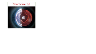
19
What are the positive findings?
What is your diagnosis?
Neovascularization along the pupillary
margin.
Neovascular glaucoma
The primary pathology is in retina
What are the causes?
a) Ischaemic central retinal vein
occlusion
b) Diabetes mellitus
c) central retinal artery occlusion and
d) ocular ischaemic syndrome
Where the primary pathology is?
What anti-glaucoma is contraindicated? And What is the medical treatment?
 Miotics should be avoided, and
 prostaglandin derivatives used with caution
 Topical atropine 1% once or twice daily will resist PS
 Topical steroids should be given if significant inflammation is present,
 Topical apraclonidine and
 oral acetazolamide maybe useful temporizing measures
What does the picture show?
Lental Opacity with
pseudoexfoliation of anterior
lens capsule
What difficulty may you overcome during cataract
surgery?
20
a) Poor dilatation of the pupil
b) Increased fragility of the zonule and lens capsule,
c) Endothelial deficiency;
If you go for phaco what other devices may you
need?
Iris hook and Capsular tension ring
What is the composition of this material?
Grey-white fibrillary amyloid-like material
What type of glaucoma may develop?
Secondary open angle
21
What are the positive
findings?
Hypermature cataract, lens
protein-containing macrophages
floating in the aqueous, and a
pseudohypopyon and KP also
What complication may develop
Secondary glaucoma (Open angle)
What is the treatment of choice?
Lens extraction is the treatment of choice, but before that
IOP has to be controlled
How you control IOP in this condition?
Steroid eye drop
Atropine eye drop
Anti-glaucoma eye drop (Brimodine+ Timolol)
Tab Acetazolamide
If not control Mannitol 20%
What is the positive findings?
What is your diagnosis?
CASE: 22
 Circumscribed nodule-like lentil is
situated 2-3 mm away from the
limbus.
 It is firm, immovable and tender.
 The conjunctiva moves freely over
the nodule.
 The conjunctiva looks purple in
colour as deep episcleral vessels
traverse it.
Diagnosis is nodular scleritis
What is the course of the diseases?
Complications
 Severe neuralgia may occur due to nerve involvement.
 Scleritis results from deeper infiltration of inflammation.
 There may be associated uveitis.
 It is usually transient lasting for several days or weeks.
 Recurrence is common.
 In episcleritis the attacks are fleeting but frequently repeated.
 It may be chronic but never ulcerates.
Treatment
 It is often difficult and unrewarding.
 Local
 Corticosteroid eye drops and ointment are applied.
 Warm compresses are very soothing.
 General
Systemic anti-inflammatory and analgesics relieve pain and control inflammation
23
What are the positive findings?
What may be the diagnosis?
 Increased C:D ratio
 Disc haemorrhages at inferior NRR
 Glaucoma
In what type glaucoma it is usually seen?
Usually NTG
In what other cases you may get it?
healthy individuals as well as patients with
systemic vascular disease.
What is the most common site of disc hge?
Most commonly inferotemporally.
24
What is the positive finding?
Trabeculectomy is glaucoma filtration surgery
that lowers IOP by creating a fistula, protected
by a superficial scleral flap, to allow aqueous
outflow from the anterior chamber to the sub-
Tenon space
Vascularized filtering bleb
What is Trabeculectomy?
a) Failure of conservative therapy
b) Avoidance of excessive polypharmacy.
c) Progressive deterioration despite
seemingly adequate IOP control
d) Primary therapy
e) Patient preference.
What are the indications?
a) Previous surgery and resulting subconjunctival fibrosis
b) Aphakia
c) Inflammation
d) Previous filtering bleb failure
e) Long-term medical therapy
f) Neovascular glaucoma
g) Intraoperative complications (e.g. iris or ciliary process incarceration in
the trabeculectomy fistula)
What are the Predisposing factors of filtering bleb failure?
What are the indications to use MMC?
 High-risk factors include:
 neovascular glaucoma,
 previous failed trabeculectomy or filtering device, and
 certain secondary glaucomas (e.g. inflammatory, post-traumatic angle
recession and iridocorneal endothelial syndrome).
 Intermediate- and lower-risk factors:
 include patients on topical medication (particularly sympathomimetics) for
over 3 years,
 previous conjunctival or cataract surgery,
 black ethnicity, and
 age under 40.
What is the type of medicine MMC? How it acts?
Mitomycin C (MMC) is an alkylating agent that inhibits proliferation of
fibroblasts and suppresses vascular ingrowth.
When MMC used intra-operative or per-operative? How long? What
concentration?
It is generally used intraoperatively .
0.2 mg/ml for 2 minutes, though a higher concentration
(e.g. 0.4 mg/ml) may be used for particularly high-risk patients;
What are the positive findings?
What may be the diagnosis?
The lens is displaced superiorly
zonule fibers are intact,
Marfan’s syndrome
Trauma
Do you think the patient has accommodation?
So, what is your probable diagnosis? Trauma or
Marfan’s
Yes, coz zonule is intact
Marfan’s but in Marfan’s subluxation is most
frequently superotemporal, but here it is in
upper quadrant but central. To make sure you
have to see other eye coz in Marfan’s it is usually
bilateral.
If it was trauma zonule was not intact
In Marfan’s what is the percentage of
subluxation?
It is about 80%
Short case: 25
What type of diseases is Marfan’s?
It is AD with variable expressivity
 Musculoskeletal features include:
 tall, thin stature with disproportionately long limbs (arm span > height),
 long fingers and toes (arachnodactyly),
 a narrow high-arched palate.
 Kyphoscoliosis,
 sternal abnormalities,
 mild joint laxity,
 muscular underdevelopment and predisposition to hernias.
 Cardiovascular lesions include
 dilatation of the aortic root,
 mitral valve prolapses and
 aortic aneurysm formation.
What are the systemic features?
What are the other ocular findings?
 angle anomaly may lead to glaucoma
 lattice retinal degeneration to retinal detachment
 hypoplasia of the dilator pupillae,
 microspherophakia, and strabismus.
What is the common complication after lens surgery?
Retinal detachment (Parsons)
In which complication, the patient may die earlier?
Rupture of the aortic aneurysm (Davidson)
Short case: 26
What is your D/D?
If it is Weill–Marchesani syndrome what may be the
features?
Homocystinuria is an AR disorder in which
decreased enzymatic metabolism of the amino acid
methionine results in systemic accumulation of
methionine and homocysteine.
Ectopia lentis (50%). Subluxation is in an inferior
direction and occurs in late childhood or early
adulthood. Microspherophakia is common, so that
pupillary block with angle closure may ensue.
Weill–Marchesani syndrome
Homocystinuria (Parson)
Trauma
Homocystinuria what type of diseases? Where the
pathology is?
How homocysteine is detected by in urine?
It is detected in urine by the cyanide nitroprusside test (Parsons)
What complications may arises?
In this disease, ectopia lentis becomes more marked with age and gives rise to glaucoma.
Such patients are poor operative risks because of the tendency to venous thromboses.
Other signs include laxity of joints and a marfanoid habitus. (Parsons)
Source Parsons
Treatment in general
If anteriorly dislocated, with inverse glaucoma, the patient must be treated as an
emergency. Lens removal is indicated after controlling the intraocular pressure.
If the lens is subluxated, the extent is assessed and refraction through the aphakic portion is
performed to give the best possible correction.
If the lens is posteriorly dislocated, with uveitis, removal is indicated; if there is no uveitis
no treatment is required.
If the vision is poor due to excessive lenticular astigmatism or presence of the lens edge in
the visual axis, removal of the lens is required.
If any of these deformities cause great visual disability, treatment by lens aspiration or
lensectomy is advisable.
Parsons
Short case: 27
What is your diagnosis?
Posterior capsular opacification
/After cataract
What is the treatment?
Posterior capsulotomy with Nd: YAG Laser
What is the meaning of Nd: YAG?
'Neodymium-doped Yttrium Aluminum Garnet
What complication may arise?
Usually it is safe but may occur pitting of IOL, IOP
may rise (if patient have glaucoma) but it is transient,
CMO (less common when capsulotomy is delayed for
6 months or more after cataract surgery), retinal tear
or detachment and IOL subluxation or dislocation.
What are the types of PCO?
 Vacuolated (pearl-type): PCO consists of proliferating swollen lens epithelial
cells, similar to the bladder (Wedl) cells seen in posterior subscapular
cataract. They are commonly termed ‘Elschnig pearls’, particularly when
grouped into clusters at the edge of a capsulotomy.
 Fibrosis-type PCO: is thought to be due to fibroblastic metaplasia of epithelial
cells, which develop contractile qualities.
 A Soemmering ring: is a whitish annular or doughnut shaped proliferation of
residual cells that classically formed almost in the periphery of the capsular
bag following older methods of cataract surgery, but is clinically uncommon
now. It may form at the edge of a capsulorrhexis or capsulotomy.
Upper left: capsular fibrosis;
Upper right: Soemmering ring
surrounding a slightly contracted
capsulotomy;
Lower: laser pitting of an IOL
Short case: 28
Factors to avoid PCO
 Non-modifiable
a) Age
b) Diabetic
c) traumatic cataracts
d) Uveitis. In these eyes, hydrophobic acrylic IOLs have been seen to provide a better
visual outcome and lower incidence of PCO than silicone, polymethyl methacrylate
(PMMA) or heparin-surface-modified PMMA IOLs
 Modifiable
a) Continuous Curvilinear Capsulorhexis. And bag size will be smaller than lens optic
b) Cortical Cleaving Hydrodissection
c) Cortical Clean Up
d) Polishing (Scraping) the Anterior Capsule
e) Square edge is
f) Haptic Designs & Angulation.

Glaucoma & lens

  • 2.
    19 What are thepositive findings? What is your diagnosis? Neovascularization along the pupillary margin. Neovascular glaucoma The primary pathology is in retina What are the causes? a) Ischaemic central retinal vein occlusion b) Diabetes mellitus c) central retinal artery occlusion and d) ocular ischaemic syndrome Where the primary pathology is?
  • 3.
    What anti-glaucoma iscontraindicated? And What is the medical treatment?  Miotics should be avoided, and  prostaglandin derivatives used with caution  Topical atropine 1% once or twice daily will resist PS  Topical steroids should be given if significant inflammation is present,  Topical apraclonidine and  oral acetazolamide maybe useful temporizing measures
  • 4.
    What does thepicture show? Lental Opacity with pseudoexfoliation of anterior lens capsule What difficulty may you overcome during cataract surgery? 20 a) Poor dilatation of the pupil b) Increased fragility of the zonule and lens capsule, c) Endothelial deficiency; If you go for phaco what other devices may you need? Iris hook and Capsular tension ring What is the composition of this material? Grey-white fibrillary amyloid-like material What type of glaucoma may develop? Secondary open angle
  • 5.
    21 What are thepositive findings? Hypermature cataract, lens protein-containing macrophages floating in the aqueous, and a pseudohypopyon and KP also What complication may develop Secondary glaucoma (Open angle) What is the treatment of choice? Lens extraction is the treatment of choice, but before that IOP has to be controlled How you control IOP in this condition? Steroid eye drop Atropine eye drop Anti-glaucoma eye drop (Brimodine+ Timolol) Tab Acetazolamide If not control Mannitol 20%
  • 6.
    What is thepositive findings? What is your diagnosis? CASE: 22  Circumscribed nodule-like lentil is situated 2-3 mm away from the limbus.  It is firm, immovable and tender.  The conjunctiva moves freely over the nodule.  The conjunctiva looks purple in colour as deep episcleral vessels traverse it. Diagnosis is nodular scleritis
  • 7.
    What is thecourse of the diseases? Complications  Severe neuralgia may occur due to nerve involvement.  Scleritis results from deeper infiltration of inflammation.  There may be associated uveitis.  It is usually transient lasting for several days or weeks.  Recurrence is common.  In episcleritis the attacks are fleeting but frequently repeated.  It may be chronic but never ulcerates. Treatment  It is often difficult and unrewarding.  Local  Corticosteroid eye drops and ointment are applied.  Warm compresses are very soothing.  General Systemic anti-inflammatory and analgesics relieve pain and control inflammation
  • 8.
    23 What are thepositive findings? What may be the diagnosis?  Increased C:D ratio  Disc haemorrhages at inferior NRR  Glaucoma In what type glaucoma it is usually seen? Usually NTG In what other cases you may get it? healthy individuals as well as patients with systemic vascular disease. What is the most common site of disc hge? Most commonly inferotemporally.
  • 9.
    24 What is thepositive finding? Trabeculectomy is glaucoma filtration surgery that lowers IOP by creating a fistula, protected by a superficial scleral flap, to allow aqueous outflow from the anterior chamber to the sub- Tenon space Vascularized filtering bleb What is Trabeculectomy? a) Failure of conservative therapy b) Avoidance of excessive polypharmacy. c) Progressive deterioration despite seemingly adequate IOP control d) Primary therapy e) Patient preference. What are the indications?
  • 10.
    a) Previous surgeryand resulting subconjunctival fibrosis b) Aphakia c) Inflammation d) Previous filtering bleb failure e) Long-term medical therapy f) Neovascular glaucoma g) Intraoperative complications (e.g. iris or ciliary process incarceration in the trabeculectomy fistula) What are the Predisposing factors of filtering bleb failure?
  • 11.
    What are theindications to use MMC?  High-risk factors include:  neovascular glaucoma,  previous failed trabeculectomy or filtering device, and  certain secondary glaucomas (e.g. inflammatory, post-traumatic angle recession and iridocorneal endothelial syndrome).  Intermediate- and lower-risk factors:  include patients on topical medication (particularly sympathomimetics) for over 3 years,  previous conjunctival or cataract surgery,  black ethnicity, and  age under 40.
  • 12.
    What is thetype of medicine MMC? How it acts? Mitomycin C (MMC) is an alkylating agent that inhibits proliferation of fibroblasts and suppresses vascular ingrowth. When MMC used intra-operative or per-operative? How long? What concentration? It is generally used intraoperatively . 0.2 mg/ml for 2 minutes, though a higher concentration (e.g. 0.4 mg/ml) may be used for particularly high-risk patients;
  • 13.
    What are thepositive findings? What may be the diagnosis? The lens is displaced superiorly zonule fibers are intact, Marfan’s syndrome Trauma Do you think the patient has accommodation? So, what is your probable diagnosis? Trauma or Marfan’s Yes, coz zonule is intact Marfan’s but in Marfan’s subluxation is most frequently superotemporal, but here it is in upper quadrant but central. To make sure you have to see other eye coz in Marfan’s it is usually bilateral. If it was trauma zonule was not intact In Marfan’s what is the percentage of subluxation? It is about 80% Short case: 25
  • 14.
    What type ofdiseases is Marfan’s? It is AD with variable expressivity  Musculoskeletal features include:  tall, thin stature with disproportionately long limbs (arm span > height),  long fingers and toes (arachnodactyly),  a narrow high-arched palate.  Kyphoscoliosis,  sternal abnormalities,  mild joint laxity,  muscular underdevelopment and predisposition to hernias.  Cardiovascular lesions include  dilatation of the aortic root,  mitral valve prolapses and  aortic aneurysm formation. What are the systemic features?
  • 15.
    What are theother ocular findings?  angle anomaly may lead to glaucoma  lattice retinal degeneration to retinal detachment  hypoplasia of the dilator pupillae,  microspherophakia, and strabismus. What is the common complication after lens surgery? Retinal detachment (Parsons) In which complication, the patient may die earlier? Rupture of the aortic aneurysm (Davidson)
  • 16.
    Short case: 26 Whatis your D/D? If it is Weill–Marchesani syndrome what may be the features? Homocystinuria is an AR disorder in which decreased enzymatic metabolism of the amino acid methionine results in systemic accumulation of methionine and homocysteine. Ectopia lentis (50%). Subluxation is in an inferior direction and occurs in late childhood or early adulthood. Microspherophakia is common, so that pupillary block with angle closure may ensue. Weill–Marchesani syndrome Homocystinuria (Parson) Trauma Homocystinuria what type of diseases? Where the pathology is?
  • 17.
    How homocysteine isdetected by in urine? It is detected in urine by the cyanide nitroprusside test (Parsons) What complications may arises? In this disease, ectopia lentis becomes more marked with age and gives rise to glaucoma. Such patients are poor operative risks because of the tendency to venous thromboses. Other signs include laxity of joints and a marfanoid habitus. (Parsons)
  • 18.
  • 19.
    Treatment in general Ifanteriorly dislocated, with inverse glaucoma, the patient must be treated as an emergency. Lens removal is indicated after controlling the intraocular pressure. If the lens is subluxated, the extent is assessed and refraction through the aphakic portion is performed to give the best possible correction. If the lens is posteriorly dislocated, with uveitis, removal is indicated; if there is no uveitis no treatment is required. If the vision is poor due to excessive lenticular astigmatism or presence of the lens edge in the visual axis, removal of the lens is required. If any of these deformities cause great visual disability, treatment by lens aspiration or lensectomy is advisable. Parsons
  • 20.
    Short case: 27 Whatis your diagnosis? Posterior capsular opacification /After cataract What is the treatment? Posterior capsulotomy with Nd: YAG Laser What is the meaning of Nd: YAG? 'Neodymium-doped Yttrium Aluminum Garnet What complication may arise? Usually it is safe but may occur pitting of IOL, IOP may rise (if patient have glaucoma) but it is transient, CMO (less common when capsulotomy is delayed for 6 months or more after cataract surgery), retinal tear or detachment and IOL subluxation or dislocation.
  • 21.
    What are thetypes of PCO?  Vacuolated (pearl-type): PCO consists of proliferating swollen lens epithelial cells, similar to the bladder (Wedl) cells seen in posterior subscapular cataract. They are commonly termed ‘Elschnig pearls’, particularly when grouped into clusters at the edge of a capsulotomy.  Fibrosis-type PCO: is thought to be due to fibroblastic metaplasia of epithelial cells, which develop contractile qualities.  A Soemmering ring: is a whitish annular or doughnut shaped proliferation of residual cells that classically formed almost in the periphery of the capsular bag following older methods of cataract surgery, but is clinically uncommon now. It may form at the edge of a capsulorrhexis or capsulotomy.
  • 22.
    Upper left: capsularfibrosis; Upper right: Soemmering ring surrounding a slightly contracted capsulotomy; Lower: laser pitting of an IOL
  • 23.
  • 24.
    Factors to avoidPCO  Non-modifiable a) Age b) Diabetic c) traumatic cataracts d) Uveitis. In these eyes, hydrophobic acrylic IOLs have been seen to provide a better visual outcome and lower incidence of PCO than silicone, polymethyl methacrylate (PMMA) or heparin-surface-modified PMMA IOLs  Modifiable a) Continuous Curvilinear Capsulorhexis. And bag size will be smaller than lens optic b) Cortical Cleaving Hydrodissection c) Cortical Clean Up d) Polishing (Scraping) the Anterior Capsule e) Square edge is f) Haptic Designs & Angulation.