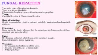
KERATITIS
- BY
NANDITA CHAKRABORTY
OPTOMETRY INTERN, L V PRASAD EYE
INSTITUTE
-Inflammation of the cornea
-Keratitis and corneal ulcer are not always synonymous.
A corneal ulcer is defined as a loss of corneal epithelium with underlying stromal infiltration
associated with signs of inflammation with or without hypopyon.
Modes of Infection
Exogenous infections: Most common, and cornea is primarily affected by virulent
organisms.
From the ocular tissues: Owing to direct anatomical continuity, the diseases of—
Conjunctiva—spread to the corneal epithelium.
Sclera—to the stroma.
Uveal tract—to the endothelium.
Endogenous infections: Rare, as the cornea is avascular. They are typically allergic in
nature.
2
• Pseudomonas aeruginosa
• Staphylococcus aureus
• Streptococci.
Symptoms:
• Acute pain, redness and lacrimation
• Photophobia
• Decreased visual acuity
• White spot on the cornea.
Signs:
• Marked blepharospasm
• Lid edema
• Ciliary congestion of the conjunctiva
• Some degree of iritis
• Hypopyon may be present
Treatment:
• Antibiotics
• Atropine sulfate (1%) eye drops, 3 times daily
• Hot fomentation is to improve circulation and to relieve pain.
• Analgesics with antacids for pain.
• Removal of local predisposing factor
• Tab acetazolamide or timolol maleate eye drop (0.5%)
3
Two main types of fungi cause keratitis:
Yeast (e.g. genus Candida),
Filamentous fungi (e.g. genera Fusarium and Aspergillus)
Types:
Candida Keratitis & Filamentous Keratitis
Mode of Infection:
Ocular trauma (often trivial in nature), mainly by agricultural and vegetable
matters.
Symptoms:
Same as for the bacterial ulcer, but the symptoms are less prominent than
an equal size bacterial ulcer.
Signs:
Dry looking, yellowish-white lesion with indistinct margin.
Immobile massive dense hypopyon
Treatment
 Scraping and debridement of the ulcer.
 Atropine eye ointment—3 times daily.
 Antifungal drugs
4
HERPES ZOSTER OPHTHALMICUS
• Caused by the varicella zoster virus
• mainly affects the elderly patients.
• immunocompromised hosts (as in HIV).
Symptoms:
• Vesicular eruptions around the eye, forehead and scalp.
• Severe pain along the distribution of the ophthalmic division of 5th nerve.
• Photophobia and lacrimation.
Signs: There are three stages:
Stage I: Acute lesions—which develops within 3 weeks.
Stage II: Chronic lesions—may persist for up to 10 years.
Stage III: Recurrent lesions—which may reappear after 10 years.
Treatment:
 Oral acyclovir: 800 mg 5 times daily for 7 days.
 Antibiotic-corticosteroid preparation
 Topical steroids
 Cycloplegic for iritis or severe keratitis
5
• Herpes Simplex Viral Keratitis
• Dendritic Keratitis
• Geographical Keratitis (Ulcer)
• Stromal Necrotic Keratitis
• Metaherpetic Keratitis (Trophic Ulcer)
• Disciform Keratitis
6
ACANTHAMOEBA KERATITIS
Modes of Infection:
Contact lens wearer
Non-contact lens wearer
Clinical Features:
 Severe pain
 Progressive chronic stromal keratitis
 Frequent development of paracentral ringshaped ulcer
 A nodular scleritis is a frequent finding.
 limbal inflammation (limbitis).
Diagnosis by calcofluor white staining of the smear for amoebic
cyst. Acanthamoeba is cultured in special media—E. coli enriched
nutrient agar plate.
Treatment:
 Propamidine isethionate (Brolene) (0.02%) drops and ointment
 Neomycin drops and ointment
 Polyhexamethylene biguanide (PHMB) (0.001%)
 Chlorhexidine (0.02%) eye drop 1 hourly
7
EXPOSURE KERATITIS
Due to exposure of the cornea when it remains
insufficiently covered by the lids.
Clinical Features:
 It ranges from minimum epithelial erosions to
severe ulceration, secondary infection and
even perforation.
 The lower-third of the cornea is commonly
affected
Treatment:
 Tarsorrhaphy
 Artificial tear at day time, Antibiotic at night
 Closure of the lids by adhesive tapes
 Soft BCL
8
PHLYCTENULAR KERATITIS
Clinical Features:
Symptoms includes Pain, photophobia, lacrimation ,
blepharospasm.
Mucopurulent discharge
The corneal phlycten is a gray nodule, slightly raised
above the surface, and an ulcer is yellow in color.
Complications includes:
Ring ulcer
Phlyctenular pannus
Perforation of the cornea
Treatment:
Topical corticosteroids, 4 times daily.
Atropine (1%) eye drop, 2 times daily.
Topical antibiotic, if secondarily infected,
4–6 times daily.
NEUROTROPHIC KERATITIS
It occurs in an anesthetic cornea.
Etiology: Congenital, Acquired.
Clinical Features:
 Punctate epithelial erosions involving the
interpalpebral area.
 Exfoliation of the epithelial cells followed by central
ulceration.
 Absense of Corneal sensation
Treatment:
 Routine treatment of corneal ulcer
 Ointments and patching
 Amniotic membrane transplantation
 Tarsorrhaphy for several months.
9
MARGINAL KERATITS
It is caused by hypersensitivity reaction to staphylococcal
exotoxins, and prevalent in patients having chronic
staphylococcal blepharitis.
Symptoms
Mild ocular irritation, Lacrimation and photophobia.
Signs
 Subepithelial infiltrates at the periphery, mostly at 4–8
o’clock position, or at 10–2 o’clock position.
 Corneal sensation is unaffected.
Treatment
 Topical corticosteroids, 3–4 times daily for a few days.
 Mild cycloplegic, like cyclopentolate or tropicamide.
 Simultaneous treatment of blepharitis to prevent
recurrence.
MOOREN’S ULCER
1. A limited form: Unilateral and usually affects the
elderly people.
2. A progressive form: Bilateral, relentlessly progressive
and affects the younger people.
Symptoms:
Severe pain, Photophobia and lacrimation
Decreased vision due to irregular astigmatism and
involvement of the visual axis in late stage.
Signs:
Ulcer usually starts at the interpalpebral area as
patches of gray infiltrates at the margin.
Healing takes places from the periphery, and the healed
area becomes vascularized, thinned and opaque.
Treatments:
Antibiotics, BCL, Cycloplegics like atropine drops. 1 0
INTERSTITIAL KERATITIS
Affecting chiefly the corneal stroma, without primary
involvement of the epithelium or endothelium.
Causes: Three causes are congenital syphilis,
tuberculosis, Cogan’s syndrome.
Signs:
ground-glass appearance.
salmon patch of Hutchinson.
Symptoms:
Initially, irritation and haziness of the Cornea,
lacrimation, photophobia and severe blepharospasm
with increased haziness of the cornea, Profound loss of
vision.
Treatment:
• Systemic penicillin
• Topical corticosteroids as drops or ointment,
• 4–6 times daily.
• Atropine (1%) ointment, 2 times daily.
-THANK YOU-
References:
Essentials of Ophthalmology- Samar Kumar Basak
Kanski’s Clinical ophthalmology
1 1

KERATITIS.pptx

  • 1.
    KERATITIS - BY NANDITA CHAKRABORTY OPTOMETRYINTERN, L V PRASAD EYE INSTITUTE
  • 2.
    -Inflammation of thecornea -Keratitis and corneal ulcer are not always synonymous. A corneal ulcer is defined as a loss of corneal epithelium with underlying stromal infiltration associated with signs of inflammation with or without hypopyon. Modes of Infection Exogenous infections: Most common, and cornea is primarily affected by virulent organisms. From the ocular tissues: Owing to direct anatomical continuity, the diseases of— Conjunctiva—spread to the corneal epithelium. Sclera—to the stroma. Uveal tract—to the endothelium. Endogenous infections: Rare, as the cornea is avascular. They are typically allergic in nature. 2
  • 3.
    • Pseudomonas aeruginosa •Staphylococcus aureus • Streptococci. Symptoms: • Acute pain, redness and lacrimation • Photophobia • Decreased visual acuity • White spot on the cornea. Signs: • Marked blepharospasm • Lid edema • Ciliary congestion of the conjunctiva • Some degree of iritis • Hypopyon may be present Treatment: • Antibiotics • Atropine sulfate (1%) eye drops, 3 times daily • Hot fomentation is to improve circulation and to relieve pain. • Analgesics with antacids for pain. • Removal of local predisposing factor • Tab acetazolamide or timolol maleate eye drop (0.5%) 3
  • 4.
    Two main typesof fungi cause keratitis: Yeast (e.g. genus Candida), Filamentous fungi (e.g. genera Fusarium and Aspergillus) Types: Candida Keratitis & Filamentous Keratitis Mode of Infection: Ocular trauma (often trivial in nature), mainly by agricultural and vegetable matters. Symptoms: Same as for the bacterial ulcer, but the symptoms are less prominent than an equal size bacterial ulcer. Signs: Dry looking, yellowish-white lesion with indistinct margin. Immobile massive dense hypopyon Treatment  Scraping and debridement of the ulcer.  Atropine eye ointment—3 times daily.  Antifungal drugs 4
  • 5.
    HERPES ZOSTER OPHTHALMICUS •Caused by the varicella zoster virus • mainly affects the elderly patients. • immunocompromised hosts (as in HIV). Symptoms: • Vesicular eruptions around the eye, forehead and scalp. • Severe pain along the distribution of the ophthalmic division of 5th nerve. • Photophobia and lacrimation. Signs: There are three stages: Stage I: Acute lesions—which develops within 3 weeks. Stage II: Chronic lesions—may persist for up to 10 years. Stage III: Recurrent lesions—which may reappear after 10 years. Treatment:  Oral acyclovir: 800 mg 5 times daily for 7 days.  Antibiotic-corticosteroid preparation  Topical steroids  Cycloplegic for iritis or severe keratitis 5
  • 6.
    • Herpes SimplexViral Keratitis • Dendritic Keratitis • Geographical Keratitis (Ulcer) • Stromal Necrotic Keratitis • Metaherpetic Keratitis (Trophic Ulcer) • Disciform Keratitis 6
  • 7.
    ACANTHAMOEBA KERATITIS Modes ofInfection: Contact lens wearer Non-contact lens wearer Clinical Features:  Severe pain  Progressive chronic stromal keratitis  Frequent development of paracentral ringshaped ulcer  A nodular scleritis is a frequent finding.  limbal inflammation (limbitis). Diagnosis by calcofluor white staining of the smear for amoebic cyst. Acanthamoeba is cultured in special media—E. coli enriched nutrient agar plate. Treatment:  Propamidine isethionate (Brolene) (0.02%) drops and ointment  Neomycin drops and ointment  Polyhexamethylene biguanide (PHMB) (0.001%)  Chlorhexidine (0.02%) eye drop 1 hourly 7
  • 8.
    EXPOSURE KERATITIS Due toexposure of the cornea when it remains insufficiently covered by the lids. Clinical Features:  It ranges from minimum epithelial erosions to severe ulceration, secondary infection and even perforation.  The lower-third of the cornea is commonly affected Treatment:  Tarsorrhaphy  Artificial tear at day time, Antibiotic at night  Closure of the lids by adhesive tapes  Soft BCL 8 PHLYCTENULAR KERATITIS Clinical Features: Symptoms includes Pain, photophobia, lacrimation , blepharospasm. Mucopurulent discharge The corneal phlycten is a gray nodule, slightly raised above the surface, and an ulcer is yellow in color. Complications includes: Ring ulcer Phlyctenular pannus Perforation of the cornea Treatment: Topical corticosteroids, 4 times daily. Atropine (1%) eye drop, 2 times daily. Topical antibiotic, if secondarily infected, 4–6 times daily.
  • 9.
    NEUROTROPHIC KERATITIS It occursin an anesthetic cornea. Etiology: Congenital, Acquired. Clinical Features:  Punctate epithelial erosions involving the interpalpebral area.  Exfoliation of the epithelial cells followed by central ulceration.  Absense of Corneal sensation Treatment:  Routine treatment of corneal ulcer  Ointments and patching  Amniotic membrane transplantation  Tarsorrhaphy for several months. 9 MARGINAL KERATITS It is caused by hypersensitivity reaction to staphylococcal exotoxins, and prevalent in patients having chronic staphylococcal blepharitis. Symptoms Mild ocular irritation, Lacrimation and photophobia. Signs  Subepithelial infiltrates at the periphery, mostly at 4–8 o’clock position, or at 10–2 o’clock position.  Corneal sensation is unaffected. Treatment  Topical corticosteroids, 3–4 times daily for a few days.  Mild cycloplegic, like cyclopentolate or tropicamide.  Simultaneous treatment of blepharitis to prevent recurrence.
  • 10.
    MOOREN’S ULCER 1. Alimited form: Unilateral and usually affects the elderly people. 2. A progressive form: Bilateral, relentlessly progressive and affects the younger people. Symptoms: Severe pain, Photophobia and lacrimation Decreased vision due to irregular astigmatism and involvement of the visual axis in late stage. Signs: Ulcer usually starts at the interpalpebral area as patches of gray infiltrates at the margin. Healing takes places from the periphery, and the healed area becomes vascularized, thinned and opaque. Treatments: Antibiotics, BCL, Cycloplegics like atropine drops. 1 0 INTERSTITIAL KERATITIS Affecting chiefly the corneal stroma, without primary involvement of the epithelium or endothelium. Causes: Three causes are congenital syphilis, tuberculosis, Cogan’s syndrome. Signs: ground-glass appearance. salmon patch of Hutchinson. Symptoms: Initially, irritation and haziness of the Cornea, lacrimation, photophobia and severe blepharospasm with increased haziness of the cornea, Profound loss of vision. Treatment: • Systemic penicillin • Topical corticosteroids as drops or ointment, • 4–6 times daily. • Atropine (1%) ointment, 2 times daily.
  • 11.
    -THANK YOU- References: Essentials ofOphthalmology- Samar Kumar Basak Kanski’s Clinical ophthalmology 1 1