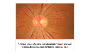
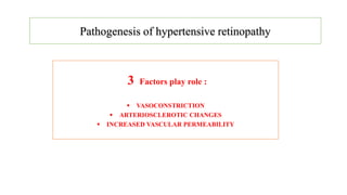
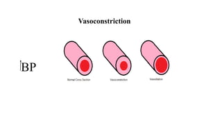
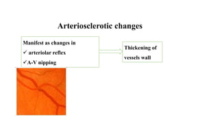
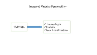
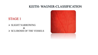
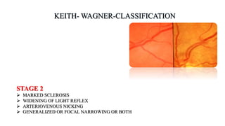
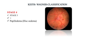
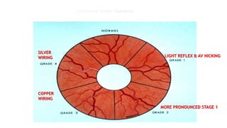
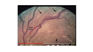
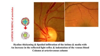
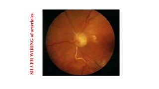
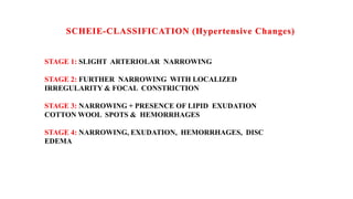
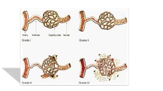
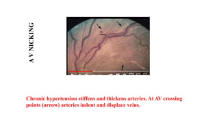
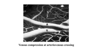
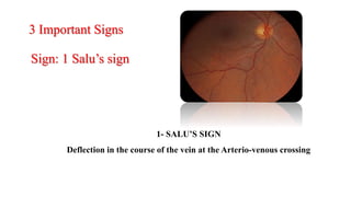
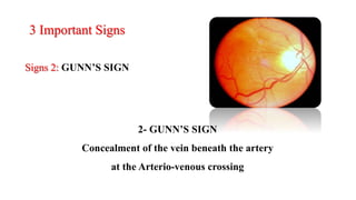
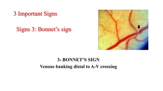
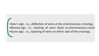
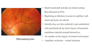
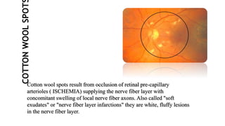
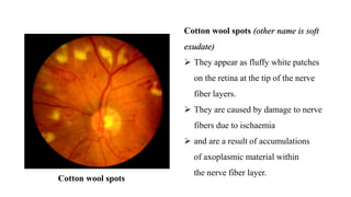
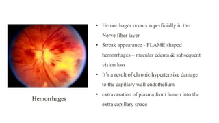
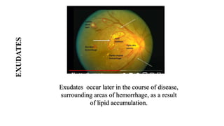
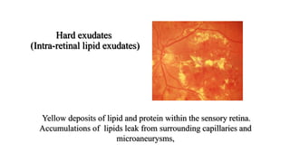
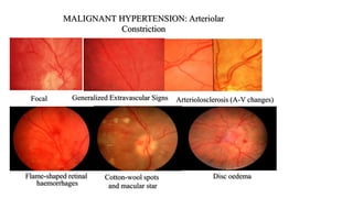
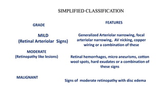
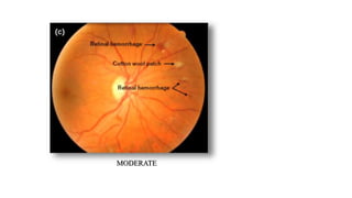
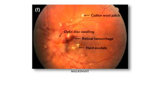
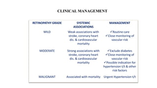
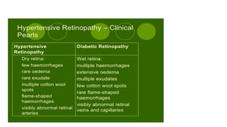
This lecture is based on medical students those are preparing for postgraduate degree namely FCPS/MS/MD/ any any subject coz hypertension is a systemic disease and by seeing the ocular fundus we can asses the general condition of blood vessels in major organ.
6. So, clinicians have to know how to
perform ophthalmoscopy
To know ophthalmoscopy is nothing, but practice,
practice and practice
7.
8.
9. 1) Arteries differ in color from veins;
Artery is lighter in colour than vein.
2) Arteries are thinner than
adjacent veins ratio A:V = 2:3
3) The central reflex is wider
in arteries than in comparably
sized veins
10. A retinal image showing the retinal artery (red) and vein
(blue) and measured caliber (cross-sectional lines)
21. Poor reliability and reproducibility
Grades do not correlate with severity of hypertension
Not sequential in nature
Grades do not correlate with prognosis,
cardiovascular events, and mortality.
25. COPPER
WIRING
of
arterioles
Hyaline thickening & lipoidal infiltration of the intima & media with
An increase in the reflected light reflex & indentation of the venous blood
Column at arteriovenous column
31. 1- SALU’S SIGN
Deflection in the course of the vein at the Arterio-venous crossing
3 Important Signs
Sign: 1 Salu’s sign
32. 3 Important Signs
Signs 2: GUNN’S SIGN
2- GUNN’S SIGN
Concealment of the vein beneath the artery
at the Arterio-venous crossing
33. 3- BONNET’S SIGN
Venous banking distal to A-V crossing
3 Important Signs
Signs 3: Bonnet’s sign
34.
35. • Small round dark red dots on retinal surface
Best detected on FFA.
• Beginning as dilations in areas in capillary wall
where pericytes are absent
• Initially they are thin walled & Later endothelial
cells proliferate & lay down layers of basement
membrane material around themselves.
• ↑in number as the degree of retinal involvement
• ↑capillary occlusion – retinal ischemia
Microaneurysms
36. COTTON
WOOL
SPOT
Cotton wool spots result from occlusion of retinal pre-capillary
arterioles ( ISCHEMIA) supplying the nerve fiber layer with
concomitant swelling of local nerve fiber axons. Also called "soft
exudates" or "nerve fiber layer infarctions" they are white, fluffy lesions
in the nerve fiber layer.
37. Cotton wool spots (other name is soft
exudate)
They appear as fluffy white patches
on the retina at the tip of the nerve
fiber layers.
They are caused by damage to nerve
fibers due to ischaemia
and are a result of accumulations
of axoplasmic material within
the nerve fiber layer.
Cotton wool spots
38. • Hemorrhages occurs superficially in the
Nerve fiber layer
• Streak appearance - FLAME shaped
hemorrhages – macular edema & subsequent
vision loss
• It’s a result of chronic hypertensive damage
to the capillary wall endothelium
• extravasation of plasma from lumen into the
extra capillary space
Hemorrhages
39. EXUDATES
Exudates occur later in the course of disease,
surrounding areas of hemorrhage, as a result
of lipid accumulation.
40. Hard exudates
(Intra-retinal lipid exudates)
Yellow deposits of lipid and protein within the sensory retina.
Accumulations of lipids leak from surrounding capillaries and
microaneurysms,
42. GRADE
SIMPLIFIED CLASSIFICATION
FEATURES
MILD
(Retinal Arteriolar Signs)
Generalized Arteriolar narrowing, focal
arteriolar narrowing, AV nicking, copper
wiring or a combination of these
MODERATE
(Retinopathy like lesions) Retinal hemorrhages, micro aneurisms, cotton
wool spots, hard exudates or a combination of
these signs
MALIGNANT
Signs of moderate retinopathy with disc edema