CRAO
•Download as PPTX, PDF•
8 likes•1,114 views

Central Retinal Artery Occlusion (CRAO) for undergraduate MBBS Students. Covers the basics of Aetiology, pathophysiology, clinical features, types, associated conditions and management of CRAO. Also encompasses salient points for PGMEE

Recommended
More Related Content
What's hot
What's hot (20)
Similar to CRAO
Similar to CRAO (20)
More from Abhishek Onkar
Recently uploaded
Recently uploaded (20)
CRAO
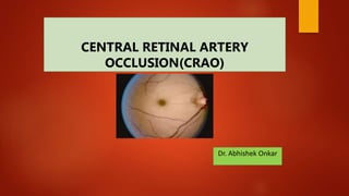
- 1. CENTRAL RETINAL ARTERY OCCLUSION(CRAO) Dr. Abhishek Onkar
- 3. Blood supply of O.N.
- 5. THE OCULAR BLOOD SUPPLY The eye receives its blood supply from the ophthalmic artery (a branch of the internal carotid artery) via the retinal artery, ciliary arteries and muscular arteries. The conjunctival circulation anastomoses anteriorly with branches from the external carotid artery. The anterior optic nerve is supplied by branches from the ciliary arteries. The retina is supplied by arterioles branching from the central retinal artery. These arterioles each supply an area of retina with little overlap. Obstruction results in ischaemia of most of the area supplied by that arteriole. The fovea is so thin that it requires no supply from the retinal circulation. It is supplied indirectly, as are the outer layers of the retina, by diffusion of oxygen and metabolites across the retinal pigment epithelium from the choroid.
- 8. CRAO An abrupt diminution of blood flow through the central retinal artery severe enough to cause ischemia of the inner retina • A branch retinal artery obstruction occurs when the site of blockage is distal to the lamina cribrosa of the optic nerve. Retinal artery obstructions selectively affect the inner retina only.
- 9. EPIDEMIOLOGY Elderly population Arteritic: > 60-70 years of age Non-arteritic: > 50 years Can be seen in young adults M:F=2:1 UNIOCULAR: 98%
- 11. Aetiology
- 12. Pathophysiology
- 13. SYMPTOMS • Monocular • Sudden loss of vision • severe • painless • Occurs acutely, possibly over the span of a few seconds. • In some cases, premonitory episodes of amaurosis fugax may be reported. Amaurosis fugax represents transient acute retinal ischemia and typically suggests an embolic source of occlusion
- 14. SIGNS 1) Visual acuity: at the time of initial presentation ranges from counting fingers to light perception • Central visual acuity may be near normal in patients who have a transient CRAO or a cilioretinal artery providing sufficient vascular supply to the fovea. • The absence of light perception is rare; therefore, in such cases, concomitant choroidal circulation deficit (e.g., due to ophthalmic artery occlusion) or optic nerve involvement should be considered • Visual acuity tends only to improve within the first week of onset with minimal chance for appreciable improvement subsequently • Visual recovery after treatment has been shown to correlate with presenting visual acuity and the duration of visual impairment
- 15. 2) RAPD 3) INTRAOCULAR PRESSURE is often normal at presentation but may become elevated in the setting of rubeosis iridis
- 16. 4) FUNDUS CHANGES • Cherry-red spot (90%) • Posterior pole retinal opacity or whitening (58%), • Box-carring of retinal arteries and veins (19% and 20% respectively) • Retinal arterial attenuation (32%) • Optic disc edema (22%), and optic nerve pallor (39%) • The retinal findings were predominantly located in the posterior pole with a normal-appearing periphery
- 18. BRAO
- 20. Cherry Red Spot • GM2 gangliosidoses (Tay-Sachs & Sandhoff) • GM1 gangliosidoses • Sialidosis • Metachromatic leukodystrophy • Niemann-Pick Type A • Farber lipogranulomatosis • CRAO • Trauma (retinal edema)
- 21. Fluorescein angiography demonstrates the slow rate of filling of the retinal circulation and the normal filling of the cilioretinal artery and choroid
- 22. Retinal emboli are visible in 20–40% of eyes with CRAO .The most common variant is a yellow, refractile cholesterol embolus (Hollenhorst plaque) Retinal emboli consist of CHOLESTEROL in 74% of cases, CALCIFIED MATERIAL in 15.5%, and PLATELET AND FIBRIN in 15.5% • 1 Cholesterol emboli (Hollenhorst plaques) are frequently asymptomatic. Calcific emboli may originate from atheromatous plaques in the ascending aorta or carotid arteries, as well as from calcified heart valves. They may cause permanent occlusion of the central retinal artery or one of its main branches. Fibrin-platelet emboli :They may cause a retinal transient ischaemic attack (TIA), with resultant amaurosis fugax, and occasionally complete obstruction.
- 24. Neuro/cardio prompt referral forestall the development of permanent neurological sequelae as patients with retinal embolization have an increased risk of developing a permanent stroke over the next few months • It should be noted that the leading cause of death in patients with retinal arterial obstruction is cardiovascular disease
- 25. The optic nerve is acutely edematous in nearly all cases of arteritic CRAO as a result of the associated anterior ischemic optic neuropathy that is typically observed in these patients. In the acute phase of nonarteritic CRAO, the disc may be normal, hyperemic, edematous, and, rarely, pale. •
- 26. EVALUATION R/o GCA • carotid Doppler imaging and echocardiography • Since the cardiac morbidity and mortality are significant in patients with retinal artery occlusion, a baseline electrocardiogram is recommended • A hypercoagulability evaluation should be considered for patients less than 50 years of age with a suggestive history (e.g.,prior thrombosis, miscarriage, or family history) or unknown embolic source • Other tests for monoclonal gammopathy, cancer, • infection, and disseminated intravascular coagulation may be ordered depending on the clinical circumstance
- 28. TREATMENT • Adoption of a supine posture might improve ocular perfusion • Ocular massage • sublingual isosorbide dinitrate • intravenous acetazolamide, • intravenousmannitol or oral glycerol, • anterior-chamber paracentesis, • Intravenous methylprednisolone, streptokinase
- 30. HBO CARBOGEN
- 31. Iris neovascularization develops after acute CRAO in approximately 18% of eyes, with a mean time interval of approximately 4- 5 weeks -typically earlier than in CRVO 3 months. Full-scatter PRP is effective in eradicating the new iris vessels in about two thirds of cases • Intravitreal injection of an anti-VEGF agent is first-line therapy for iris, trabecular meshwork, or optic disc neovascularization
- 32. Treatment of carotid disease In patients with a localized stenosis of the artery, endarterectomy significantly reduces the risk of subsequent stroke. • If endarterectomy is contraindicated, medical treatment with drugs that reduce platelet stickiness (aspirin, dipyridamole) or anticoagulants may be used to reducing the frequency of transient ischaemic attacks and the risk of a major stroke.
- 33. FOLLOW-UP • The patient should be seen by an ophthalmologist in 3–4 weeks and again a month later in order to detect incipient neovascularization, particularly of the anterior segment. PROGNOSIS • Patients with visualized retinal artery emboli, whether or not obstruction is present, have a 56% mortality rate over 9 years, compared to 27% for an age-matched population without retinal artery emboli. • Life expectancy of patients with CRAO is 5.5 years compared to 15.4 years for an age-matched population without CRAO