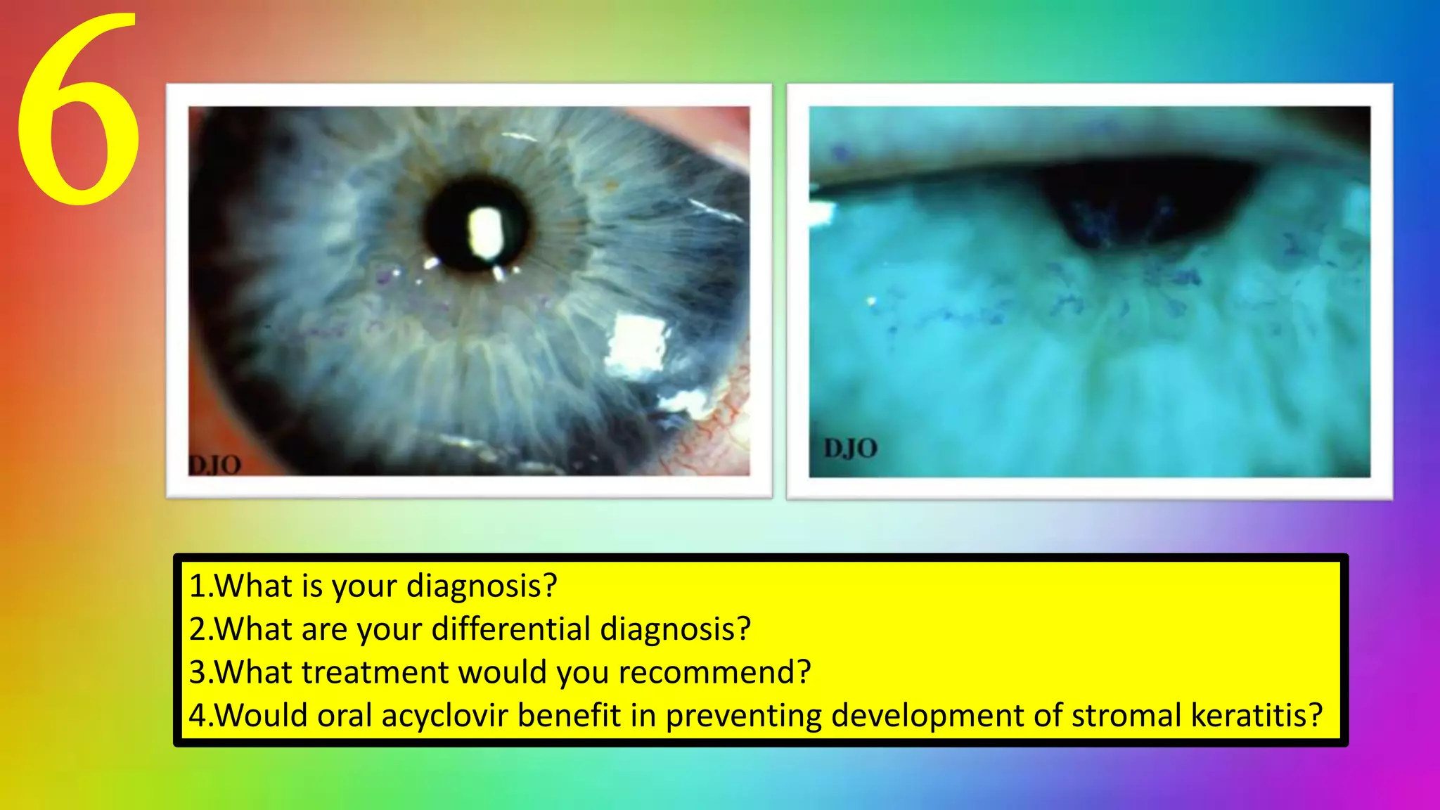
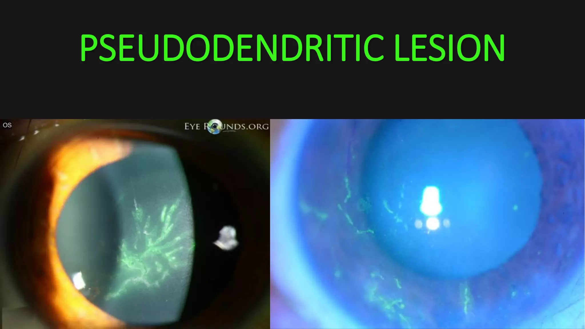
CORNEA
QUIZ
1
1.Describe finding on this picture
2.What is your diagnosis?
3.Malignant transformation should be suspected in which appearance?
2
1.What condition could be benefit from using the device?
3
1.Describe what you think is abnormal in this test
2.What is your diagnosis in this finding?
3.What is your definite treatment in this case?
4.Can this patient undergo phacoemulsification for cataract surgery?
Patient underwent endothelial keratoplasty procedure,
this picture reveal an examination in day 1 after surgery
1.Describe finding
2.What is your management?
4
5
1.What happened to this patient?
2.Which are the complications for patient who underwent this operation?
3.What is the most common complication after this operation?
6
1.What is your diagnosis?
2.What are your differential diagnosis?
3.What treatment would you recommend?
4.Would oral acyclovir benefit in preventing development of stromal keratitis?
This is too easy
for me, DUH!!
7
1.Describe finding
2.What is your management?
8
1.What is your diagnosis?
2.What are the risk factors causing this condition?
9
Patient underwent LASIK operation, a few months later he/she began to have a decreased vision
1.What is your diagnosis?
2.What is your management?
10
1.Describe finding
2.What is your diagnosis?
3.What is the usual presentation for this condition?
ANSWERS
1
1.Describe finding on this picture
2.What is your diagnosis?
3.Malignant transformation should be suspected in which appearance?
• 1.Describe finding on this picture
• Multiple flat, brown patches of noncystic pigmentation on superficial
conjunctiva
• 2.What is your diagnosis?
• Primary acquired melanosis (PAM)
• 3.Malignant transformation should be suspected in which
appearance?
• Nodularity
• Enlargement
• Increased vascularity
Type History Color Appearance Laterality Specific
features
Nevus Longtime presence,
usually from
childhood
Light brown, may
be non-pigmented
during childhood
Slightly raised and
cystic, with well-
defined margins
Mostly
unilateral
Changes
pigmentation
with puberty and
pregnancy
PAM New pigmentation
on ocular surface
Varies from light
brown to darker
brown; rarely non-
pigmented
Flat, diffuse and
noncircumscribed
Unilateral Waxing and
waning of size
and pigmentation
Conjunctival
melanoma
Arises de novo,
from nevus or from
areas of PAM with
atypia
Dark brown in most
cases; can be
amelanotic
Elevated lesion,
thickened, nodular
may be amelanotic or
mixed pigmentation
Unilateral High vascularity
with feeder
vessel
Conjunctival melanomanevus
PAM
Differential diagnosis
Primary Acquired Melanosis
• Acquired melanosis may be
benign, precancerous or cancerous
• Characteristic
• Later in life (30-40 years of age)
• Unilateral
• Almost always Caucasians
• 15-17% of benign PAM will convert to
cancer within 30 years
sign
• Sudden development of irregular diffuse, flat, grayish black, brown or
tan bulbar pigmentation
• May extend onto palpebral conjunctiva
• Without cyst**
• Malignant transformation should be suspected with elevation,
nodularity, increased vascularity
treatment
• Monitor closely (photo) every 3-6 months
• Refer for biopsy
• Dilated fundus exam to r/o choroidal melanoma
PAM without atypia
• Benign proliferation of normal melanocytes confined to the basal
layers of conjunctiva
PAM with atypia
• Pre-malignant condition with 50% chance of malignant
transformation within 5 years
• Characterized by melanocytes involving all layers of conjunctiva
2
1.What condition could be benefit from using the device?
• 1.What condition could be benefit from using the device?
• Meibomian gland dysfunction
• Dry eye
• Contact lens intolerance
• blepharitis
MGD
• Meibomian glands or glandulae
tarsals are large sebaceous glands
which secrete lipids
• Chronic, diffuse abnormality of
the meibomian glands commonly
characterized by terminal duct
obstruction and may result in
alteration of the tear film,
symptoms of eye irritation and
other ocular surface disorders
Risk factors
• Aging
• Deficiency of sex hormones
(androgens)
• Sjogren’s syndrome
• Stevens-Johnson Syndrome
• Psoriasis
• Atopy
• Polycystic ovary syndrome
• Hypertension
• Chronic blepharitis
• CL wear
• Eyelid tattooing
• Demodex folliculorum infection
• Isotretinoin for acne
• Anti-histamines
3
1.Describe what you think is abnormal in this test
2.What is your diagnosis in this finding?
3.What is your definite treatment in this case?
4.Can this patient undergo phacoemulsification for cataract surgery?
1. Describe what you think is abnormal in this test
1. Cell Density = 1763
2. Guttata presentation
3. CV = 44%
2. What is your diagnosis in this finding?
1. Fuchs endothelial dystrophy
3. What is your definite treatment in this case?
1. DMEK
2. DSAEK
3. PKP
4. Can this patient undergo phacoemulsification for cataract surgery?
1. Yes but with caution of using too high energy
DDX PPMD
Vesicle-like lesions at the level of Descemet's membrane and endothelium are the hallmark lesions of PPMD.
They appear as transparent cystic lesions surrounded by gray halos and commonly occur in lines or clusters. .
Band lesions, sometimes called "snail tracks," are classically horizontal lesions with parallel, scalloped, non-tapering
edges at the level of the posterior cornea. Below are examples of these lesions in 2 separate patients.
DDX PPMD
DDX ICE (chandler variant)
Fine hammered silver appearance of posterior cornea
Pleomorphism in size and shape
Dark areas within the cells
Loss of clear hexagonal margins
Endothelial cell count
• CD is a measurement of cell density in mm2 and decreases with age.
• A low CD value for a particular age may indicate that the endothelium is depleting faster than
normal.
• CV represents the coefficient, or degree, of variation in the sizes of the
endothelial cells (polymegethism).
• By measuring the variation in size between endothelial cells, the system can measure how
much cell loss is occurring. A CV less than 40 is normal.
• HEX indicates the variability in hexagonal cell shape over time.
• Hexagonality above 50% is suggested to be normal.
• Endothelial cell photography is a covered procedure under Medicare
when reasonable and necessary for patients who meet one or more
of the following medical criteria
• slit lamp evidence of endothelial dystrophy (corneal guttata).
• slit lamp evidence of corneal edema (unilateral or bilateral).
• about to undergo a secondary intraocular lens implantation.
• had previous intraocular surgery and require cataract surgery.
• about to undergo a surgical procedure associated with a higher risk to the
corneal endothelium (i.e., phacoemulsification or refractive surgery, with
evidence of posterior polymorphous dystrophy of the cornea or irido-corneal-
endothelium syndrome).
• about to be fitted with extended wear contact lenses after intraocular
surgery.
Patient underwent endothelial keratoplasty procedure,
this picture reveal an examination in day 1 after surgery
1.Describe finding
2.What is your management?
4
Patient underwent endothelial keratoplasty procedure, this picture
reveal an examination in day 1 after surgery
• 1.Describe finding
• Donor graft dislocation
• 2.What is your management?
• Reposition with bubble injection
Endothelial keratoplasty complication
during postoperative phase
1. Pupillary block
2. Dislocation of donor graft
3. Epithelial ingrowth
4. Interface problem
5. Decreased postoperative visual acuity
6. Cataract progression
7. Primary graft failure
8. Corneal graft rejection
9. Endothelial cell loss
Pupillary block
• Inadequate intraoperative air
release
• Patient inadvertently leans his or
her head forward
• Pupillary block may occur due to
migration of the air behind the iris
• The acute rise in pressure
produces pain and can exacerbate
underlying glaucoma
• An inferior iridectomy may
prevent pupillary block
Dislocation of the donor graft
• Rate of occurrence depend on surgeon’s experience
• 4% with experienced surgeon
• 35-50% with novice surgeon (those who have had fewer than 10 cases)
• Occurs primarily within the first 24 hours
• Occasionally inadvertent trauma from eye rubbing or a sudden blow to the eye
• The dislocation is managed with re-injection of air
Epithelial ingrowth
• May be seen first as a white deposit within the
interface
• May be relatively stable and asymptomatic
• Rare case, may lead to graft failure
• The source of the ingrowth may be host
surface epithelial cells implanted within the
eye during placement of the donor tissue or
donor epithelial cells inadvertently left in place
and implanted following eccentric
trephination beyond the microkeratome
dissection
• Most case can be observed without further
intervention
• In the atypical case that lead to graft failure, a
second DSEK or PK leads to a good prognosis
without recurrent ingrowth
Interface problems
• Infections can occur as pathogens pass through vent incision, due to donor
tissue’s contamination or from ocular surface into an interface
• Interface opacification may occur because of retention of fibers, incomplete
removal of Descemet membrane, calcareous deposition and persistent interface
fluid
• Textural interface opacity
• Elongated type (lacy honeycomb pattern of deposits intervening clear zones)
• Punctate type (small discrete deposits)
Decreased postoperative VA
• Preexisting basement membrane changes may cause superficial irregularity or
subepithelial fibrosis
• Corneal debridement or superficial keratectomy may be necessary
• Light scattering due to anterior stromal haze
• Alteration of the posterior corneal curvature
Progression of cataracts
• Phakic DSEK induces progression
of cataracts
• Narrow anterior chambers
(<3.0mm)
• Combined DSEK and cataract
extraction has been recommended
in patients older then 50 years or
presented with mild to moderate
cataract
Primary graft failure
• Rate 3-12%
• Higher rate depend on surgeon’s learning curve
• Lower rate depend on surgeon with more experienced
• Less tissue manipulation
• Less endothelium trauma
Corneal graft rejection
• Lower rate than PK
• Average rejection rate of 1-10%
• Lower incidence may be related
to the lack of corneal sutures
• Presented with multiple KP
scattered across the cornea
Endothelial cell loss
• Manipulation of tissue
• Microkeratome dissection
• Placement
• Air bubble
• 5-years follow up case series of 95 eyes, mean cell loss was only 53%
comparing to PK with cell loss up to 70%
5
1.What happened to this patient?
2.Which are the complications for patient who underwent this operation?
3.What is the most common complication after this operation?
• 1.What happened to this patient?
• Radial keratotomy
• 2.What are the complications for patient who underwent this
operation?
• Undercorrection
• Overcorrection
• Hyperopic shift
• Diurnal fluctuation of RF error
• 3.What is the most common complication after this operation?
• Hyperopic shift
Radial keratotomy
• A refractive surgical procedure to
correct myopia that was
developed in 1974 by Svyatoslav
Fyodorov
Radial keratotomy
• Incision are made with a diamond
knife
• Penetrate only the superficial
corneal stroma are less effective
than those reaching deep into the
cornea
• Roughly equivalent to 90% of
corneal depth based on thickness
Postsurgical healing
• Healing corneal wounds consist of newly
abutting corneal stroma, fibroblastic cells
and irregular fibrous connective tissue
• Closer to the wound surface lies the
epithelial plug
• A bed of the cells that form the normal
corneal epithelium which have fallen into
the wound
• As the cells migrate from the depth of the
plug up to the surface, some die before
reaching it, forming breaches in the
otherwise healthy epithelial layers
• May be prone to infection in late onset
complications
• Corneal perforation
• Incisions and decentration
• Overcorrection
• Undercorrection
• Astigmatism
• Premature presbyopia
• CL intolerance
• Stromal melting
• Endothelial cell loss
• Infectious keratitis
• Endophthalmitis
• Cataract
• Traumatic rupture of
keratotomy scars
• Epithelial ingrowth
• Iridocyclitis
• RD and maculopathy
Corneal perforations
• Microperforation
• Inferior and temporal cornea
• Incidence of small ruptures in DM is 2-10%
• Macroperforation
• Incidence is 0-0.45%
• Factors contribute to corneal perforations
• Inexperienced with knife blade
• Centripetal incisions
• IOP elevation during incision
• Recutting incisions
• Prolonged corneal dehydration intraoperatively
• Corneal thickness measurement error
• Unexpected movement of the patient
Corneal perforation
• Self-sealing perforation are treated with cycloplegia ( to dilate the pupil and
prevent adherence of the iris to the perforation site)
• Topical aqueous suppressants
• Topical ATB
• Ocular shield
• Eye patching is not recommended
• In macroperforation, interrupted 10-0 nylon suture are place to seal the wound
and the eye should be patched if the AC is depth
overcorrection
• Risk factors
• Radial incision prolonged to the limbus
• Multiple enhancement procedures
• Peripheral re-deepening operation
• Absence of preoperative cycloplegic refraction
• CL wear postoperatively
• Postoperatively ocular massage
• Can occur immediately after surgery when edema and the effect of
the incisions cause gaping and greater flattening of the cornea
• It usually resolves spontaneously within a few weeks
undercorrection
• More frequent in high myopes
• Occur because of inaccuracy in refraction with insufficient surgery
6
1.What is your diagnosis?
2.What are your differential diagnosis?
3.What treatment would you recommend?
4.Would oral acyclovir benefit in preventing development of stromal keratitis?
• 1.What is your diagnosis?
• Herpes simplex keratitis with dendritic lesions and terminal bulbs
• 2.What are your differential diagnosis?
• Herpes zoster keratitis
• Healing recurrent erosion
• Soft CL wear
• 3.What treatment would you recommend?
• Trifluorothymidine 1% (Viroptic) 9 times per day.
• Alternatively, 3% vidarabine ointment (Vira A) may be used 5 times a day.
• The patient should be examined about 48 hours after starting treatment to evaluate therapeutic
efficacy
• 4.Would oral acyclovir benefit in preventing development of stromal keratitis?
• NO
• According to HEDS, oral acyclovir not significant prevent HSV epithelial keratitis from developing
stromal keratitis and irits
TRUE DENDRITIC LESION
PSEUDODENDRITIC LESION
Px; resolve spontaneously 90%
• การให้ antiviral ไม่ช่วยป้องกัน stromal keratitis, recurrent แต่อาจช่วยกัน herpetic neuropathy
• topical trifluridine1% 8/d, vidarabine3% ointment 5/d, acyclovir3% ควรหยุดใน 10-14d เพื่อกัน toxic
• oral acyclovir (400mg x 5), valacyclovir (1g x 2) ได้ผลดีเหมือน topical และไม่มี toxic ต่อ cornea แต่แพง
• oral valacyclovir ได้ผลดี แต่ต้องระวังใน IRต่าเช่น HIV เพราะทาให้เกิด TTP, HUS
• debridement ช่วย resolution เร็ว
• topical ATBs
• ห้ามใช้ topical steroid
HEDS study results
• Stromal keratitis : topical steroid was significantly decrease stromal inflammation and
shortened duration of stromal keratitis
• Stromal keratitis : oral acyclovir has no benefit if add on topical trifluridine and topical
steroid
• HSV iritis : oral acyclovir 400mg x 5times/day for 10 weeks may be in benefit but non
significant result due to low number of subject
• Oral acyclovir is not significant in preventing HSV epithelial keratitis from developing
stromal keratitis and iritis
• Low dose acyclovir 400mg oral bid for 12 months significant decrease recurrent rate of
HSV infection 50%
• No result confirm trigger factor for recurrent HSV infection (stress, sunlight, systemic
infection, men, CL wear, eye injury)
7
1.Describe finding
2.What is your management?
• 1.Describe finding
• Khodadoust line
• Donor graft edema and haziness
• 2.What is your management?
• Corticosteroid eyedrops in the mainstay of therapy
• As often as every 15 minutes to 2 hours depending on the severity of the
episodes
PK graft rejection
• Epithelial rejection
• Subepithelial rejection
• Stromal rejection
• Endothelial rejection
Epithelial rejection
• Immune response may be directed entirely at the donor epithelium
• Lymphocytes cause an elevated, linear epithelial ridge that advances
centripetally
• Rate of 10% of patient experiencing rejection
• Seen in 1-13 months
Subepithelial rejection
• May cause no symptoms
• May accompany with AC reaction
• Subepithelial infiltrates can best be seen with broad, tangential light
• If treated, leave no sequelae
Stromal rejection
• Not common
• Stromal infiltrates
• Neovascularization
• Typically noninfiltrative keratolysis
within the graft-host interface not
extending into the peripheral recipient
stroma
• If prolonged, stroma can become
necrotic
Endothelial rejection
• Most common form
• 8-37%
• Most serious form of corneal
transplant rejection
• Leading to loss of a significant
number of endothelial cells which
results in graft failure
• Presented with fine KP, corneal
edema, khodadoust line, AC reaction
• Patient have symptoms of corneal
edema such as photophobia,
redness, irritation, halos around
lights ang foggy vision
Treatment
• Topical steroids is the mainstay of therapy
• Dexamethasone 0.1% or prednisolone 1.0% are used q 15 min up to 2 hours
depending on the severity of episode
• Difluprednate ophthalmic emulsion 0.05% can also be used with less frequency in
dosage
• Close follow up
• Monitor increased IOP
• Topical steroids oinment may be used on occasion
• Periocular injection of corticosteroids for severe rejection episodes or
noncompliant patients
• In fulminant case, systemic corticosteroids may be used orally or intravenously
8
1.What is your diagnosis?
2.What are the risk factors causing this condition?
• 1.What is your diagnosis?
• Spheroidal degeneration
• Climatic droplet keratopathy
• 2.What are the risk factors causing this condition?
• Actinic exposure
• Dust and wind exposure
Ddx BAND KERATOPATHY
Spheroidal degeneration
• Translucent, golden brown, spheroid-like deposits in the
subepithelium, bowman layer, or superficial stroma
• Have many names
• Climatic droplet keratopathy
• Bietti nodular dystrophy
• Labrador keratopathy
• Primary spheroidal degeneration
• Bilateral and initially located in the nasal and temporal cornea
• May extend to conjunctiva
• Unrelated to other ocular disease
• Secondary spheroidal degeneration
• Ocular injury, Ocular inflammation
• All cases show extracellular, proteinaceous, hyaline deposits with elastotic
degeneration
• Combined factors : genetic predisposition, actinic exposure, age,
environmental trauma (dust and wind)
Spheroidal degeneration
treatment
• Lubrication is recommended to address uneven layering of the tear
film over affected area
• Central involvement requires superficial keratectomy or
Phototherapeutic keratectomy using excimer laser
9
Patient underwent LASIK operation, a few months later he/she began to have a decreased vision
1.What is your diagnosis?
2.What is your management?
• Patient underwent LASIK operation, a few months later he/she began
to have a decreased vision
• 1.What is your diagnosis?
• Epithelial ingrowth
• 2.What is your management?
• lifting the flap and scraping the epithelial cells from the stromal bed and
undersurface of the flap, typically followed by placed of a bandage contact
lens.
Epithelial ingrowth
• Ingrowth of epithelium into the corneal flap interface is a relatively uncommon
complication of LASIK.
• The incidence of visually significant epithelial ingrowth is about 1% in primary cases and
2% on enhancement cases in microkeratome-assisted flap creation.
• The incidence may be less with femtosecond-assisted flap creation.
• Ingrowth of these cells into the corneal stromal interface is usually asymptomatic,
however, these cells may lead to decreased vision due to irregular corneal astigmatism,
direct intrusion of cells into the visual axis, or lead to melting of the overlying flap.
Treatment is generally needed in instances where there is decreased vision or threat for
a flap melt.
Signs
• Nests of cells at flap interface, usually
close to the edge of the flap
• Fibrosis in area of longstanding
epithelial ingrowth
• White plaque of cells in flap interface
• Irregular astigmatism
• Fluorescein staining at border of flap
where cellular invasion may originate
Symptoms
• Decreased vision, dryness or foreign
body sensation, light sensitivity,
glare, haloes.
Probst/Machat grading
• Grade 1: thin ingrowth, 1-2 cells thick, limited to within 2 mm of flap edge, transparent,
difficult to detect, well-delineated white line along advancing edge, no associated flap
changes, nonprogressive. (No treatment required)
• Grade 2: thicker ingrowth, discrete cells evident within nest, at least 2 mm from flap
edge, individual cells translucent, easily seen on slit lamp, no demarcation line along
nest, corneal flap edge rolled or grey, no flap edge melting or erosion, usually
progressive. (Requires non-urgent treatment within 2-3 weeks)
• Grade 3: pronounced ingrowth, several cells thick, greater than 2 mm from flap edge,
ingrowth areas appear opaque, obvious on slit lamp, white geographic areas of necrotic
epithelial cells without a demarcation line, corneal flap margins rolled with thickened
white-greyish appearance. Progression results in large areas of flap melting from
collagenase release from necrotic epithelium. Confluent haze develops peripheral to the
flap edge as flap pulls away, leaving exposed stromal bed in contact with surface
epithelium. (Urgent treatment required with close follow-up due to frequent
recurrences)
1 2
3
General treatment
• Grade 1 ingrowth may be observed.
• Grades 2 and 3 require treatment.
• Treatment involves removing the invading epithelial cells from the
interface and achieving closure of the flap edge to prevent recurrent
invasion of epithelium into the flap stromal interface space.
Medical therapy
• Topical antibiotics and steroids are given postoperatively as per
routine after LASIK surgery.
• A bandage contact lens may be placed to improve comfort.
• Glasses and contact lenses may be used to improve vision at least
until the patient is treated surgically.
Surgery
• Surgery should be considered as per the Probst/Machat grading criteria.
• The general treatment for removing epithelial ingrowth is lifting the flap and scraping the
epithelial cells from the stromal bed and undersurface of the flap, typically followed by
placed of a bandage contact lens.
• The recurrence rate of lifting and scraping the cells alone has been reported to be as high
as 44%.
• Adjuvant treatments such as ethanol, mitomycin, phototherapeutinc keratectomy (PTK)
have been described for recurrent epithelial ingrowth, however, these may cause
adverse effects.
• Suturing the flap after removal of the epithelial cells to create a tight apposition
between the flap and the stromal bed has been shown to reduce the recurrence rate
without the adverse effects of the listed adjunctive treatments.
Surgery
• Adjunctive gluing of the flap after epithelial debribement in recurrent
ingrowth cases to improve flap adhesion to the stromal bed has also been
reported to have favorable outcomes.
• Additionally, Nd:YAG laser is another method used to destroy epithelial
cells. This option may be especially useful when cells are encroaching the
visual axis.
• There are also reports of using amniotic membrane as an adjuvant therapy
for the prevention of epithelial in-growth in patients who require
placement of sutures in the visual axis. The membrane has intrinsic
elasticity and if sutured tightly to the episclera, it can serve as a gentle
pressure patch over the flap.
10
1.Describe finding
2.What is your diagnosis?
3.What is the usual presentation for this condition?
• 1.Describe finding
• Glasslike branching lines in the stroma
• 2.What is your diagnosis?
• Lattice corneal dystrophy
• 3.What is the usual presentation for this condition?
• Recurrent corneal epithelial erosion usually start at 4th decade of life, leading
to progressive scarring and visual loss
DDX INTERSTITIAL KERATITIS GHOST VESSELS
DDX CROCODILE SHAGREEN DEGENERATION
Lattice corneal dystrophy
(LCD1)(classic type)
• Classic LCD, LCD type 1, Biber-
Haab-Dimmer
• Autosomal dominant
• Genetics : Locus 5q31; gene
TGFBI
pathology
• Amyloid deposits concentrated most heavily in the anterior stroma
• May accumulate in the subepithelial area, giving rise to poor
epithelial-stromal adhesion
• Epithelial atrophy and disruption, with degeneration of basal
epithelial cells and focal thinning or absence of the bowman layer
increase progressively with age
• Eosinophilic layer between epithelial basement membrane and
bowman layer develops
Amyloid stains rose to orange-red with
congo red dye and metachromatically
with crystal violet dye
Clinical finding
• Glasslike branching lines in the
stroma
• Refractile lines, central and
subepithelial ovoid white dots, and
diffuse anterior stromal haze appear
early in life
• Lattice lines, are best seen against a
red reflex or with retro-illumination
• Recurrent epithelial erosions occur
often
• Stromal haze and epithelial surface
irregularity may decrease vision
• Recurrent erosions are managed with therapeutic contact lenses,
superficial keratectomy, or PTK
• Sever cases of lattice dystrophy with vision loss are treated with DALK
or PK
• Recurrence may occur in corneal graft
• More recurrence rate comparing to granular or macular dystrophy
treatment
THANK YOU FOR YOUR COOPERATION

cornea photo quiz

  • 1.
  • 2.
    1 1.Describe finding onthis picture 2.What is your diagnosis? 3.Malignant transformation should be suspected in which appearance?
  • 3.
    2 1.What condition couldbe benefit from using the device?
  • 4.
    3 1.Describe what youthink is abnormal in this test 2.What is your diagnosis in this finding? 3.What is your definite treatment in this case? 4.Can this patient undergo phacoemulsification for cataract surgery?
  • 5.
    Patient underwent endothelialkeratoplasty procedure, this picture reveal an examination in day 1 after surgery 1.Describe finding 2.What is your management? 4
  • 6.
    5 1.What happened tothis patient? 2.Which are the complications for patient who underwent this operation? 3.What is the most common complication after this operation?
  • 7.
    6 1.What is yourdiagnosis? 2.What are your differential diagnosis? 3.What treatment would you recommend? 4.Would oral acyclovir benefit in preventing development of stromal keratitis?
  • 8.
    This is tooeasy for me, DUH!!
  • 9.
  • 10.
    8 1.What is yourdiagnosis? 2.What are the risk factors causing this condition?
  • 11.
    9 Patient underwent LASIKoperation, a few months later he/she began to have a decreased vision 1.What is your diagnosis? 2.What is your management?
  • 12.
    10 1.Describe finding 2.What isyour diagnosis? 3.What is the usual presentation for this condition?
  • 13.
  • 14.
    1 1.Describe finding onthis picture 2.What is your diagnosis? 3.Malignant transformation should be suspected in which appearance?
  • 15.
    • 1.Describe findingon this picture • Multiple flat, brown patches of noncystic pigmentation on superficial conjunctiva • 2.What is your diagnosis? • Primary acquired melanosis (PAM) • 3.Malignant transformation should be suspected in which appearance? • Nodularity • Enlargement • Increased vascularity
  • 16.
    Type History ColorAppearance Laterality Specific features Nevus Longtime presence, usually from childhood Light brown, may be non-pigmented during childhood Slightly raised and cystic, with well- defined margins Mostly unilateral Changes pigmentation with puberty and pregnancy PAM New pigmentation on ocular surface Varies from light brown to darker brown; rarely non- pigmented Flat, diffuse and noncircumscribed Unilateral Waxing and waning of size and pigmentation Conjunctival melanoma Arises de novo, from nevus or from areas of PAM with atypia Dark brown in most cases; can be amelanotic Elevated lesion, thickened, nodular may be amelanotic or mixed pigmentation Unilateral High vascularity with feeder vessel
  • 17.
  • 18.
    Primary Acquired Melanosis •Acquired melanosis may be benign, precancerous or cancerous • Characteristic • Later in life (30-40 years of age) • Unilateral • Almost always Caucasians • 15-17% of benign PAM will convert to cancer within 30 years
  • 19.
    sign • Sudden developmentof irregular diffuse, flat, grayish black, brown or tan bulbar pigmentation • May extend onto palpebral conjunctiva • Without cyst** • Malignant transformation should be suspected with elevation, nodularity, increased vascularity
  • 20.
    treatment • Monitor closely(photo) every 3-6 months • Refer for biopsy • Dilated fundus exam to r/o choroidal melanoma
  • 21.
    PAM without atypia •Benign proliferation of normal melanocytes confined to the basal layers of conjunctiva
  • 22.
    PAM with atypia •Pre-malignant condition with 50% chance of malignant transformation within 5 years • Characterized by melanocytes involving all layers of conjunctiva
  • 23.
    2 1.What condition couldbe benefit from using the device?
  • 24.
    • 1.What conditioncould be benefit from using the device? • Meibomian gland dysfunction • Dry eye • Contact lens intolerance • blepharitis
  • 33.
    MGD • Meibomian glandsor glandulae tarsals are large sebaceous glands which secrete lipids • Chronic, diffuse abnormality of the meibomian glands commonly characterized by terminal duct obstruction and may result in alteration of the tear film, symptoms of eye irritation and other ocular surface disorders
  • 36.
    Risk factors • Aging •Deficiency of sex hormones (androgens) • Sjogren’s syndrome • Stevens-Johnson Syndrome • Psoriasis • Atopy • Polycystic ovary syndrome • Hypertension • Chronic blepharitis • CL wear • Eyelid tattooing • Demodex folliculorum infection • Isotretinoin for acne • Anti-histamines
  • 40.
    3 1.Describe what youthink is abnormal in this test 2.What is your diagnosis in this finding? 3.What is your definite treatment in this case? 4.Can this patient undergo phacoemulsification for cataract surgery?
  • 41.
    1. Describe whatyou think is abnormal in this test 1. Cell Density = 1763 2. Guttata presentation 3. CV = 44% 2. What is your diagnosis in this finding? 1. Fuchs endothelial dystrophy 3. What is your definite treatment in this case? 1. DMEK 2. DSAEK 3. PKP 4. Can this patient undergo phacoemulsification for cataract surgery? 1. Yes but with caution of using too high energy
  • 42.
    DDX PPMD Vesicle-like lesionsat the level of Descemet's membrane and endothelium are the hallmark lesions of PPMD. They appear as transparent cystic lesions surrounded by gray halos and commonly occur in lines or clusters. .
  • 43.
    Band lesions, sometimescalled "snail tracks," are classically horizontal lesions with parallel, scalloped, non-tapering edges at the level of the posterior cornea. Below are examples of these lesions in 2 separate patients. DDX PPMD
  • 44.
    DDX ICE (chandlervariant) Fine hammered silver appearance of posterior cornea Pleomorphism in size and shape Dark areas within the cells Loss of clear hexagonal margins
  • 45.
    Endothelial cell count •CD is a measurement of cell density in mm2 and decreases with age. • A low CD value for a particular age may indicate that the endothelium is depleting faster than normal. • CV represents the coefficient, or degree, of variation in the sizes of the endothelial cells (polymegethism). • By measuring the variation in size between endothelial cells, the system can measure how much cell loss is occurring. A CV less than 40 is normal. • HEX indicates the variability in hexagonal cell shape over time. • Hexagonality above 50% is suggested to be normal.
  • 47.
    • Endothelial cellphotography is a covered procedure under Medicare when reasonable and necessary for patients who meet one or more of the following medical criteria • slit lamp evidence of endothelial dystrophy (corneal guttata). • slit lamp evidence of corneal edema (unilateral or bilateral). • about to undergo a secondary intraocular lens implantation. • had previous intraocular surgery and require cataract surgery. • about to undergo a surgical procedure associated with a higher risk to the corneal endothelium (i.e., phacoemulsification or refractive surgery, with evidence of posterior polymorphous dystrophy of the cornea or irido-corneal- endothelium syndrome). • about to be fitted with extended wear contact lenses after intraocular surgery.
  • 49.
    Patient underwent endothelialkeratoplasty procedure, this picture reveal an examination in day 1 after surgery 1.Describe finding 2.What is your management? 4
  • 50.
    Patient underwent endothelialkeratoplasty procedure, this picture reveal an examination in day 1 after surgery • 1.Describe finding • Donor graft dislocation • 2.What is your management? • Reposition with bubble injection
  • 51.
    Endothelial keratoplasty complication duringpostoperative phase 1. Pupillary block 2. Dislocation of donor graft 3. Epithelial ingrowth 4. Interface problem 5. Decreased postoperative visual acuity 6. Cataract progression 7. Primary graft failure 8. Corneal graft rejection 9. Endothelial cell loss
  • 52.
    Pupillary block • Inadequateintraoperative air release • Patient inadvertently leans his or her head forward • Pupillary block may occur due to migration of the air behind the iris • The acute rise in pressure produces pain and can exacerbate underlying glaucoma • An inferior iridectomy may prevent pupillary block
  • 53.
    Dislocation of thedonor graft • Rate of occurrence depend on surgeon’s experience • 4% with experienced surgeon • 35-50% with novice surgeon (those who have had fewer than 10 cases) • Occurs primarily within the first 24 hours • Occasionally inadvertent trauma from eye rubbing or a sudden blow to the eye • The dislocation is managed with re-injection of air
  • 55.
    Epithelial ingrowth • Maybe seen first as a white deposit within the interface • May be relatively stable and asymptomatic • Rare case, may lead to graft failure • The source of the ingrowth may be host surface epithelial cells implanted within the eye during placement of the donor tissue or donor epithelial cells inadvertently left in place and implanted following eccentric trephination beyond the microkeratome dissection • Most case can be observed without further intervention • In the atypical case that lead to graft failure, a second DSEK or PK leads to a good prognosis without recurrent ingrowth
  • 56.
    Interface problems • Infectionscan occur as pathogens pass through vent incision, due to donor tissue’s contamination or from ocular surface into an interface • Interface opacification may occur because of retention of fibers, incomplete removal of Descemet membrane, calcareous deposition and persistent interface fluid • Textural interface opacity • Elongated type (lacy honeycomb pattern of deposits intervening clear zones) • Punctate type (small discrete deposits)
  • 57.
    Decreased postoperative VA •Preexisting basement membrane changes may cause superficial irregularity or subepithelial fibrosis • Corneal debridement or superficial keratectomy may be necessary • Light scattering due to anterior stromal haze • Alteration of the posterior corneal curvature
  • 58.
    Progression of cataracts •Phakic DSEK induces progression of cataracts • Narrow anterior chambers (<3.0mm) • Combined DSEK and cataract extraction has been recommended in patients older then 50 years or presented with mild to moderate cataract
  • 59.
    Primary graft failure •Rate 3-12% • Higher rate depend on surgeon’s learning curve • Lower rate depend on surgeon with more experienced • Less tissue manipulation • Less endothelium trauma
  • 60.
    Corneal graft rejection •Lower rate than PK • Average rejection rate of 1-10% • Lower incidence may be related to the lack of corneal sutures • Presented with multiple KP scattered across the cornea
  • 61.
    Endothelial cell loss •Manipulation of tissue • Microkeratome dissection • Placement • Air bubble • 5-years follow up case series of 95 eyes, mean cell loss was only 53% comparing to PK with cell loss up to 70%
  • 62.
    5 1.What happened tothis patient? 2.Which are the complications for patient who underwent this operation? 3.What is the most common complication after this operation?
  • 63.
    • 1.What happenedto this patient? • Radial keratotomy • 2.What are the complications for patient who underwent this operation? • Undercorrection • Overcorrection • Hyperopic shift • Diurnal fluctuation of RF error • 3.What is the most common complication after this operation? • Hyperopic shift
  • 64.
    Radial keratotomy • Arefractive surgical procedure to correct myopia that was developed in 1974 by Svyatoslav Fyodorov
  • 65.
    Radial keratotomy • Incisionare made with a diamond knife • Penetrate only the superficial corneal stroma are less effective than those reaching deep into the cornea • Roughly equivalent to 90% of corneal depth based on thickness
  • 66.
    Postsurgical healing • Healingcorneal wounds consist of newly abutting corneal stroma, fibroblastic cells and irregular fibrous connective tissue • Closer to the wound surface lies the epithelial plug • A bed of the cells that form the normal corneal epithelium which have fallen into the wound • As the cells migrate from the depth of the plug up to the surface, some die before reaching it, forming breaches in the otherwise healthy epithelial layers • May be prone to infection in late onset
  • 67.
    complications • Corneal perforation •Incisions and decentration • Overcorrection • Undercorrection • Astigmatism • Premature presbyopia • CL intolerance • Stromal melting • Endothelial cell loss • Infectious keratitis • Endophthalmitis • Cataract • Traumatic rupture of keratotomy scars • Epithelial ingrowth • Iridocyclitis • RD and maculopathy
  • 68.
    Corneal perforations • Microperforation •Inferior and temporal cornea • Incidence of small ruptures in DM is 2-10% • Macroperforation • Incidence is 0-0.45% • Factors contribute to corneal perforations • Inexperienced with knife blade • Centripetal incisions • IOP elevation during incision • Recutting incisions • Prolonged corneal dehydration intraoperatively • Corneal thickness measurement error • Unexpected movement of the patient
  • 69.
    Corneal perforation • Self-sealingperforation are treated with cycloplegia ( to dilate the pupil and prevent adherence of the iris to the perforation site) • Topical aqueous suppressants • Topical ATB • Ocular shield • Eye patching is not recommended • In macroperforation, interrupted 10-0 nylon suture are place to seal the wound and the eye should be patched if the AC is depth
  • 71.
    overcorrection • Risk factors •Radial incision prolonged to the limbus • Multiple enhancement procedures • Peripheral re-deepening operation • Absence of preoperative cycloplegic refraction • CL wear postoperatively • Postoperatively ocular massage • Can occur immediately after surgery when edema and the effect of the incisions cause gaping and greater flattening of the cornea • It usually resolves spontaneously within a few weeks
  • 72.
    undercorrection • More frequentin high myopes • Occur because of inaccuracy in refraction with insufficient surgery
  • 73.
    6 1.What is yourdiagnosis? 2.What are your differential diagnosis? 3.What treatment would you recommend? 4.Would oral acyclovir benefit in preventing development of stromal keratitis?
  • 74.
    • 1.What isyour diagnosis? • Herpes simplex keratitis with dendritic lesions and terminal bulbs • 2.What are your differential diagnosis? • Herpes zoster keratitis • Healing recurrent erosion • Soft CL wear • 3.What treatment would you recommend? • Trifluorothymidine 1% (Viroptic) 9 times per day. • Alternatively, 3% vidarabine ointment (Vira A) may be used 5 times a day. • The patient should be examined about 48 hours after starting treatment to evaluate therapeutic efficacy • 4.Would oral acyclovir benefit in preventing development of stromal keratitis? • NO • According to HEDS, oral acyclovir not significant prevent HSV epithelial keratitis from developing stromal keratitis and irits
  • 75.
  • 76.
  • 77.
    Px; resolve spontaneously90% • การให้ antiviral ไม่ช่วยป้องกัน stromal keratitis, recurrent แต่อาจช่วยกัน herpetic neuropathy • topical trifluridine1% 8/d, vidarabine3% ointment 5/d, acyclovir3% ควรหยุดใน 10-14d เพื่อกัน toxic • oral acyclovir (400mg x 5), valacyclovir (1g x 2) ได้ผลดีเหมือน topical และไม่มี toxic ต่อ cornea แต่แพง • oral valacyclovir ได้ผลดี แต่ต้องระวังใน IRต่าเช่น HIV เพราะทาให้เกิด TTP, HUS • debridement ช่วย resolution เร็ว • topical ATBs • ห้ามใช้ topical steroid
  • 78.
    HEDS study results •Stromal keratitis : topical steroid was significantly decrease stromal inflammation and shortened duration of stromal keratitis • Stromal keratitis : oral acyclovir has no benefit if add on topical trifluridine and topical steroid • HSV iritis : oral acyclovir 400mg x 5times/day for 10 weeks may be in benefit but non significant result due to low number of subject • Oral acyclovir is not significant in preventing HSV epithelial keratitis from developing stromal keratitis and iritis • Low dose acyclovir 400mg oral bid for 12 months significant decrease recurrent rate of HSV infection 50% • No result confirm trigger factor for recurrent HSV infection (stress, sunlight, systemic infection, men, CL wear, eye injury)
  • 79.
  • 80.
    • 1.Describe finding •Khodadoust line • Donor graft edema and haziness • 2.What is your management? • Corticosteroid eyedrops in the mainstay of therapy • As often as every 15 minutes to 2 hours depending on the severity of the episodes
  • 81.
    PK graft rejection •Epithelial rejection • Subepithelial rejection • Stromal rejection • Endothelial rejection
  • 82.
    Epithelial rejection • Immuneresponse may be directed entirely at the donor epithelium • Lymphocytes cause an elevated, linear epithelial ridge that advances centripetally • Rate of 10% of patient experiencing rejection • Seen in 1-13 months
  • 83.
    Subepithelial rejection • Maycause no symptoms • May accompany with AC reaction • Subepithelial infiltrates can best be seen with broad, tangential light • If treated, leave no sequelae
  • 84.
    Stromal rejection • Notcommon • Stromal infiltrates • Neovascularization • Typically noninfiltrative keratolysis within the graft-host interface not extending into the peripheral recipient stroma • If prolonged, stroma can become necrotic
  • 85.
    Endothelial rejection • Mostcommon form • 8-37% • Most serious form of corneal transplant rejection • Leading to loss of a significant number of endothelial cells which results in graft failure • Presented with fine KP, corneal edema, khodadoust line, AC reaction • Patient have symptoms of corneal edema such as photophobia, redness, irritation, halos around lights ang foggy vision
  • 86.
    Treatment • Topical steroidsis the mainstay of therapy • Dexamethasone 0.1% or prednisolone 1.0% are used q 15 min up to 2 hours depending on the severity of episode • Difluprednate ophthalmic emulsion 0.05% can also be used with less frequency in dosage • Close follow up • Monitor increased IOP • Topical steroids oinment may be used on occasion • Periocular injection of corticosteroids for severe rejection episodes or noncompliant patients • In fulminant case, systemic corticosteroids may be used orally or intravenously
  • 87.
    8 1.What is yourdiagnosis? 2.What are the risk factors causing this condition?
  • 88.
    • 1.What isyour diagnosis? • Spheroidal degeneration • Climatic droplet keratopathy • 2.What are the risk factors causing this condition? • Actinic exposure • Dust and wind exposure
  • 89.
  • 90.
    Spheroidal degeneration • Translucent,golden brown, spheroid-like deposits in the subepithelium, bowman layer, or superficial stroma • Have many names • Climatic droplet keratopathy • Bietti nodular dystrophy • Labrador keratopathy
  • 91.
    • Primary spheroidaldegeneration • Bilateral and initially located in the nasal and temporal cornea • May extend to conjunctiva • Unrelated to other ocular disease • Secondary spheroidal degeneration • Ocular injury, Ocular inflammation • All cases show extracellular, proteinaceous, hyaline deposits with elastotic degeneration • Combined factors : genetic predisposition, actinic exposure, age, environmental trauma (dust and wind) Spheroidal degeneration
  • 92.
    treatment • Lubrication isrecommended to address uneven layering of the tear film over affected area • Central involvement requires superficial keratectomy or Phototherapeutic keratectomy using excimer laser
  • 93.
    9 Patient underwent LASIKoperation, a few months later he/she began to have a decreased vision 1.What is your diagnosis? 2.What is your management?
  • 94.
    • Patient underwentLASIK operation, a few months later he/she began to have a decreased vision • 1.What is your diagnosis? • Epithelial ingrowth • 2.What is your management? • lifting the flap and scraping the epithelial cells from the stromal bed and undersurface of the flap, typically followed by placed of a bandage contact lens.
  • 95.
    Epithelial ingrowth • Ingrowthof epithelium into the corneal flap interface is a relatively uncommon complication of LASIK. • The incidence of visually significant epithelial ingrowth is about 1% in primary cases and 2% on enhancement cases in microkeratome-assisted flap creation. • The incidence may be less with femtosecond-assisted flap creation. • Ingrowth of these cells into the corneal stromal interface is usually asymptomatic, however, these cells may lead to decreased vision due to irregular corneal astigmatism, direct intrusion of cells into the visual axis, or lead to melting of the overlying flap. Treatment is generally needed in instances where there is decreased vision or threat for a flap melt.
  • 96.
    Signs • Nests ofcells at flap interface, usually close to the edge of the flap • Fibrosis in area of longstanding epithelial ingrowth • White plaque of cells in flap interface • Irregular astigmatism • Fluorescein staining at border of flap where cellular invasion may originate
  • 97.
    Symptoms • Decreased vision,dryness or foreign body sensation, light sensitivity, glare, haloes.
  • 98.
    Probst/Machat grading • Grade1: thin ingrowth, 1-2 cells thick, limited to within 2 mm of flap edge, transparent, difficult to detect, well-delineated white line along advancing edge, no associated flap changes, nonprogressive. (No treatment required) • Grade 2: thicker ingrowth, discrete cells evident within nest, at least 2 mm from flap edge, individual cells translucent, easily seen on slit lamp, no demarcation line along nest, corneal flap edge rolled or grey, no flap edge melting or erosion, usually progressive. (Requires non-urgent treatment within 2-3 weeks) • Grade 3: pronounced ingrowth, several cells thick, greater than 2 mm from flap edge, ingrowth areas appear opaque, obvious on slit lamp, white geographic areas of necrotic epithelial cells without a demarcation line, corneal flap margins rolled with thickened white-greyish appearance. Progression results in large areas of flap melting from collagenase release from necrotic epithelium. Confluent haze develops peripheral to the flap edge as flap pulls away, leaving exposed stromal bed in contact with surface epithelium. (Urgent treatment required with close follow-up due to frequent recurrences)
  • 99.
  • 100.
    General treatment • Grade1 ingrowth may be observed. • Grades 2 and 3 require treatment. • Treatment involves removing the invading epithelial cells from the interface and achieving closure of the flap edge to prevent recurrent invasion of epithelium into the flap stromal interface space.
  • 101.
    Medical therapy • Topicalantibiotics and steroids are given postoperatively as per routine after LASIK surgery. • A bandage contact lens may be placed to improve comfort. • Glasses and contact lenses may be used to improve vision at least until the patient is treated surgically.
  • 102.
    Surgery • Surgery shouldbe considered as per the Probst/Machat grading criteria. • The general treatment for removing epithelial ingrowth is lifting the flap and scraping the epithelial cells from the stromal bed and undersurface of the flap, typically followed by placed of a bandage contact lens. • The recurrence rate of lifting and scraping the cells alone has been reported to be as high as 44%. • Adjuvant treatments such as ethanol, mitomycin, phototherapeutinc keratectomy (PTK) have been described for recurrent epithelial ingrowth, however, these may cause adverse effects. • Suturing the flap after removal of the epithelial cells to create a tight apposition between the flap and the stromal bed has been shown to reduce the recurrence rate without the adverse effects of the listed adjunctive treatments.
  • 103.
    Surgery • Adjunctive gluingof the flap after epithelial debribement in recurrent ingrowth cases to improve flap adhesion to the stromal bed has also been reported to have favorable outcomes. • Additionally, Nd:YAG laser is another method used to destroy epithelial cells. This option may be especially useful when cells are encroaching the visual axis. • There are also reports of using amniotic membrane as an adjuvant therapy for the prevention of epithelial in-growth in patients who require placement of sutures in the visual axis. The membrane has intrinsic elasticity and if sutured tightly to the episclera, it can serve as a gentle pressure patch over the flap.
  • 104.
    10 1.Describe finding 2.What isyour diagnosis? 3.What is the usual presentation for this condition?
  • 105.
    • 1.Describe finding •Glasslike branching lines in the stroma • 2.What is your diagnosis? • Lattice corneal dystrophy • 3.What is the usual presentation for this condition? • Recurrent corneal epithelial erosion usually start at 4th decade of life, leading to progressive scarring and visual loss
  • 106.
  • 107.
  • 108.
    Lattice corneal dystrophy (LCD1)(classictype) • Classic LCD, LCD type 1, Biber- Haab-Dimmer • Autosomal dominant • Genetics : Locus 5q31; gene TGFBI
  • 109.
    pathology • Amyloid depositsconcentrated most heavily in the anterior stroma • May accumulate in the subepithelial area, giving rise to poor epithelial-stromal adhesion • Epithelial atrophy and disruption, with degeneration of basal epithelial cells and focal thinning or absence of the bowman layer increase progressively with age • Eosinophilic layer between epithelial basement membrane and bowman layer develops
  • 110.
    Amyloid stains roseto orange-red with congo red dye and metachromatically with crystal violet dye
  • 111.
    Clinical finding • Glasslikebranching lines in the stroma • Refractile lines, central and subepithelial ovoid white dots, and diffuse anterior stromal haze appear early in life • Lattice lines, are best seen against a red reflex or with retro-illumination • Recurrent epithelial erosions occur often • Stromal haze and epithelial surface irregularity may decrease vision
  • 112.
    • Recurrent erosionsare managed with therapeutic contact lenses, superficial keratectomy, or PTK • Sever cases of lattice dystrophy with vision loss are treated with DALK or PK • Recurrence may occur in corneal graft • More recurrence rate comparing to granular or macular dystrophy treatment
  • 113.
    THANK YOU FORYOUR COOPERATION

Editor's Notes

  • #36 MGD is a highly complex disease condition that is associated with or caused by several host, microbial, hormonal, metabolic and environmental factors. The pathways involved in the pathophysiology of MGD proposed by the 2011 International Workshop on Meibomian Gland Dysfunction are shown in Figure 4.[4][17]A comprehensive double vicious circle which takes DED and MGD pathophysiologies into consideration has been proposed recently.[17] As depicted in the Figure 4, obstruction due to increased viscosity of meibum or hyperkeratinization Meibomian gland ductal system leads to decreased secretion of meibum affecting the tear film’s stability leading to dry eyes. A number factors such as aging process, alterations in sex hormones and the expression of their receptors, nutritional status, reduced blinking of eyes, medications and infections and disease conditions such as seborrheic dermatitis influence physical and functional status of Meibomian glands leading to loss of function, morphologic changes, atrophy and gland drop out.[16][13][18][19]Significant alterations in the expression of 400 genes have been reported in meibomian glands in MGD.[20] Furthermore, these changes also impact the composition of meibum leading to instability of tear film.[21][22][23][24]Staph aureus incidence was higher and Coagulase negative staphylococcus, Corynebacterium and streptococci incidence was lower in patients with mild and moderate to severe disease in lid margin swabs after gland expression.[