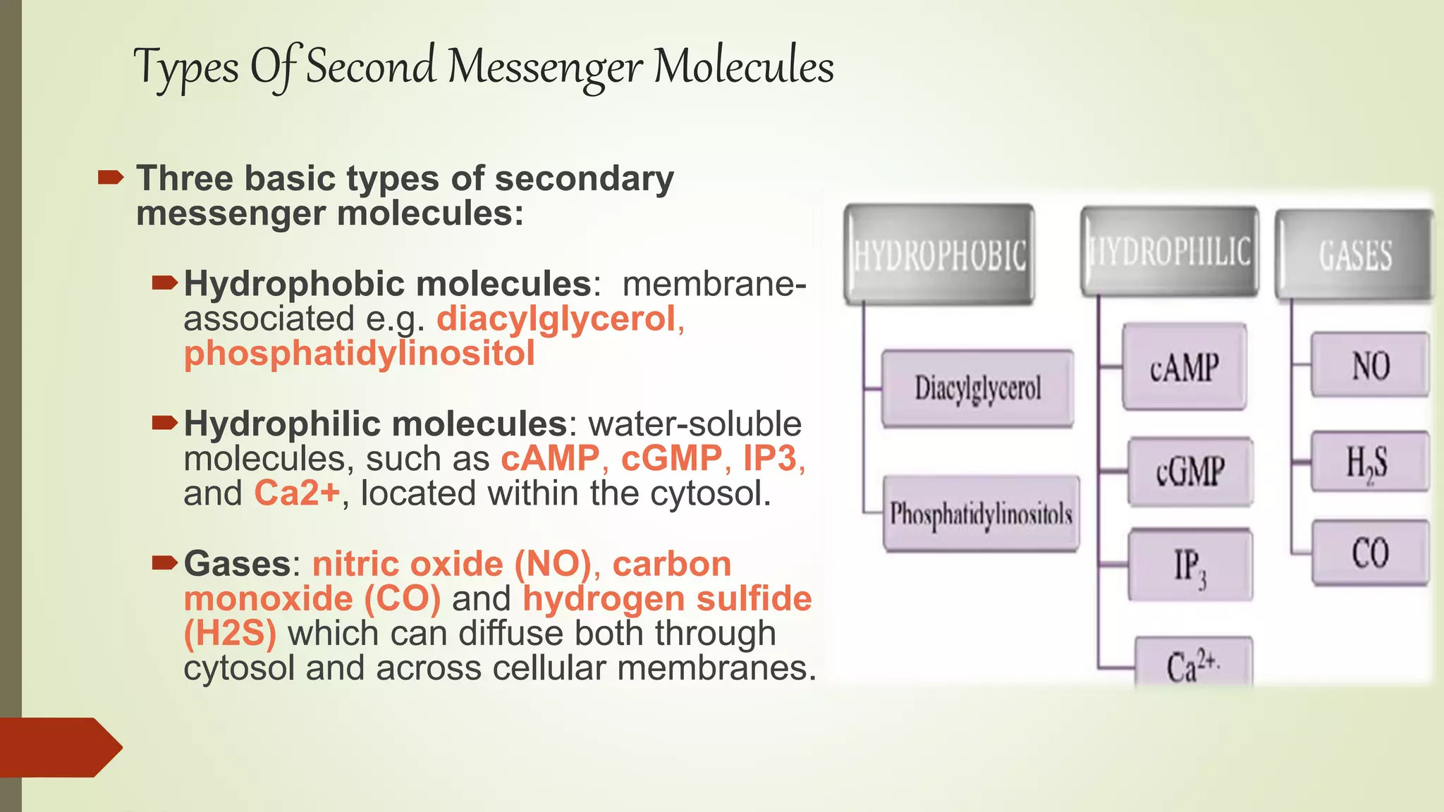
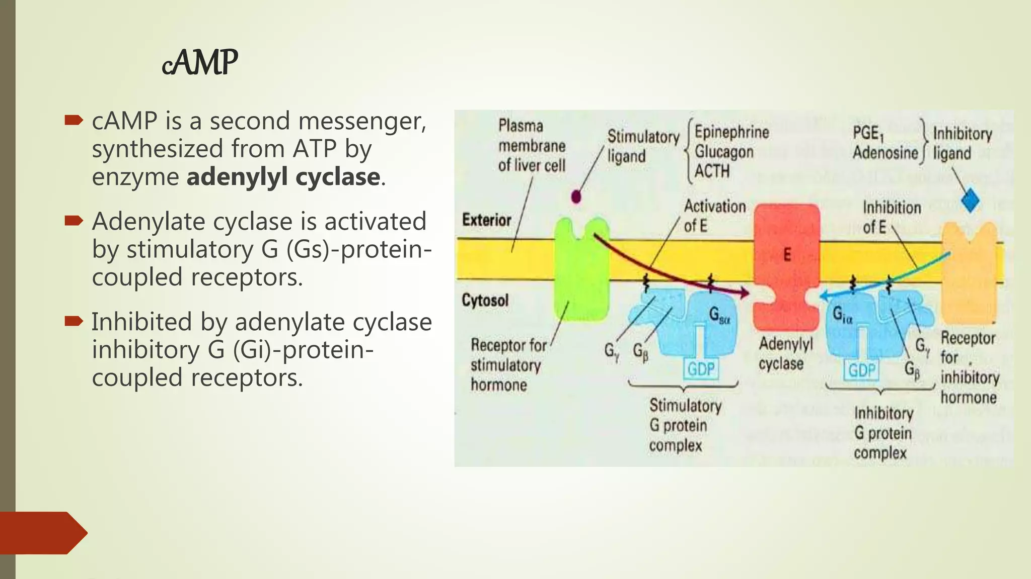
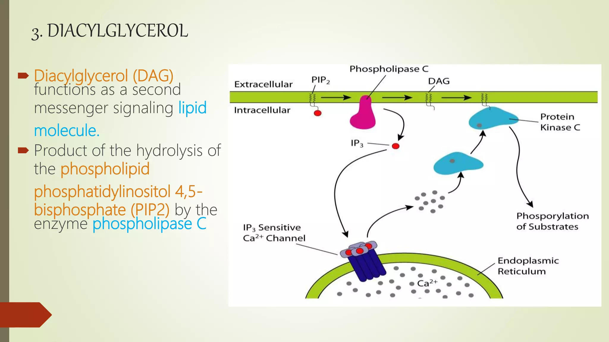
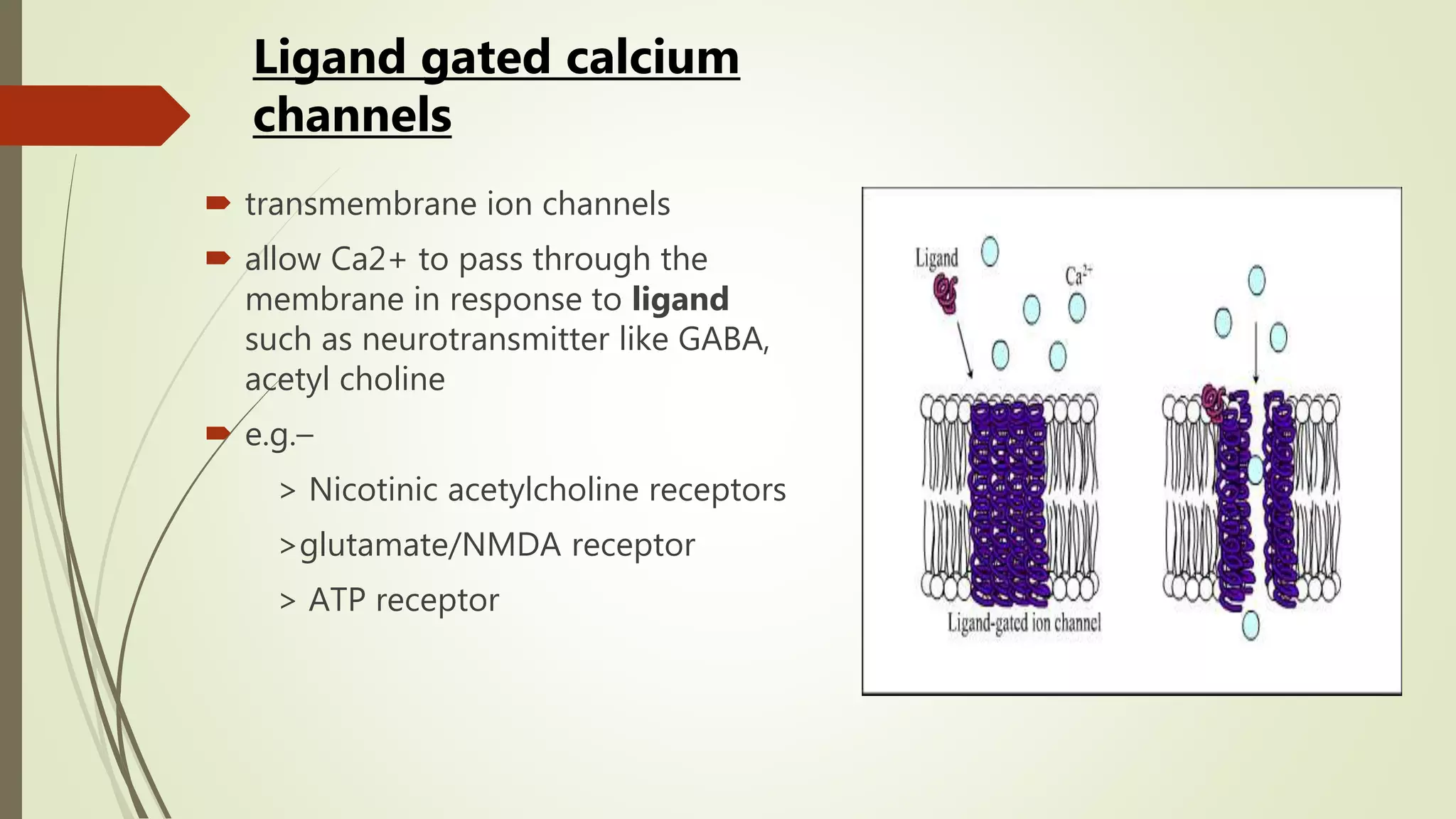
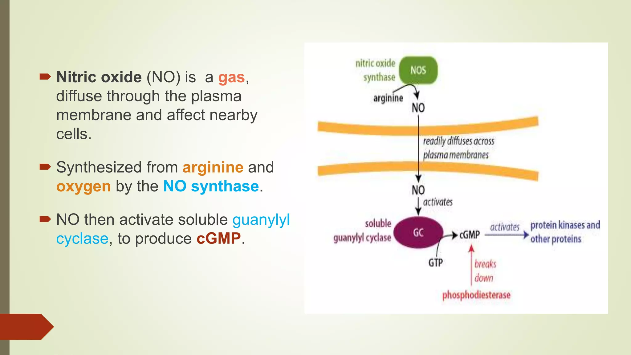
Secondary messengers are intracellular signaling molecules that are released within cells in response to extracellular signaling molecules known as first messengers like hormones and neurotransmitters. Common examples include cyclic AMP, inositol trisphosphate, diacylglycerol, and calcium ions. These secondary messengers help amplify and propagate intracellular signals by binding to target proteins and modulating their activity, often through phosphorylation. They allow cells to mount robust physiological responses despite the extracellular signals not directly crossing the cell membrane.
secondary messengers and intracellular signalingGHAZALA ZIA
Introduction of different types of primary and secondary messengers including hydrophilic, hydrophobic and gaseous.
it also describes the intracellular pathway of cyclic amp, jak stat and mapk in a very simple language.
G Proteins - Dr. P. Saranraj, Assistant Professor, Department of Microbiology, Sacred Heart College (Autonomous), Tirupattur, Vellore District, Tamil Nadu, India.
Cyclic di GMP signaling network in Legionella pneumophilaasslev
This presentation sums up a 3 years long research aimed to study the involvement of the ubiquitous bacterial cyclic di-GMP signaling network in Legionella\’s host infection and intracellular multiplication.
This Slide gives you a idea about the subject Cellular and Molecular pharmacology where the cell signalling, secondary messengers and its intracellular signalling pathways has been celarly explained
1.Receptors Link to other Enzymatic Activity.
2.Pathway of Intracellular Signal Transduction.
3.The Cyclic AMP pathway4.Cyclic GMP pathway
5.Phospholipids and Ca2+
6.The PI3-Kinase /Akt and mTOR pathways.
7.MAP Kinase Pathway.
Cell signaling / Signal Transduction / Transmembrane signaling.
It is the process by which cells communicate with their environment and respond to external stimuli.
When a signaling molecule(ligand) binds to its receptor, it alters the shape or activity of the receptor, triggering a change inside of the cell such as alteration in the activity of a gene / cell division. Thus the original Intercellular Signal is converted into an Intracellular Signal that triggers as a response.
New Directions in Targeted Therapeutic Approaches for Older Adults With Mantl...i3 Health
i3 Health is pleased to make the speaker slides from this activity available for use as a non-accredited self-study or teaching resource.
This slide deck presented by Dr. Kami Maddocks, Professor-Clinical in the Division of Hematology and
Associate Division Director for Ambulatory Operations
The Ohio State University Comprehensive Cancer Center, will provide insight into new directions in targeted therapeutic approaches for older adults with mantle cell lymphoma.
STATEMENT OF NEED
Mantle cell lymphoma (MCL) is a rare, aggressive B-cell non-Hodgkin lymphoma (NHL) accounting for 5% to 7% of all lymphomas. Its prognosis ranges from indolent disease that does not require treatment for years to very aggressive disease, which is associated with poor survival (Silkenstedt et al, 2021). Typically, MCL is diagnosed at advanced stage and in older patients who cannot tolerate intensive therapy (NCCN, 2022). Although recent advances have slightly increased remission rates, recurrence and relapse remain very common, leading to a median overall survival between 3 and 6 years (LLS, 2021). Though there are several effective options, progress is still needed towards establishing an accepted frontline approach for MCL (Castellino et al, 2022). Treatment selection and management of MCL are complicated by the heterogeneity of prognosis, advanced age and comorbidities of patients, and lack of an established standard approach for treatment, making it vital that clinicians be familiar with the latest research and advances in this area. In this activity chaired by Michael Wang, MD, Professor in the Department of Lymphoma & Myeloma at MD Anderson Cancer Center, expert faculty will discuss prognostic factors informing treatment, the promising results of recent trials in new therapeutic approaches, and the implications of treatment resistance in therapeutic selection for MCL.
Target Audience
Hematology/oncology fellows, attending faculty, and other health care professionals involved in the treatment of patients with mantle cell lymphoma (MCL).
Learning Objectives
1.) Identify clinical and biological prognostic factors that can guide treatment decision making for older adults with MCL
2.) Evaluate emerging data on targeted therapeutic approaches for treatment-naive and relapsed/refractory MCL and their applicability to older adults
3.) Assess mechanisms of resistance to targeted therapies for MCL and their implications for treatment selection
Lung Cancer: Artificial Intelligence, Synergetics, Complex System Analysis, S...Oleg Kshivets
RESULTS: Overall life span (LS) was 2252.1±1742.5 days and cumulative 5-year survival (5YS) reached 73.2%, 10 years – 64.8%, 20 years – 42.5%. 513 LCP lived more than 5 years (LS=3124.6±1525.6 days), 148 LCP – more than 10 years (LS=5054.4±1504.1 days).199 LCP died because of LC (LS=562.7±374.5 days). 5YS of LCP after bi/lobectomies was significantly superior in comparison with LCP after pneumonectomies (78.1% vs.63.7%, P=0.00001 by log-rank test). AT significantly improved 5YS (66.3% vs. 34.8%) (P=0.00000 by log-rank test) only for LCP with N1-2. Cox modeling displayed that 5YS of LCP significantly depended on: phase transition (PT) early-invasive LC in terms of synergetics, PT N0—N12, cell ratio factors (ratio between cancer cells- CC and blood cells subpopulations), G1-3, histology, glucose, AT, blood cell circuit, prothrombin index, heparin tolerance, recalcification time (P=0.000-0.038). Neural networks, genetic algorithm selection and bootstrap simulation revealed relationships between 5YS and PT early-invasive LC (rank=1), PT N0—N12 (rank=2), thrombocytes/CC (3), erythrocytes/CC (4), eosinophils/CC (5), healthy cells/CC (6), lymphocytes/CC (7), segmented neutrophils/CC (8), stick neutrophils/CC (9), monocytes/CC (10); leucocytes/CC (11). Correct prediction of 5YS was 100% by neural networks computing (area under ROC curve=1.0; error=0.0).
CONCLUSIONS: 5YS of LCP after radical procedures significantly depended on: 1) PT early-invasive cancer; 2) PT N0--N12; 3) cell ratio factors; 4) blood cell circuit; 5) biochemical factors; 6) hemostasis system; 7) AT; 8) LC characteristics; 9) LC cell dynamics; 10) surgery type: lobectomy/pneumonectomy; 11) anthropometric data. Optimal diagnosis and treatment strategies for LC are: 1) screening and early detection of LC; 2) availability of experienced thoracic surgeons because of complexity of radical procedures; 3) aggressive en block surgery and adequate lymph node dissection for completeness; 4) precise prediction; 5) adjuvant chemoimmunoradiotherapy for LCP with unfavorable prognosis.
Title: Sense of Taste
Presenter: Dr. Faiza, Assistant Professor of Physiology
Qualifications:
MBBS (Best Graduate, AIMC Lahore)
FCPS Physiology
ICMT, CHPE, DHPE (STMU)
MPH (GC University, Faisalabad)
MBA (Virtual University of Pakistan)
Learning Objectives:
Describe the structure and function of taste buds.
Describe the relationship between the taste threshold and taste index of common substances.
Explain the chemical basis and signal transduction of taste perception for each type of primary taste sensation.
Recognize different abnormalities of taste perception and their causes.
Key Topics:
Significance of Taste Sensation:
Differentiation between pleasant and harmful food
Influence on behavior
Selection of food based on metabolic needs
Receptors of Taste:
Taste buds on the tongue
Influence of sense of smell, texture of food, and pain stimulation (e.g., by pepper)
Primary and Secondary Taste Sensations:
Primary taste sensations: Sweet, Sour, Salty, Bitter, Umami
Chemical basis and signal transduction mechanisms for each taste
Taste Threshold and Index:
Taste threshold values for Sweet (sucrose), Salty (NaCl), Sour (HCl), and Bitter (Quinine)
Taste index relationship: Inversely proportional to taste threshold
Taste Blindness:
Inability to taste certain substances, particularly thiourea compounds
Example: Phenylthiocarbamide
Structure and Function of Taste Buds:
Composition: Epithelial cells, Sustentacular/Supporting cells, Taste cells, Basal cells
Features: Taste pores, Taste hairs/microvilli, and Taste nerve fibers
Location of Taste Buds:
Found in papillae of the tongue (Fungiform, Circumvallate, Foliate)
Also present on the palate, tonsillar pillars, epiglottis, and proximal esophagus
Mechanism of Taste Stimulation:
Interaction of taste substances with receptors on microvilli
Signal transduction pathways for Umami, Sweet, Bitter, Sour, and Salty tastes
Taste Sensitivity and Adaptation:
Decrease in sensitivity with age
Rapid adaptation of taste sensation
Role of Saliva in Taste:
Dissolution of tastants to reach receptors
Washing away the stimulus
Taste Preferences and Aversions:
Mechanisms behind taste preference and aversion
Influence of receptors and neural pathways
Impact of Sensory Nerve Damage:
Degeneration of taste buds if the sensory nerve fiber is cut
Abnormalities of Taste Detection:
Conditions: Ageusia, Hypogeusia, Dysgeusia (parageusia)
Causes: Nerve damage, neurological disorders, infections, poor oral hygiene, adverse drug effects, deficiencies, aging, tobacco use, altered neurotransmitter levels
Neurotransmitters and Taste Threshold:
Effects of serotonin (5-HT) and norepinephrine (NE) on taste sensitivity
Supertasters:
25% of the population with heightened sensitivity to taste, especially bitterness
Increased number of fungiform papillae
- Video recording of this lecture in English language: https://youtu.be/lK81BzxMqdo
- Video recording of this lecture in Arabic language: https://youtu.be/Ve4P0COk9OI
- Link to download the book free: https://nephrotube.blogspot.com/p/nephrotube-nephrology-books.html
- Link to NephroTube website: www.NephroTube.com
- Link to NephroTube social media accounts: https://nephrotube.blogspot.com/p/join-nephrotube-on-social-media.html
ARTIFICIAL INTELLIGENCE IN HEALTHCARE.pdfAnujkumaranit
Artificial intelligence (AI) refers to the simulation of human intelligence processes by machines, especially computer systems. It encompasses tasks such as learning, reasoning, problem-solving, perception, and language understanding. AI technologies are revolutionizing various fields, from healthcare to finance, by enabling machines to perform tasks that typically require human intelligence.
These simplified slides by Dr. Sidra Arshad present an overview of the non-respiratory functions of the respiratory tract.
Learning objectives:
1. Enlist the non-respiratory functions of the respiratory tract
2. Briefly explain how these functions are carried out
3. Discuss the significance of dead space
4. Differentiate between minute ventilation and alveolar ventilation
5. Describe the cough and sneeze reflexes
Study Resources:
1. Chapter 39, Guyton and Hall Textbook of Medical Physiology, 14th edition
2. Chapter 34, Ganong’s Review of Medical Physiology, 26th edition
3. Chapter 17, Human Physiology by Lauralee Sherwood, 9th edition
4. Non-respiratory functions of the lungs https://academic.oup.com/bjaed/article/13/3/98/278874
263778731218 Abortion Clinic /Pills In Harare ,sisternakatoto
263778731218 Abortion Clinic /Pills In Harare ,ABORTION WOMEN’S CLINIC +27730423979 IN women clinic we believe that every woman should be able to make choices in her pregnancy. Our job is to provide compassionate care, safety,affordable and confidential services. That’s why we have won the trust from all generations of women all over the world. we use non surgical method(Abortion pills) to terminate…Dr.LISA +27730423979women Clinic is committed to providing the highest quality of obstetrical and gynecological care to women of all ages. Our dedicated staff aim to treat each patient and her health concerns with compassion and respect.Our dedicated group ABORTION WOMEN’S CLINIC +27730423979 IN women clinic we believe that every woman should be able to make choices in her pregnancy. Our job is to provide compassionate care, safety,affordable and confidential services. That’s why we have won the trust from all generations of women all over the world. we use non surgical method(Abortion pills) to terminate…Dr.LISA +27730423979women Clinic is committed to providing the highest quality of obstetrical and gynecological care to women of all ages. Our dedicated staff aim to treat each patient and her health concerns with compassion and respect.Our dedicated group of receptionists, nurses, and physicians have worked together as a teamof receptionists, nurses, and physicians have worked together as a team wwww.lisywomensclinic.co.za/
These lecture slides, by Dr Sidra Arshad, offer a quick overview of physiological basis of a normal electrocardiogram.
Learning objectives:
1. Define an electrocardiogram (ECG) and electrocardiography
2. Describe how dipoles generated by the heart produce the waveforms of the ECG
3. Describe the components of a normal electrocardiogram of a typical bipolar leads (limb II)
4. Differentiate between intervals and segments
5. Enlist some common indications for obtaining an ECG
Study Resources:
1. Chapter 11, Guyton and Hall Textbook of Medical Physiology, 14th edition
2. Chapter 9, Human Physiology - From Cells to Systems, Lauralee Sherwood, 9th edition
3. Chapter 29, Ganong’s Review of Medical Physiology, 26th edition
4. Electrocardiogram, StatPearls - https://www.ncbi.nlm.nih.gov/books/NBK549803/
5. ECG in Medical Practice by ABM Abdullah, 4th edition
6. ECG Basics, http://www.nataliescasebook.com/tag/e-c-g-basics
2. secondary MESSENGERS
Secondary messengers, intracellular signaling molecules released
by the cell to trigger physiological changes
Amplifying components of intracellular signal transduction cascades.
Examples of secondary messengers include cyclic AMP, cyclic
GMP, inositol trisphosphate, diacylglycerol, and calcium .
Releases in response to exposure to extracellular signaling
molecules/ligands the first messengers, such as
neurotransmitters, hormones (epinephrine, growth hormone and
serotonin).
3. The first messengers such as peptide hormones,
neurotransmitters usually do not physically cross the
phospholipid bilayers.
First messengers need to be transduced into
secondary messengers, so that the extracellular signal
may be propagated intracellularly.
Secondary messengers greatly amplify the strength of
the signal.
Activate or inhibit the target enzymes of the pathway.
secondary MESSENGERS
4. History of secondary messengers
Earl Wilbur Sutherland Jr. discovered
secondary messengers won the 1971 Nobel
Prize in Medicine
He saw that epinephrine stimulate
glycogenolysis in liver cells, but
epinephrine alone would not convert
glycogen to glucose
He found that epinephrine had to trigger a
secondary messenger, cyclic AMP for the
liver to convert glycogen to glucose.
Earl Wilbur Sutherland Jr.
1915-1974
5. After receiving his M.D degree from Washington University Medical
School ,He started his research career in Biochemistry department of
Washington University of Medical School
At that time he came in contact with Arthur Kornberg, Edwin Krebs,
C.DeDuve, Victor Najjar
Sutherland collaborated with DeDuve on the origin and distribution
of a hyperglycemic-glycogenolytic factor present in commercial
insulin preparations
And concluded that it came from the a-cells of the islets of
Langerhans, later renamed glucagon
Sutherland than studied two parallel line of work-
The enzyme phosphorylase which initiate the breakdown the
glycogen in liver and muscle
How epinephrine and glucagon stimulated the release of glucose
from glycogen in the liver
6. Liver slices take as a test system for the action of glucagon and
epinephrine, increased the glucose output when added in vitro
In a control incubation the phosphorylase activity of the liver slices
showed a large drop
When epinephrine and glucagon are added the phosphorylate
was restored
At that time Krebs and Fisher studying the reactivation of inactive
rabbit muscle phosphorylase
Shown that this occurred with ATP and Mg2+ or Mn2+ and a
enzyme, a kinase, was necessary for this reaction
With this information they add hormones to inactive liver
phosphorylase in the presence of Mg2+ and ATP
They observed activation of phosphorylase by epinephrine and
glucagon if they use relatively crude liver homogenate
7. But if they centrifuged the extracts to remove the cellular debris, the
hormone action disappeared
They were able to show that if the particulate fraction alone
incubated with hormones, a heat-stable factor was produced that
could in turn activate phosphorylase.
The next step to isolate and identify the heat-stable factor
This is a difficult task for isolate because it is rapidly destroyed by
phosphodiesterase
This is the history of discovery adenosine-3',5'-phosphoric acid,
generally referred to as cyclic AMP.
9. Types Of Second Messenger Molecules
Three basic types of secondary
messenger molecules:
Hydrophobic molecules: membrane-
associated e.g. diacylglycerol,
phosphatidylinositol
Hydrophilic molecules: water-soluble
molecules, such as cAMP, cGMP, IP3,
and Ca2+, located within the cytosol.
Gases: nitric oxide (NO), carbon
monoxide (CO) and hydrogen sulfide
(H2S) which can diffuse both through
cytosol and across cellular membranes.
10.
11. cAMP
cAMP is a second messenger,
synthesized from ATP by
enzyme adenylyl cyclase.
Adenylate cyclase is activated
by stimulatory G (Gs)-protein-
coupled receptors.
Inhibited by adenylate cyclase
inhibitory G (Gi)-protein-
coupled receptors.
12. PKA REGULATION by cAMP
The most common downstream effector of
cAMP is Protein kinase A(PKA).
PKA is normally inactive as tetrameric
holoenzyme(two catalytic and two
regulatory units).
The regulatory unit always block the
catalytic center of catalytic unit.
Two cAMP molecules bind to each PKA
regulatory subunit.
The regulatory subunit dissociate from the
catalytic subunit.
The free catalytic subunits interact with
proteins to phosphorylate Ser or Thr
residues, either increases or decreases the
activity of the protein.
13. Protein synthesis-PKA directly
activate CREB, which bind the cAMP
response element(CRE)and altering
the transcription.
15. Cells maintain signaling specificity
Protein scaffold complexes are key mechanism that
integrate cAMP signaling with other pathways
and signaling events.
AKAPs act as scaffold proteins, they bind PKA and
physically tether these multi-protein complexes to
specific locations, such as the nucleus and other
compartments in cells.
ANCHORAGE
16. INACTIVATION
feedback mechanism using phosphodiesterase.
Phosphodiesterase quickly converts cAMP to AMP, thus reducing the
amount of cAMP that can activate protein kinase A.
17. PDE4 Promotes Inflammation by Degrading cAMP Within Immune
Cells
Phosphodiesterase 4 (PDE4) is the predominant cAMP-degrading
enzyme expressed in inflammatory cells.
cAMP helps regulate T cell function.
cAMP helps maintain immune homeostasis by suppressing the
release of proinflammatory mediators (eg, TNF-α, IL-17, and IFN-
γ)
cAMP promote the release of anti-inflammatory mediators (eg, IL-
10) by immune cells.
cAMP activated PKA, which translocates into the nucleus and
activates transcription factor CREB (cAMP response element
binding protein).
18. Decrease in PDE4 increases cAMP, leads to increased transcription of
genes that have CRE sites, including the gene for IL-10, which is an anti-
inflammatory mediator.
In contrast, cAMP elevation would inhibit expression of genes driven by
the transcription factor nuclear factor κ B (NF-κB)
Decreasing intracellular cAMP, PDE4 could prevent PKA from modulating
pro- and anti-inflammatory mediators released by a cell
19. Glucagon stimulates liver cells to start
glycogenolysis through GPCR.
Leading to the activation of adenylyl
cyclase and the formation of cAMP.
The cAMP binds to protein kinase A
(PKA), activating it.
PKA in turn phosphorylates other
downstream target proteins including
phosphorylase kinase (PhosK) and
glycogen synthase (GS).
Role of cAMP in Glycogen breakdown
20. 2. INOSITOL TRIPHOSPHATE
Inositol triphosphate (IP3) is a
lipid-derived secondary
messenger.
A product of the hydrolysis of the
phospholipid
phosphatidylinositol 4,5-
bisphosphate (PIP2) by the
enzyme phospholipase C
21. Being water-soluble molecule
IP3 diffuses rapidly through the
cytosol.
At endoplasmic reticulum(ER),
it binds to and opens IP3 -
gated Ca2+ channels in the ER
membrane.
Ca2+ stored in the ER is released
through the open channels
resulting in increased
concentration of Ca2+ in the
cytosol.
2. INOSITOL TRIPHOSPHATE
22. FUNCTION of IP3
Blocks Polyspermy in Sea Urchin
Fertilization -Fusion of egg’s and sperm’ cellular membrane
At this point egg is extremely susceptible to attack by other sperm attempting to
fertilize, so immediate action must be taken
It can be achieved by two process- fast block and slow block
In slow block, binding and fusion of the sperm’s membrane to the egg’s membrane
creates a cascade of events that enable the Gq Pathway.
First Phospholipase C is activated.
PLC cleaves Phosphatidylinositol 4,5 Bisphosphate to create two compounds: 1)
Inositol Triphosphate (IP3) and 2) Diacylglycerol (DAG).
IP3 diffuses to the ER, where it opens Ca2+ channels.
The release of Ca2+ ions stimulates the Cortical Granule Response.
CG’s release four main compounds: Proteases, Hyaluronic acid, Peroxidases, Hyaline
Hyaline
23.
24. IP3 ROLE in PATHOPHYSIOLOGY
HUNTINGTON’S DISEASE
neurons in the brain degenerate.
Affects medium spiny neurons (MSN) presents in stratium
The cytosolic protein Huntingtin (Htt) has an additional 35 glutamine
residues added to its amino terminal region.
This modified form of Htt is called Httexp.
Httexp makes Type 1 IP3 receptors more sensitive to IP3, which leads
to the release of too much Ca2+ from the ER.
The release of Ca2+ from the ER causes an increase in the cytosolic
and mitochondrial concentrations of Ca2+.
This increase in Ca2+ is thought to be the cause of GABAergic MSN
degradation.
25. 3. DIACYLGLYCEROL
Diacylglycerol (DAG)
functions as a second
messenger signaling lipid
molecule.
Product of the hydrolysis of
the phospholipid
phosphatidylinositol 4,5-
bisphosphate (PIP2) by the
enzyme phospholipase C
26. Diacylglycerol remains within the plasma membrane, activate
serine/threonine protein kinase called protein kinase C (PKC), so
named because it is Ca2+ dependent.
The initial rise in cytosolic Ca2+ induced by IP3 alters the PKC so that
it translocates from the cytosol to the cytoplasmic face of the plasma
membrane.
There it is activated by the combination of Ca2+, diacylglycerol, and
the phospholipid phosphatidylserine
Activated PKC phosphorylates target proteins like glucose
transporter, HMG-CoA reductase, cytochromeP450 etc.
3. DIACYLGLYCEROL
27. 4. CALCIUM IONS
Once calcium enter the cytoplasm exert allosteric regulatory effects on
many enzymes and proteins (toxic in excess)
Low cytoplasmic Ca++ at rest (10–100 nM).
To maintain this low concentration, Ca2+ is actively pumped from the
cytosol to the extracellular space and into the endoplasmic
reticulum (ER)
Certain proteins of the cytoplasm and organelles act as buffers by
binding Ca2+.
Acts as a secondary messenger by signal transduction pathways such
as via G protein-coupled receptors.
Signaling occurs when the cell is stimulated to release calcium ions
(Ca2+) from intracellular stores, or when calcium enters the cell
through plasma membrane ion channels.
28. Sources of Ca2+ :
Extracellular compartment, nerve, cardiac
and smooth muscle cells
Three types of plasma-membrane
localized calcium channels :
Voltage-dependent calcium channels :
At physiological condition VDCCs are
closed (resting membrane potential)
The concentration of calcium ions are
several times higher outside of the cell
than inside .
Action potential depolarizes plasma
membrane, which results in the opening
of VDCCs and calcium ion rush into the
cell.
29. transmembrane ion channels
allow Ca2+ to pass through the
membrane in response to ligand
such as neurotransmitter like GABA,
acetyl choline
e.g.–
> Nicotinic acetylcholine receptors
>glutamate/NMDA receptor
> ATP receptor
Ligand gated calcium
channels
30. Stored-operated calcium channels :
Located in the plasma membrane of all non-
excitable cells (myocytes, endocrine cells etc.)
They are major source of intracellular calcium
Although it was initially considered to function only
in non excitable cells, growing evidence now points
towards a central role for in excitable cells too.
31. Intracellular compartment:
Calcium is stored in higher concentrations
in endoplasmic reticulum and sarcoplasmic
sarcoplasmic reticulum
In sarcoplasmic reticulum they are bound
with calsequestrin.
Calsequestrin is highly acidic, containing up
to 50 Ca(2+)-binding sites, formed simply
by clustering of two or more acidic protein.
Two forms of calsequestrin have been
identified i.e. Cardiac form (Calsequestrin-
and slow skeletal and fast skeletal form
(calsequestrin-1)
32. Ca2+ Sensors
Calmodulin
Effects of Ca2+ are mediated through
ubiquitous Ca2+ sensing protein,
calmodulin(CaM).
CaM is a 17 kDa Ca2+-binding protein
Composed of, N- and C-terminal lobe
tethered by a loop, allows CaM to adopt a
variety of conformations
Each lobe of CaM contains a pair of EF-
hand motifs .
Each EF-hand motif allows calmodulin to
sense intracellular calcium levels by binding
up to four Ca2+ ions.
33. Calmodulin
Activated by Ca2+ binding, it undergoes
conformational change that permits
Ca2+/calmodulin to bind various target
proteins
The protein responds in an almost switch
like manner to increasing concentrations of
Ca2+.
A tenfold increase in Ca2+ concentration
typically causes a fiftyfold increase in
calmodulin activation
When an activated molecule of
Ca2+/calmodulin binds to its target,
calmodulin further changes its conformation.
34. Whenever the concentration of Ca2+ in
the cytosol rises,Ca2+/calmodulin,
activates the plasma membrane Ca2+-
pump that uses ATP hydrolysis to pump
Ca2+ out of cells.
35. CaM-kinases
Ca2+/calmodulin, plays a great role in protein phosphorylations,
catalyzed by a family of serine/threonine protein kinases called
Ca2+/calmodulin-dependent kinases (CaM-kinases).
CaM-kinases phosphorylate gene regulatory proteins, such as the
CREB protein, and in this way activate or inhibit the transcription of
specific genes.
One of the best-studied CaM-kinases is CaM-kinase II, found in most
animal cells, especially enriched in the nervous system.
It is highly concentrated in synapses.
36. CaM-kinase II function as a molecular memory device, switching to an
active state when exposed to Ca2+/calmodulin and remain active even
after the Ca2+ signal has decayed.
This is because the kinase phosphorylates itself (autophosphorylation).
In its autophosphorylated state, the enzyme remains active even in the
absence of Ca2+
The enzyme maintains this activity until serine/threonine protein
phosphatases inhibit the autophosphorylation and shut the kinase off.
CaM-kinase II activation serve as a memory trace and seems to have a role
in some types of memory and learning in the vertebrate nervous system.
Mutant mice that lack a brain-specific form of the enzyme have specific
defects in their ability to remember where things are.
40. NFAT(NUCLEAR FACTOR OF ACTIVATED T CELLS)
ACTIVATION
In unstimulated cells, phosphorylated
NFAT is located in the cytosol.
Ca2+/calmodulin complex binds to and
activates calcineurin, a protein-serine
phosphatase.
Activated calcineurin then
dephosphorylates phosphate residues
on cytosolic NFAT
NFAT, exposing a nuclear localization
sequence that allows NFAT activity and
expression of gene essential for T cell
activation
41. 5. NITRIC OXIDE
HISTORY
NO functions as a messenger molecule began with an accidental
observation
It had been known for many years that acetylcholine acts in the body to
the smooth muscle cells of blood vessels, but the response could not be
duplicated in vitro
When portions of a major blood vessel such as the aorta were incubated in
physiologic concentrations of acetylcholine in vitro, the preparation usually
showed little or no response
In the late 1970s, Robert Furchgott, a pharmacologist at New York State
medical center, was studying the in vitro response of pieces of rabbit aorta
various agents
In his earlier studies, Furchgott used strips of aorta that had been dissected
from the organ.
42. Furchgott switched from strips of aortic tissue to aortic rings and aortic
rings responded to acetylcholine by undergoing relaxation
The strips had failed to display the relaxation response because the
endothelial layer that lines the aorta had been rubbed away during the
dissection
This surprising finding suggested that the endothelial cells were somehow
involved in the response by the adjacent muscle cells.
Acetylcholine binds to receptors on the surface of endothelial cells,
leading to the production and release of an agent that diffuses through
the cell’s plasma membrane and causes the muscle cells to relax.
The diffusible agent was identified in 1986 as nitric oxide by Louis
Ignarro and Salvador Moncada
43. Nitric oxide (NO) is a gas,
diffuse through the plasma
membrane and affect nearby
cells.
Synthesized from arginine and
oxygen by the NO synthase.
NO then activate soluble guanylyl
cyclase, to produce cGMP.
44. The function of NO is the dilation
of blood vessels.
The acetylcholine
(neurotransmitter) acts on
endothelial cells to stimulate NO
synthesis.
NO, diffuses to neighboring
smooth muscle cells where it
interacts with the guanylyl
cyclase.
This increase enzymatic activity
resulting in the synthesis of cGMP.
The cGMP then induces muscle
relaxation and blood vessel
dilation.
45. NITROGLYCERINE ACT AS VASODILATER
A BRIEF HISTORY
Nitroglycerin is an oily liquid that may explode
when subjected to heat, shock or flame.
Alfred Nobel developed the use of nitroglycerin
as a blasting explosive by mixing the
nitroglycerin with inert absorbents such as
diatomaceous earth
Named them as dynamite and patented it in
1867.
Dr. William Murrell experimented with the use of
nitroglycerin to relieve angina pectoris and to
reduce the blood pressure.
46. A few months before his death in 1896, Alfred Nobel was prescribed
nitroglycerine for this heart condition
He said to his friend that "Isn't it the irony of fate that I have been
prescribed nitro-glycerin, to be taken internally !
They call it Trinitrin, so as not to scare the chemist and the public, so
that it also called as glyceryl trinitrin
48. REFERANCE
Kaestner, Lars. "Calcium Signalling." (2013): n. pag. Web.
<http://dx.doi.org/10.1016/j.cell.2007.11.028>.
Fujisawa, H. "Regulation of the Activities of Multifunctional Ca2 /Calmodulin-Dependent Protein
Kinases." Journal of Biochemistry 129.2 (2001): 193-99. Web. <10.1007/s00018-008-8086-2>.
Carnegie, Graeme K., Christopher K. Means, and John D. Scott. "A-kinase Anchoring Proteins:
From Protein Complexes to Physiology and Disease." IUBMB Life 61.4 (2009): 394-406. Web.
<10.1002/iub.168>.
Alberts, Bruce. Molecular Biology of the Cell, 5th Edition. New York: Garland Science, 2008. Print.
Editor's Notes
The first messengers such as peptide hormones, neurotransmitters typically are hydrophilic molecules, these first messengers may not physically cross the phospholipid bilayer cell membrane to initiate changes within the cell directly—unlike steroid hormones, which usually do .
1) Hydrophobic molecules: diacylglycerol, phosphatidylinositol, membrane-associated diffuse from the plasma membrane into the intermembrane space where they can reach and regulate membrane-associated effector proteins
How can each GPCR transmit a specific signal even though so many different GPCRs are using the same second messenger?
CREB (cAMP response element-binding protein) is a cellular transcription factor
A crucial element in signal transduction is the specificity
One family of diverse, well-studied scaffolding proteins are the A-kinase anchoring proteins (AKAPs). These anchoring proteins form multi-protein complexes that integrate cAMP signaling with other pathways and signaling events.
The phosphorylation of PhosK leads to its activation. Phosphorylation of GS
causes its inhibition stopping the formation of glycogen. The activated PhosK then phosphorylates the next
kinase in the chain, glycogen phosphorylase kinase (GPhos). Phosphorylation of GPhos activates the enzyme
leading to the release of glucose subunits from glycogen.
The contents of the granules contain proteases, mucopolysaccharides, hyalin, and peroxidases.
Regulation of store-operated channels by intracellular Ca2+-buffering organelles. Store-operated channels (SOC) are activated by a signal from a specialized compartment of the endoplasmic reticulum. As depicted in the figure, this compartment may not coincide exactly with the major InsP3-sensitive component of the endoplasmic reticulum. The Ca2+ buffering capabilities of the endoplasmic reticulum and mitochondria facilitate SOC activation at least in part by buffering entering Ca2+ near the mouth of the channel, and reducing negative-feedback inhibition of the channels. In addition, location of mitochondria in close proximity to InsP3 receptors on the endoplasmic reticulum buffers the release of Ca2+ and thereby reduces Ca2+ inhibition of release through InsP3 receptor channels. Finally, energized mitochondria appear capable of positively regulating SOC function by a mechanism that is independent of Ca2+buffering.
When activated by Ca2+ binding, it
undergoes a conformational change. Because two or more Ca2+ ions must bind
before calmodulin adopts its active conformation, the protein responds in an
almost switchlike manner to increasing concentrations of Ca2+
The regulatory
region of many gene contain a short DNA sequence, called the cyclic
AMP response element (CRE). A specific gene regulatory protein
called CRE-binding (CREB) protein recognizes this sequence.
calcium to release from the sarcoplasmic reticulum of the cells. The calcium ions then flow into the cytoplasm and bind to the troponin and tropomyosin molecules in the actin filaments of the muscle cells. The binding of calcium allows the troponin to change shape and expose the actin-myosin binding site. Myosin then connects to actin, and the formed complex forms a cross bridge that opens and closes and allows the muscle cells to contract
Myosin light chain kinase
Nuclear factor of activated T cells
ANGINA-CHEST PAIN(ATHERSCLEROSIS)
NITATE SUCH AS NITROGLYCERINE
Mechanism of action of nitrates. Organic nitrates have the chemical structure RNO2. The nitro group is reduced to form NO in by a mitochondrial enzyme (aldehyde dehydrogenase-2). Free nitrite is released, which is converted to NO. NO activates guanylyl cylcase (GC) by interacting with its heme group. Activated GC converts GTP to cGMP. cGMP activates a phosphatase which dephosphorylates myosin light chains, resulting in decreased interaction between actin & myosin filaments, and relaxation. cGMP is normally short lived due to metabolism by intracellular phosphodiesterase (PDE). Drugs such as sildenafil can inhibit PDE, resulting in a potentially dangerous intensification of the vaso-relaxant effect of nitrates. The inset (top right) illustrates the rapid onset of effect of nitroglycerin (NTG) on a segment of human vein that has been contracted by norepinephrine.