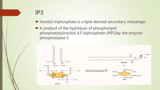
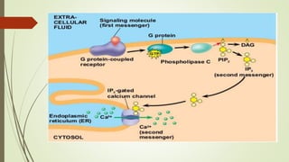
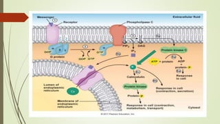
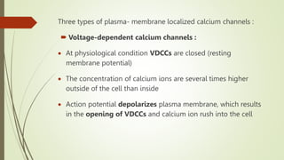
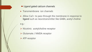
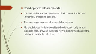
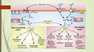
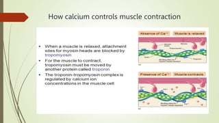
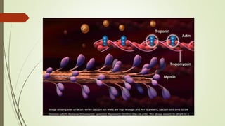
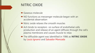
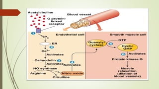
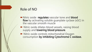
Secondary Messengers
IP3,DAG,CA2+ & NO
Presented by: Miss. Rubina U. Watangi. M. Pharm
Department of Pharmacology
Subject: Molecular Pharmacology
Contents
1.IP3
 Functions of IP3
 Role in pathophysiology
2.DAG
 Physiological role
3.Calcium ion
 Role of Calmodulin
4.Nitric oxide
 Role of NO
IP3
 Inositol triphosphate is a lipid derived secondary messenger
 A product of the hydrolysis of phospholipid
phosphatidylinositol 4,5 biphosphate (PIP2)by the enzyme
phospholipase C
FUNCTION of IP3
To transfer a chemical signal received by the cell,
such as from a hormone, neurotransmitters, growth
factors and hypertrophic stimuli such as Ang II
(Angiotensin-II), Beta-adrenergic receptor agonists
IP3 ROLE in PATHOPHYSIOLOGY
HUNTINGTON’S DISEASE
 Neurons in the brain degenerate.
 Affects medium spiny neurons (MSN) presents in stratium
 The cytosolic protein Huntingtin (Htt) has an additional 35 glutamine
residues added to its amino terminal region
 This modified form of Htt is called Httexp
 Httexp makes Type 1 IP3 receptors more sensitive to IP3, which leads to
the release of too much Ca2+ from the ER
 The release of Ca2+ from the ER causes an increase in the cytosolic and
mitochondrial concentrations of Ca2+
 The increase in Ca2+ is thought to be the cause of GABAergic MSN
degradation
DIACYLGLYCEROL
 Diacylglycerol (DAG) functions as a second messenger signalling lipid
molecule
 Product of the hydrolysis of the phospholipid phosphatidylinositol 4,5-
bisphosphate (PIP2) by the enzyme phospholipase C
 DIACYLGLYCEROL
 Diacylglycerol remains within the plasma membrane, activate
serine/threonine protein kinase called protein kinase C(PKC), so named
because it is Ca2+ dependent
 The initial rise in cytosolic Ca2+ induced by IP3 alters the PKC so that it
translocate from the cytosol to the cytoplasmic face to the plasma
membrane.
 There it is activated by the combination of Ca2+, diacylglycerol, and the
phospholipid phosphatidylserine
 Activated PKC phosphorylates target proteins like glucose transporter,
HMG-CoA reductase, cytochromeP450 etc.
Physiological role
 Diacylglycerol (DAG) is a lipid signal messenger and plays a
physiological role in β-cells.
 Defective glucose homeostasis increases DAG synthesis,
 DAG may also contribute to β-cell dysfunction in type 2
diabetes.
 The primary function of DAG is to activate protein kinase C
(PKC),
CALCIUM IONS
 Acts as a secondary messenger by signal transduction
pathways such as via G protein- coupled receptors.
 Signalling occurs when the cell is stimulated to release
calcium ions (Ca2+) from intracellular stores, or when calcium
enters the cell through plasma membrane ion channels
Control of Calcium Levels
 Intracellular calcium levels are controlled by an assortment of
channels, pumps, transporters, buffers, and effector moieties
 Release of calcium from internal stores represents a major
source of signal calcium for many cells.
 The principal calcium stores are the ER, sarcoplasmic
reticulum (SR), Golgi
 Calcium release channels are present on the membranes of
these organelles and gate the flux of calcium from the ER/SR
lumen into the cytosol.
Three types of plasma- membrane localized calcium channels :
 Voltage-dependent calcium channels :
 At physiological condition VDCCs are closed (resting
membrane potential)
 The concentration of calcium ions are several times higher
outside of the cell than inside
 Action potential depolarizes plasma membrane, which results
in the opening of VDCCs and calcium ion rush into the cell
 Ligand gated calcium channels
 Transmembrane ion channels
 Allow Ca2+ to pass through the membrane in response to
ligand such as neurotransmitter like GABA, acetyl choline
e.g.-
 Nicotinic acetylcholine receptor
 Glutamate / NMDA receptor
 ATP receptor
 Stored-operated calcium channels :
 Located in the plasma membrane of all non-excitable cells
(myocytes, endocrine cells etc.)
 They are major sources of intracellular calcium
 Although it was initially considered to function only in non
excitable cells, growing evidence now points towards a central
role for in excitable cells too
Calmodulin
 Calmodulin (CaM) (an abbreviation for calcium-modulated
protein)
 It is a multifunctional intermediate calcium-binding
messenger protein expressed in all eukaryotic cells.
 It is an intracellular target of the secondary messenger Ca2+
 The binding of Ca2+ is required for the activation of
calmodulin.
 Once bound to Ca2+, calmodulin acts as part of a calcium
signal transduction pathway by modifying its interactions with
various target proteins such as kinases
Calmodulin is Activated by Ca2+ binding undergoes
conformational change that permits Ca2+/calmodulin to bind
targets proteins
A tenfold increase in Ca2+ concentration typically causes a fiftyfold
increase in calmodulin activation
When an activated molecule of Ca2+/calmodulin binds to its target,
Calmodulin further changes its conformation
Whenever the concentration of Ca2+ in the cytosol rises,
Ca2+/calmodulin, activates the plasma membrane Ca2+- pump
uses ATP hydrolysis to pump Ca2+ out of cells or storing it in ER
(holo)
Role in metabolism
 Calmodulin plays an important role in the activation of
phosphorylase kinase, which ultimately leads to glucose being
cleaved from glycogen by glycogen phosphorylase.
 Calmodulin also plays an important role in lipid metabolism by
affecting Calcitonin.
Role in memory
 Ca2+/calmodulin-dependent protein kinase II (CaMKII) plays a crucial
role in a type of synaptic plasticity called long-term potentiation
(LTP) and requires the presence of calcium/calmodulin.
Role in smooth muscle contraction
 Calmodulin plays an important role in contraction of smooth muscle
How calcium controls muscle contraction
NITRIC OXIDE
 Gaseous molecule
 NO functions as messenger molecule began with an
accidental observation
 Nitric oxide relaxes the smooth muscles
 Ach binds to receptors on surface of endothelial cells
production and release of an agent diffuses through the cell’s
plasma membrane and causes muscle to relax
 The diffusible agent was identified in 1986 as NITRIC OXIDE
by Louis Ignarro and Salvador Moncada
Role of NO
Nitric oxide regulates vascular tone and blood
flow by activating soluble guanylate cyclase (sGC) in
the vascular smooth muscle
 Nitric oxide dilates blood vessels, raising blood
supply and lowering blood pressure.
 Nitric oxide controls mitochondrial Oxygen
consumption by inhibiting Cytochrome C oxidase.
Secondary messengers.pptx

Secondary messengers.pptx

  • 1.
    Secondary Messengers IP3,DAG,CA2+ &NO Presented by: Miss. Rubina U. Watangi. M. Pharm Department of Pharmacology Subject: Molecular Pharmacology
  • 2.
    Contents 1.IP3  Functions ofIP3  Role in pathophysiology 2.DAG  Physiological role 3.Calcium ion  Role of Calmodulin 4.Nitric oxide  Role of NO
  • 3.
    IP3  Inositol triphosphateis a lipid derived secondary messenger  A product of the hydrolysis of phospholipid phosphatidylinositol 4,5 biphosphate (PIP2)by the enzyme phospholipase C
  • 5.
    FUNCTION of IP3 Totransfer a chemical signal received by the cell, such as from a hormone, neurotransmitters, growth factors and hypertrophic stimuli such as Ang II (Angiotensin-II), Beta-adrenergic receptor agonists
  • 6.
    IP3 ROLE inPATHOPHYSIOLOGY HUNTINGTON’S DISEASE  Neurons in the brain degenerate.  Affects medium spiny neurons (MSN) presents in stratium  The cytosolic protein Huntingtin (Htt) has an additional 35 glutamine residues added to its amino terminal region  This modified form of Htt is called Httexp  Httexp makes Type 1 IP3 receptors more sensitive to IP3, which leads to the release of too much Ca2+ from the ER  The release of Ca2+ from the ER causes an increase in the cytosolic and mitochondrial concentrations of Ca2+  The increase in Ca2+ is thought to be the cause of GABAergic MSN degradation
  • 7.
    DIACYLGLYCEROL  Diacylglycerol (DAG)functions as a second messenger signalling lipid molecule  Product of the hydrolysis of the phospholipid phosphatidylinositol 4,5- bisphosphate (PIP2) by the enzyme phospholipase C
  • 8.
     DIACYLGLYCEROL  Diacylglycerolremains within the plasma membrane, activate serine/threonine protein kinase called protein kinase C(PKC), so named because it is Ca2+ dependent  The initial rise in cytosolic Ca2+ induced by IP3 alters the PKC so that it translocate from the cytosol to the cytoplasmic face to the plasma membrane.  There it is activated by the combination of Ca2+, diacylglycerol, and the phospholipid phosphatidylserine  Activated PKC phosphorylates target proteins like glucose transporter, HMG-CoA reductase, cytochromeP450 etc.
  • 9.
    Physiological role  Diacylglycerol(DAG) is a lipid signal messenger and plays a physiological role in β-cells.  Defective glucose homeostasis increases DAG synthesis,  DAG may also contribute to β-cell dysfunction in type 2 diabetes.  The primary function of DAG is to activate protein kinase C (PKC),
  • 11.
    CALCIUM IONS  Actsas a secondary messenger by signal transduction pathways such as via G protein- coupled receptors.  Signalling occurs when the cell is stimulated to release calcium ions (Ca2+) from intracellular stores, or when calcium enters the cell through plasma membrane ion channels
  • 12.
    Control of CalciumLevels  Intracellular calcium levels are controlled by an assortment of channels, pumps, transporters, buffers, and effector moieties  Release of calcium from internal stores represents a major source of signal calcium for many cells.  The principal calcium stores are the ER, sarcoplasmic reticulum (SR), Golgi  Calcium release channels are present on the membranes of these organelles and gate the flux of calcium from the ER/SR lumen into the cytosol.
  • 13.
    Three types ofplasma- membrane localized calcium channels :  Voltage-dependent calcium channels :  At physiological condition VDCCs are closed (resting membrane potential)  The concentration of calcium ions are several times higher outside of the cell than inside  Action potential depolarizes plasma membrane, which results in the opening of VDCCs and calcium ion rush into the cell
  • 14.
     Ligand gatedcalcium channels  Transmembrane ion channels  Allow Ca2+ to pass through the membrane in response to ligand such as neurotransmitter like GABA, acetyl choline e.g.-  Nicotinic acetylcholine receptor  Glutamate / NMDA receptor  ATP receptor
  • 15.
     Stored-operated calciumchannels :  Located in the plasma membrane of all non-excitable cells (myocytes, endocrine cells etc.)  They are major sources of intracellular calcium  Although it was initially considered to function only in non excitable cells, growing evidence now points towards a central role for in excitable cells too
  • 17.
    Calmodulin  Calmodulin (CaM)(an abbreviation for calcium-modulated protein)  It is a multifunctional intermediate calcium-binding messenger protein expressed in all eukaryotic cells.  It is an intracellular target of the secondary messenger Ca2+  The binding of Ca2+ is required for the activation of calmodulin.  Once bound to Ca2+, calmodulin acts as part of a calcium signal transduction pathway by modifying its interactions with various target proteins such as kinases
  • 18.
    Calmodulin is Activatedby Ca2+ binding undergoes conformational change that permits Ca2+/calmodulin to bind targets proteins A tenfold increase in Ca2+ concentration typically causes a fiftyfold increase in calmodulin activation When an activated molecule of Ca2+/calmodulin binds to its target, Calmodulin further changes its conformation Whenever the concentration of Ca2+ in the cytosol rises, Ca2+/calmodulin, activates the plasma membrane Ca2+- pump uses ATP hydrolysis to pump Ca2+ out of cells or storing it in ER
  • 19.
  • 20.
    Role in metabolism Calmodulin plays an important role in the activation of phosphorylase kinase, which ultimately leads to glucose being cleaved from glycogen by glycogen phosphorylase.  Calmodulin also plays an important role in lipid metabolism by affecting Calcitonin. Role in memory  Ca2+/calmodulin-dependent protein kinase II (CaMKII) plays a crucial role in a type of synaptic plasticity called long-term potentiation (LTP) and requires the presence of calcium/calmodulin. Role in smooth muscle contraction  Calmodulin plays an important role in contraction of smooth muscle
  • 21.
    How calcium controlsmuscle contraction
  • 23.
    NITRIC OXIDE  Gaseousmolecule  NO functions as messenger molecule began with an accidental observation  Nitric oxide relaxes the smooth muscles  Ach binds to receptors on surface of endothelial cells production and release of an agent diffuses through the cell’s plasma membrane and causes muscle to relax  The diffusible agent was identified in 1986 as NITRIC OXIDE by Louis Ignarro and Salvador Moncada
  • 25.
    Role of NO Nitricoxide regulates vascular tone and blood flow by activating soluble guanylate cyclase (sGC) in the vascular smooth muscle  Nitric oxide dilates blood vessels, raising blood supply and lowering blood pressure.  Nitric oxide controls mitochondrial Oxygen consumption by inhibiting Cytochrome C oxidase.

Editor's Notes

  • #5 There are multiple isoforms of ryanodine receptors: RyR1 is primarily expressed in skeletal muscle RyR2 is primarily expressed in myocardium (heart muscle) RyR3 is expressed more widely, but especially in the brain.
  • #7 The huntingtin gene, also called the HTT or HD gene, is the IT15 gene, which codes for a protein called the huntingtin protein.
  • #10 opening of voltage gated calcium channels causing insulin granule fusion and exocytosis. -PKC has been reported to act as both a positive and negative regulator of insulin secretion -the role of PKC in insulin secretion is controversial
  • #12 Calcium is an extremely versatile intracellular messenger that controls a wide range of cellular functions by regulating the activity of a vast number of target proteins The cellular effects of calcium are mediated either by direct binding to a target protein, or stimulation of calcium sensors that detect changes in calcium concentration
  • #13 Alterations in ER homeostasis, including severe Ca2+ depletion, loss of luminal Ca2+ causes ER stress and activates an unfolded protein response, which depending on the duration and severity of the stress, can re establish normal ER function or lead to cell death.
  • #14 Sources of Ca2+ : Extracellular compartment, nerve, cardiac and smooth muscle cells
  • #16 The “Journal of Cell Biology” reported that when a cell undergoes a stressful situation it can kill itself by releasing calcium from its endoplasmic reticulum. This form of cell suicide, or programmed cell death, is known as apoptosis.
  • #17 Ryanodine receptors are located in SR/ER membrane and responsible for release calcium from intracellular stores ROC receptor operated channel VOC voltage operated channel Duchenne muscular dystrophy-muscle weakness tends to appear in early childhood
  • #20 Calmodulin further Activates MLCK ,MLC phosphorylated ,contraction Inactivated MLCK ,MLC dephosphorylated,relaxation
  • #21 Calcitonin is a polypeptide hormone that lowers blood Ca2+ levels and activates G protein cascades that leads to the generation of cAMP. The actions of calcitonin can be blocked by inhibiting the actions of calmodulin, suggesting that calmodulin plays a crucial role in the activation of calcitonin.
  • #22 Troponin, or the troponin complex, is a complex of three regulatory proteins (troponin C, troponin I, and troponin T) that is integral to muscle contraction Blood troponin levels may be used as a diagnostic marker for stroke
  • #23 Calcium binds to troponin which displaces tropomyocin Exposing myosin binding sites on actin Allows myosin to attach to a binding sites on acting forming cross bridge
  • #25 Half life 40sec (transient signal )