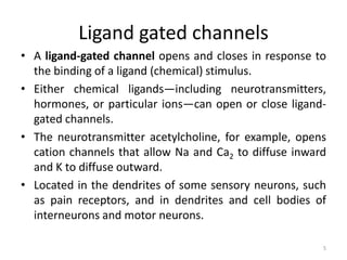
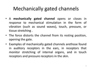
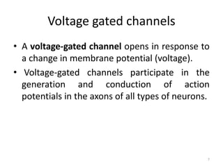
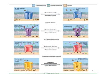
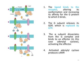
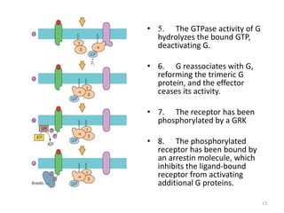
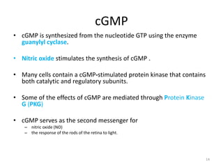
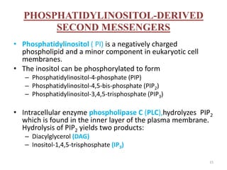
CALL ON ➥9907093804 🔝 Call Girls Hadapsar ( Pune) Girls Service
4.ion channels & secondary messengers
1. Ion channels & secondary
messengers
Anu K R
Lecturer
Dept.of Pharmacology
ELIMS College of Pharmacy
2. Ion channels
• Passage to communicate through.
• When ion channels are open, they allow specific ions to move
across the plasma membrane, down their electrochemical
gradient—a concentration (chemical) difference plus an
electrical difference.
• Ions move from areas of higher concentration to areas of
lower concentration (the chemical part of the gradient). Also,
positively charged cations move toward a negatively charged
area, and negatively charged anions move toward a positively
charged area (the electrical aspect of the gradient).
• As ions move, they create a flow of electrical current that can
change the membrane potential.
2
3. • Ion channels open and close due to the presence
of “gates.”
• The gate is a part of the channel protein that can
seal the channel pore shut or move aside to open
the pore.
• The electrical signals produced by neurons and
muscle fibers rely on four types of ion channels:
leak channels,
ligand-gated channels,
mechanically gated channels, and
voltage-gated channels:
3
4. Leak channels
• The gates of which randomly alternate between
open and closed positions .
• More potassium ion (K) leak channels than sodium
ion (Na) leak channels, and they are more leakier
too.
• Thus, the membrane’s permeability to K is much
higher than its permeability to Na.
• Found in nearly all cells, including the dendrites, cell
bodies, and axons of all types of neurons.
4
5. Ligand gated channels
• A ligand-gated channel opens and closes in response to
the binding of a ligand (chemical) stimulus.
• Either chemical ligands—including neurotransmitters,
hormones, or particular ions—can open or close ligand-
gated channels.
• The neurotransmitter acetylcholine, for example, opens
cation channels that allow Na and Ca2 to diffuse inward
and K to diffuse outward.
• Located in the dendrites of some sensory neurons, such
as pain receptors, and in dendrites and cell bodies of
interneurons and motor neurons.
5
6. Mechanically gated channels
• A mechanically gated channel opens or closes in
response to mechanical stimulation in the form of
vibration (such as sound waves), touch, pressure, or
tissue stretching .
• The force distorts the channel from its resting position,
opening the gate.
• Examples of mechanically gated channels arethose found
in auditory receptors in the ears, in receptors that
monitor stretching of internal organs, and in touch
receptors and pressure receptors in the skin.
6
7. Voltage gated channels
• A voltage-gated channel opens in response to
a change in membrane potential (voltage).
• Voltage-gated channels participate in the
generation and conduction of action
potentials in the axons of all types of neurons.
7
9. Secondary messengers
• Molecules that relay signals from receptors on
the cell surface to target molecules inside the
cell i.e. Cytoplasm or nucleus.
• Causing some kind of change in the activity of
the cell.
9
10. 10
• Short lived
• Elevated concentration of second messenger
leads to rapid alteration in the activity of one or
more cellular enzymes
• Removal or degradation of second messenger
terminate the cellular response
• Four classes of second messengers
– Cyclic nucleotides(cAMP,cGMP)
– Membrane lipid derivatives(IP3,DAG)
– Ca2+
– Nitric oxide/carbon monoxide
11. cAMP
• cAMP is a second messenger that is synthesized from ATP by the
action of the enzyme adenylyl cyclase.
• Binding of the hormone to its receptor activates a G protein which,
in turn, activates adenylyl cyclase.
• Leads to appropriate response in the cell by either (or both):
– using Protein Kinase A (PKA) — a cAMP-dependent protein kinase that
phosphorylates target proteins;
– cAMP binds to a protein called CREB (cAMP response element binding
protein), and the resultant complex controls transcription of genes.
• Eg.of cAMP action - adrenaline, glucagon, LH
11
12. 1. The ligand binds to the
receptor, altering its
conformation and increasing
its affinity for the G protein
to which it binds.
2. The G subunit releases its
GDP, which is replaced by
GTP.
3. The α subunit dissociates
from the G complex and
binds to an effector (in this
case adenylyl cyclase),
activating the effector.
4. Activated adenylyl cyclase
produces cAMP.
12
13. • 5. The GTPase activity of G
hydrolyzes the bound GTP,
deactivating G.
• 6. G reassociates with G,
reforming the trimeric G
protein, and the effector
ceases its activity.
• 7. The receptor has been
phosphorylated by a GRK
• 8. The phosphorylated
receptor has been bound by
an arrestin molecule, which
inhibits the ligand-bound
receptor from activating
additional G proteins.
13
14. cGMP
• cGMP is synthesized from the nucleotide GTP using the enzyme
guanylyl cyclase.
• Nitric oxide stimulates the synthesis of cGMP .
• Many cells contain a cGMP-stimulated protein kinase that contains
both catalytic and regulatory subunits.
• Some of the effects of cGMP are mediated through Protein Kinase
G (PKG)
• cGMP serves as the second messenger for
– nitric oxide (NO)
– the response of the rods of the retina to light.
14
15. PHOSPHATIDYLINOSITOL-DERIVED
SECOND MESSENGERS
• Phosphatidylinositol ( PI) is a negatively charged
phospholipid and a minor component in eukaryotic cell
membranes.
• The inositol can be phosphorylated to form
– Phosphatidylinositol-4-phosphate (PIP)
– Phosphatidylinositol-4,5-bis-phosphate (PIP2)
– Phosphatidylinositol-3,4,5-trisphosphate (PIP3)
• Intracellular enzyme phospholipase C (PLC),hydrolyzes PIP2
which is found in the inner layer of the plasma membrane.
Hydrolysis of PIP2 yields two products:
– Diacylglycerol (DAG)
– Inositol-1,4,5-trisphosphate (IP3)
15
16. DIACYLGLYCEROL
• Diacylglycerol stimulates protein kinase C activity by
greatly increasing the affinity of the enzyme for
calcium ions.
• Protein kinase C phosphorylates specific serine and
threonine residues in target proteins.
• Known target proteins include calmodulin, the glucose
transporter, HMG-CoA reductase, cytochrome P450
etc.
16
17. INOSITOL TRIPHOSPHATE, IP3
This soluble molecule diffuses through the cytosol and binds
to receptors on the endoplasmic reticulum causing the release
of calcium ions (Ca2+) into the cytosol.
The rise in intracellular calcium triggers the response.
Example: the calcium rise is needed for NF-AT (the "nuclear
factor of activated T cells") to turn on the appropriate genes in
the nucleus.
17
18. Mode of action
• Peptide and protein hormones like vasopressin, TSH, and
neurotransmitters like GABA bind to GPCRs
• This activate the intracellular enzyme phospholipase C (PLC).
• PLC in turn cleaves PIP2 to yield two products – DAG and IP3.
• Both of these products act as second messengers.
• So, the cleavage of PIP2 by PLC is the functional equivalent of the
synthesis of cAMP by adenylyl cyclase.
18
20. Nitric oxide
• Nitric oxide (NO) acts as a second messenger because it is a
free radical that can diffuse through the plasma membrane
and affect nearby cells.
• It is synthesised from arginine and oxygen by the NO
synthase.
• It activates soluble guanylyl cyclase, which when activated
produces another second messenger, cGMP.
• It is toxic in high concentrations , but is the cause of many
other functions like relaxation of blood vessels, apoptosis
etc.
20
21. Calcium ions
• Many cells respond to extracellular stimuli by altering their
intracellular calcium concentration.
• Ca++ acts as a second messenger in two ways:
– it binds to an effector molecule, such as an enzyme, activating it;
– it binds to an intermediary cytosolic calcium binding protein such as
calmodulin.
• The binding of Ca++ causes profound conformational changes in
calmodulin that increase calmodulin`s affinity for its effector
molecules.
• Calmodulin, when activated, causes contraction of smooth muscles.
21
22. Reference
• Tortora and Derrickson
• Karp, Gerald. Cell and Molecular biology, 6th edition, John Wiley and Sons,
Inc.
• Rastogi S.C, Cell and Molecular biology, 3rd edition (2010), New Age
International (P) Limited, publishers.
• Twyman R.M, Advanced Molecular Biology (2003), Viva Books Private
Limited, New Delhi.
• http://users.rcn.com/jkimball.ma.ultranet/BiologyPages/S/Second_messe
ngers.html
• http://en.wikipedia.org/wiki/Second_messenger_system
22