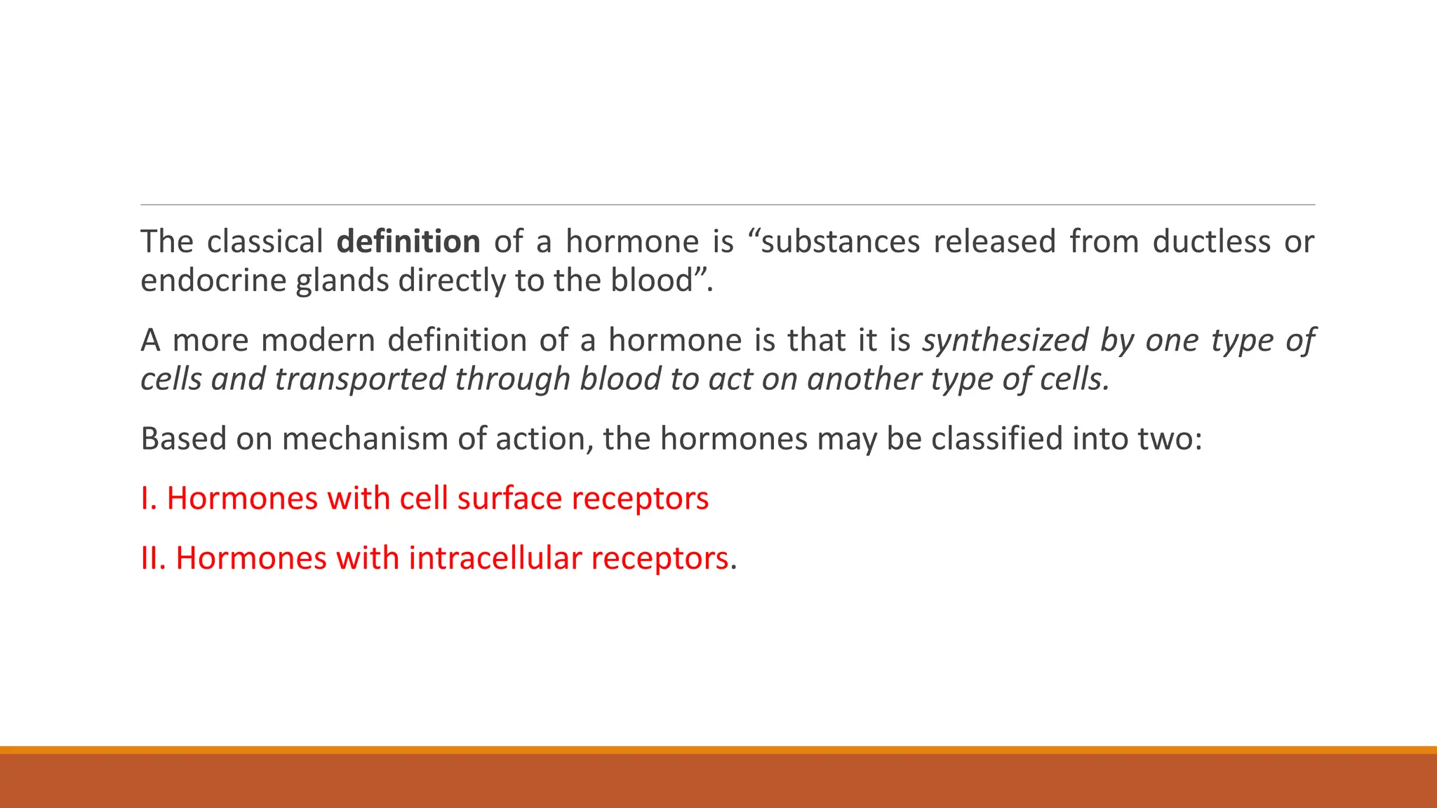
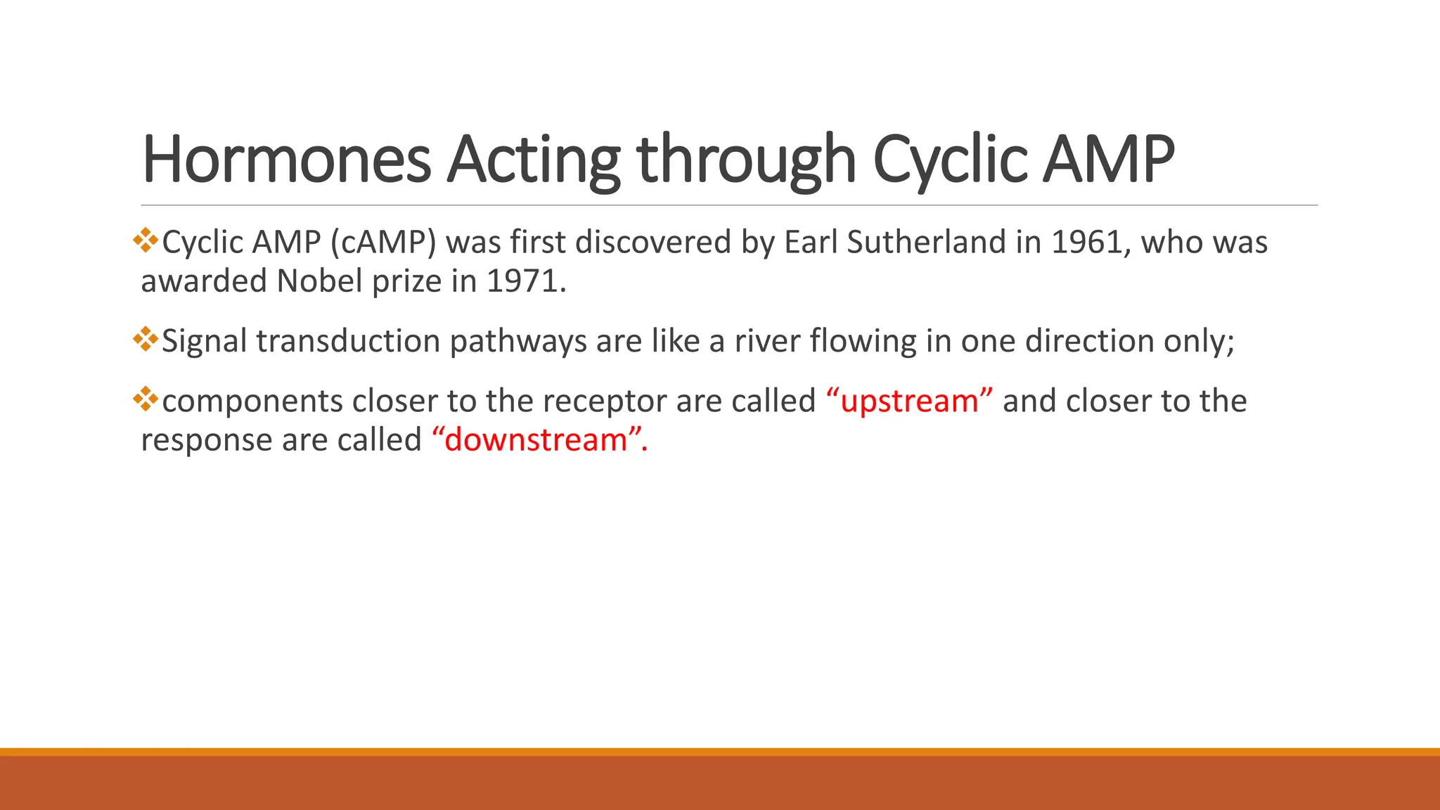
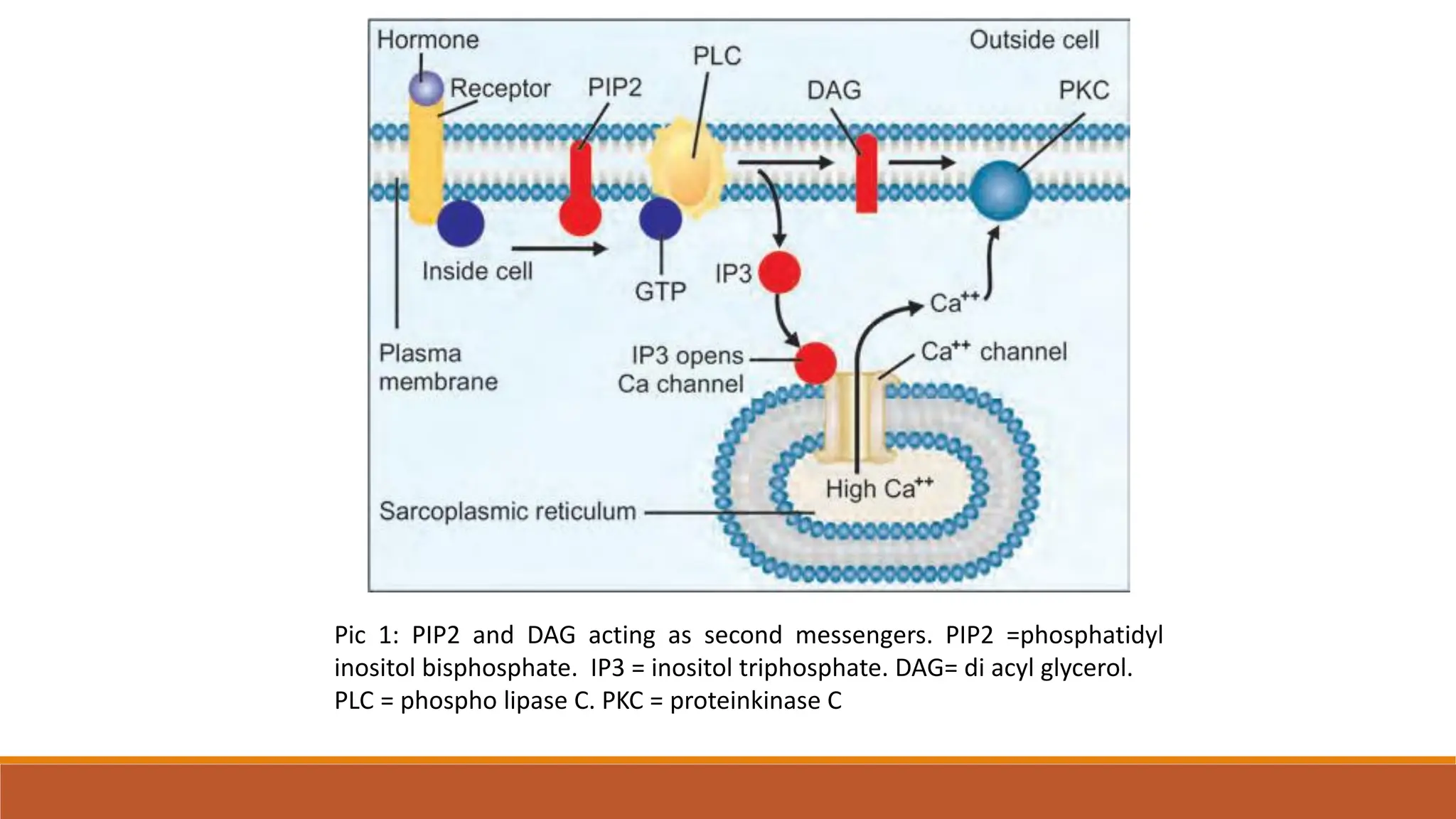
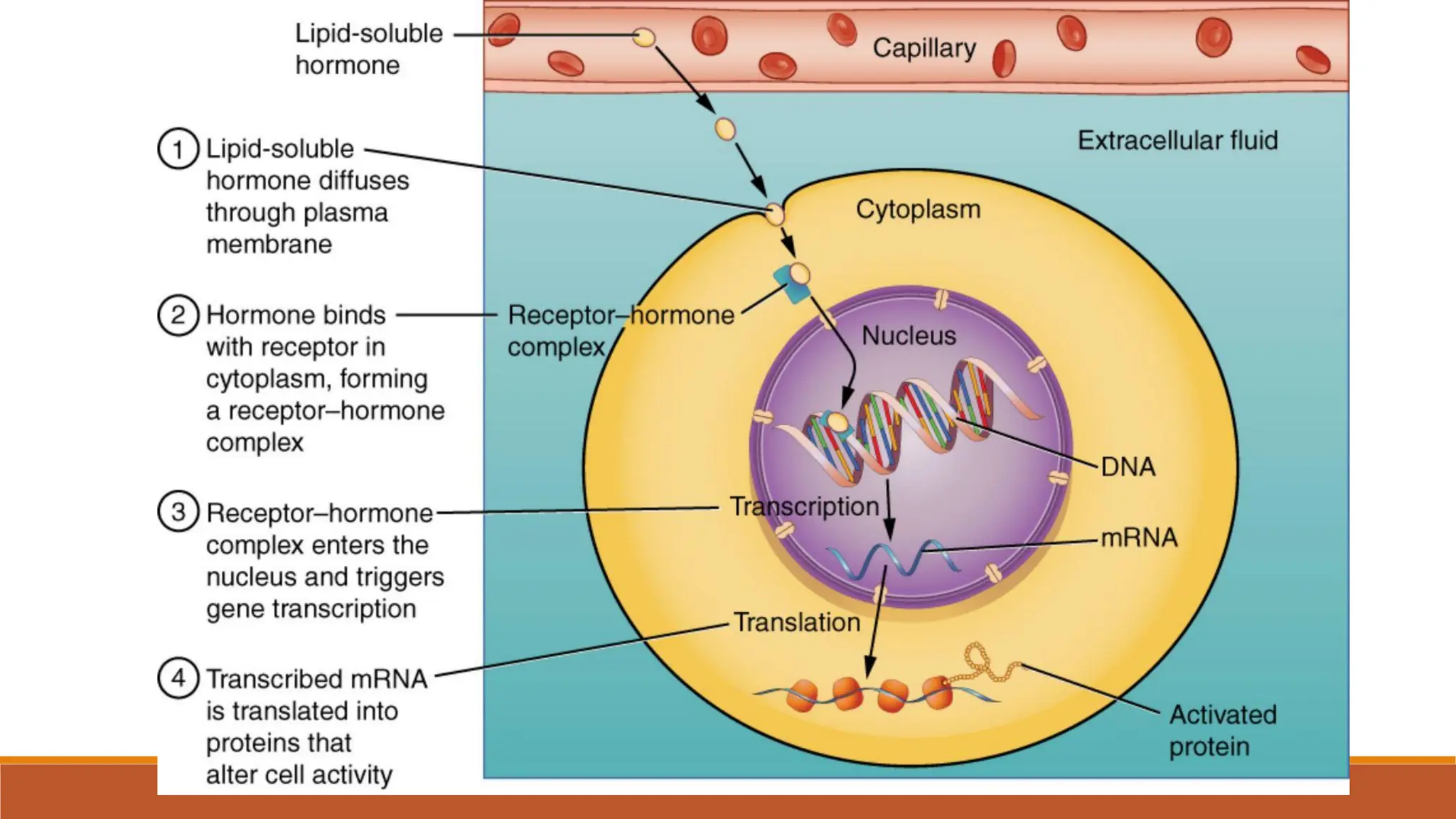
3. The classical definition of a hormone is “substances released from ductless or
endocrine glands directly to the blood”.
A more modern definition of a hormone is that it is synthesized by one type of
cells and transported through blood to act on another type of cells.
Based on mechanism of action, the hormones may be classified into two:
I. Hormones with cell surface receptors
II. Hormones with intracellular receptors.
4.
5. Mecahnism of action Examples
Hormones bind with cell
surface receptors with cAMP
as secondary messenger
ADH,FSH,LH,TSH,MSH,Glucago
n
Hormones bind with cell
surface receptors with cGMP
as secondary messenger
ANF (atrial natriuretic factor)
Hormones having cell surface
receptors, second messenger is
calcium or PIP2
TRH,CCK,Gastrin,Vasopresin,
Oxytocin
6. Hormones Acting through Cyclic AMP
âť–Cyclic AMP (cAMP) was first discovered by Earl Sutherland in 1961, who was
awarded Nobel prize in 1971.
âť–Signal transduction pathways are like a river flowing in one direction only;
❖components closer to the receptor are called “upstream” and closer to the
response are called “downstream”.
7. Signal Transduction through G-Protein
âť–Action is through G-protein coupled receptors (GPCRs).
âť–Binding of different types of signal molecules to G-protein coupled receptors is a general
mechanism of signal transduction.
âť–The GPCRs are transmembrane proteins with 7 helical segments spanning the membrane.
âť–When any ligand binds, the GPCRs activate heterotrimeric GTP binding regulatory proteins (G-
proteins).
âť–The G-protein in turn will interact with effector proteins which may be enzymes or ion channel
proteins, which result in the desired effect.
âť–Different types of G-proteins are present in the cells that are coupled with different receptors
and activating different effector proteins.
8. The extracellular messenger, the hormone (H) combines with the specific receptor (R) on the
plasma membrane
The H-R complex activates the regulatory component of the protein designated as G-protein or
nucleotide regulatory protein.
G-proteins are so named, because they can bind GTP and GDP. GDP-GTP exchange is mediated
by the GEF (Guanine nucleotide exchange factor).
The G- protein is a trimeric membrane protein consisting of alpha, beta and gamma subunits.
9.
10. G-protein Activates Adenyl Cyclase
âť–When the hormone receptor complex is formed, the activated
receptor stimulates the G-protein, which carries the excitation signal
to adenylate cyclase .
âť–The hormone is not passed through the membrane; but only the
signal is passed; hence this mechanism is called signal transduction.
The adenyl cyclase is embedded in the plasma membrane
11.
12. Inactivation of G- proteins
âť–The active G-alpha-GTP is immediately inactivated by GTPase. The G-alpha-
GDP form is inactive
âť–The activation is switched off when the GTP is hydrolyzed to GDP by the
GTPase activity of the alpha subunit .
âť–This is a built-in mechanism for deactivation.
13.
14. Cyclic AMP
❖Adenyl cyclase or adenylate cyclase converts ATP to cAMP (3’,5’-cyclic AMP), and
phosphodiesterase hydrolyzes cAMP to 5’ AMP.
âť–Cyclic AMP is a second messenger produced in the cell in response to activation of
adenylate cyclase by active G-protein. During hormonal stimulation, cyclic AMP
level in the cell increases several times.
âť–The level of cyclic AMP in the cell is regulated by its rate of production by
adenylate cyclase (AC) and hydrolyzis by phosphodiesterase (PDE).
âť–Therefore, cellular level of cyclic AMP can be increased by inhibition of PDE. For
example insulin activates PDE, decreasing the cellular level of cAMP while caffeine
and theophylline inhibit PDEs increasing cAMP levels.
15.
16. Second Messenger Activates PKA
âť–The cAMP (second messenger), in turn, activates the enzyme, PKA (Cyclic AMP
dependent protein kinase).
âť–Cyclic AMP binds to the regulatory subunits of PKA so that the catalytic
subunits having kinase activity can phosphorylate proteins.
âť–The cascade amplification effect is seen in this series of activation reactions.
This PKA is a tetrameric molecule having two regulatory (R) and two catalytic (C)
subunits (R2C2).
âť–This complex has no activity. But cAMP binds to the regulatory subunit and
dissociates the tetramer into regulatory and catalytic subunits . The catalytic
subunit is now free to act.
17.
18. Kinase Phosphorylates the Enzymes
âť–The catalytic subunit then transfers a phosphate group from ATP to different
enzyme proteins.
âť–Phosphorylation usually takes place on the OH groups of serine, threonine or
tyrosine residues of the substrates.
âť–Hence, these kinases are called Ser/Thr kinases. The enzymes may be activated
or inactivated by this phosphorylation. This is an example of covalent
modification.
âť–Glycogen phosphorylase and hormone sensitive lipase are controlled by cyclic
AMP.
19. There are Many G-proteins
About 30 different G-proteins are identified, each being used for different signal
transduction pathways. The G-protein, which stimulates adenyl cyclase, is called
Gs (G-stimulatory) and the opposite group is called Gi (G-inhibitory). An
example of inhibitory G-protein is the inhibition of adenylate kinase. The alpha
subunit of the Gs and Gi are different, but beta and gamma are the same.
20.
21. There are Many Protein Kinases
More than thousand protein kinases are now known. Some
important hormone responsive protein kinases are, cAMP-
dependent kinases, epidermal growth factor-dependent tyrosine
kinase, insulin-dependent tyrosine kinase. All the known effects of
cAMP in eukaryotic cells result from activation of protein kinases,
which are serine/threonine kinases.
22. Calcium-based Signal Transduction
âť–Calcium is an important intracellular regulator of cell function like
contraction of muscles, secretion of hormones and
neurotransmitters, cell division and regulation of gene regulation.
âť–Rapid but transient increase in cytosolic calcium result from either
opening of calcium channels in the plasma membrane or calcium
channels in the ER.
âť–The released calcium can be rapidly taken-up by ER to terminate
the response.
23. Even small increase in cytosolic free calcium can have maximal effect on calcium
regulated cellular functions. There are mainly 3 types of calcium transport systems:
a. Voltage gated calcium channels
b. Sodium/calcium antiport transporter
c. Calcium transporting ATPase.
The calcium transporting ATPase transporter accumulates calcium within the lumen
of ER (sarcoplasmic reticulum) in muscle.
These calcium ions can be released into the cytoplasm by an inositol triphosphate
(IP3) gated calcium channel or by a ligand gated calcium release channel (ryanodine
receptor).
24. âť–When cytosolic calcium increases, binding regulatory proteins, activation of
several calcium binding regulatory proteins occurs.
âť–Calmodulin is expressed in various tissues and mediates the regulatory actions
of calcium ions.
âť–Calcium binding causes conformational change in calmodulin resulting in
interaction with kinases, phosphatases, NOS, etc.
25. Hormones can increase the cytosolic calcium level
by the following mechanisms:
A. By altering the permeability of the membrane.
B. The action of Ca/H+-ATPase pump which extrudes calcium in exchange for H+.
C. By releasing the intracellular calcium stores.
D. Calmodulin, the calcium dependent regulatory protein within the cell has four calcium
binding sites. When calcium binds there is a conformational change to the calmodulin, which
has a role in regulating various kinases. Calmodulin is a 17 kDa protein which has structural and
functional similarity with the muscle protein troponin C.
26. . Examples of enzymes or functional proteins regulated by calmodulin are:
âť–Adenyl cyclase
âť–calcium-dependent protein kinases
âť–calcium-magnesium-ATPase
âť–cyclic nucleotide phosphodiesterase
âť–nitric oxide synthase
âť–phosphorylase kinase
27.
28. Hormones Acting through PIP2 Cascade
âť–The major player in this type of signal transduction is phospholipase C that
hydrolyses phosphatidyl inositol in membrane lipids to 1,4,5-Inositol
triphosphate (IP3) and Diacyl Glycerol (DAG) that act as second messengers.
âť–PIP3 (Phosphatidyl Inositol 3,4,5- phosphate) is another second messenger
produced by the action of a phosphoinositide kinase.
âť–The phospholipase C may be activated either by G-proteins or calcium ions.
âť–DAG can also be generated by the action of phospholipase D that produces
phosphatidic acid which is hydrolyzed to DAG.
29. âť–The binding of hormones like serotonin to cell surface receptor
triggers the activation of the enzyme phospholipase-C which
hydrolyzes the phosphatidyl inositol to diacylglycerol.
âť–IP3 can release Ca++ from intracellular stores, such as from
endoplasmic reticulum and from sarcoplasmic reticulum.
âť–The elevated intracellular calcium then triggers processes like
smooth muscle contraction, glycogen breakdown and exocytosis.
30.
31. âť–PIP3 can be formed by the action of PI3-kinases that are activated through
growth factors and cytokine mediated receptor tyrosine kinases.
âť–PIP3 which is a lipid second messenger has a role in regulation of cell motility,
membrane trafficking and cell survival signaling pathways.
âť–The major mediator of PIP3 action is PKB (Protein kinase B) which has a role in
glucose transport, glycogen metabolism and cell death signaling pathways.
âť–Active PKB/Akt is the major mediator of PIP3 action. It represses the activity of
cell death signaling pathways..
32. Pic 1: PIP2 and DAG acting as second messengers. PIP2 =phosphatidyl
inositol bisphosphate. IP3 = inositol triphosphate. DAG= di acyl glycerol.
PLC = phospho lipase C. PKC = proteinkinase C
33. Role of Cyclic GMP
âť–Cyclic GMP (cGMP) is another important second Messenger involved in
contractile function of smooth muscles, visual signal transduction and
maintenance of blood volume.
âť– Cyclic GMP degradation is catalyzed by membrane bound PDEs
âť–It is formed from GTP by the action of guanyl cyclase. Several compounds have
been found to increase the concentration of cGMP by activating guanyl cyclase.
âť–Cyclic GMP activates cGMP-dependent protein kinase G (PKG), which
phosphorylates important effector proteins that can regulate calcium dependent
contraction or motility by modulating calcium influx. An example is smooth
muscle myosin, leading to relaxation and vasodilatation
34.
35. NO and cGMP
âť–NO (Nitric oxide) is the major activator of guanylate cyclase. NO in turn is produced by the
action of NOS (Nitric oxide synthase) in tissues like vascular endothelial cells.
âť–NO can easily diffuse through the membrane and activate guanylate cyclase. Increased level of
cyclic GMP in smooth muscle triggers rapid and sustained relaxation of the smooth muscles.
âť–The vasodilatation resulting from NO induced increase in cGMP has great physiological and
pharmacological significance. The drugs that act via NO release are nitroprusside, nitrites (used
in angina as coronary vasodilators) and sildenaphil citrate (Viagra).
âť–Even though nitroglycerin was used to relieve angina for the last few decades, its role as an
exogenous NO donor has been described only recently.
36.
37. Hormones with Intracellular Receptors
The hormones in this group include the steroid hormones and thyroid
hormones. They diffuse through the plasma membrane and bind to the
receptors in the cytoplasm
The hormone receptor (HR) complex is formed in the cytoplasm. The complex is
then translocated to the nucleus. Steroid hormone receptor proteins have a
molecular weight of about 80–100 kD. Each monomer binds to a single steroid
molecule at a hydrophobic site, but on binding to genes they dimerize
38.
39. âť–In the nucleus, the HR binds to the hormone response elements (HRE) or steroid response
elements (SRE).
âť–The SRE acts as an enhancer element and when stimulated by the hormone, would increase
the transcriptional activity.
âť–The newly formed mRNA is translated to specific protein, which brings about the metabolic
effects.
âť–Binding to the SRE sequence leads to dimerization of the receptor.
âť–Steroid hormones influence gene expression, so that the rate of transcription is increased.
âť–The stability of mRNA is also increased.
âť–This would lead to induction of protein synthesis.